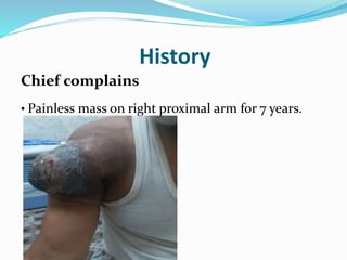
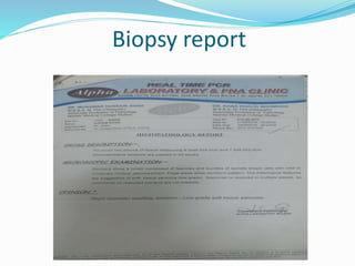
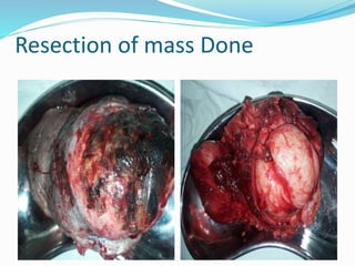
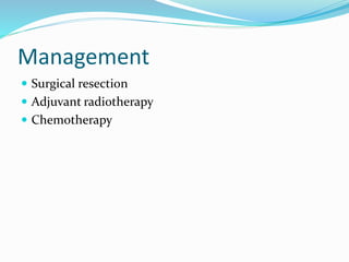
Dr Badal khan
PGR Ortho Unit III
BMCH Quetta
PATIENT PARTICULARS
Name. Karam khan
Age.60 years
Sex. Male
Occupation: Labourer
Address. Killa saifullah
Date of admission 1st July 2018
History
Chief complains
• Painless mass on right proximal arm for 7 years.
History of present illness
ļ‚—According to patient he was alright
until 7 years back then he noticed a
swelling on proximal arm, initially it
was small in size, gradually it increased
in size and became huge within 4
months.
ļ‚—There is no hx of trauma, fever, weight
loss and pain.
Systemic Inquiries
ļ‚— GPE, CVS, CNS, Respiratory, GIT & Urinary systems
are normal.
ļ‚— Systemic symptoms such as fever, weight loss, and
night sweat are absent.
Past medical & Surgical History
He has operated three times for same mass.
He has received 3 dose of chemotherapy and radiation in 2017.
Family History
•Married
•5 sons & 2 daughter
Personal History
ļ‚— Non smoker
ļ‚— Sleep Normal
ļ‚— Appetite normal
ļ‚— Bowel habits normal
Socioeconomic History
ļ‚— He belongs to a poor family.
ļ‚— There are 9 members of family and 3 rooms for them.
General Physical & Systemic
Examination
ļ‚— No anemia, pallor and weight loss.
Focused Examination
LOOK
ļ‚— Round shaped mass on anterolateral aspect of right
proximal arm , skin over the swelling is lost and
necrosed .
ļ‚— Margins of mass is red and there is no dilated vein
around .
ļ‚— Previous surgical scar is present.
ļ‚— No visible muscle wasting of arm, forearm & wrist.
Focused Examination…..
FEEL
ļ‚— Local skin temperature is warm as compared to normal
side.
ļ‚— Tenderness –ve
ļ‚— Distal pulses normal.
ļ‚— Sensations intact
Focused Examination…..
Movement
Shoulder Joint
ļ‚— All movements Normal
Elbow Joint
ļ‚— All movements Normal
Wrist Joint
ļ‚— All movements normal
Neurological Examination
ļ‚— Sensation Normal
ļ‚— Reflexes Normal
ļ‚— Motor Normal
Labs
Blood CP
ļ‚— Hb..13mg/dl
ESR
Normal
CRP
0.4 mg/dl (normal)
Radiographs
ļ‚— On x-ray there is soft
tissue opacity and no any
bony involvement.
MRI
BIOPSY
Histopathology show low grade soft tissue sarcoma.
Biopsy report
Diagnosis
Recurrent soft tissue sarcoma
Management Plan
ļ‚— Wide excision with reconstruction of defect.
Resection of mass Done
Soft tissue sarcoma
Introduction
Soft tissue sarcomas are malignant tumor that
originate in soft tissues of body.
Common sites.
ļ‚— Extremity 43 %
ļ‚— Visceral 19 %
ļ‚— Retroperitoneal 15 %
ļ‚— Trunk or thoracic 10 %
ļ‚— Others 13 %
Etiologies
ļ‚— Radiation exposure.
ļ‚— Chronic lymphedema.
ļ‚— Trauma.
ļ‚— Chemical exposure e.g. arsenic, polyvinyl chloride.
ļ‚— Infections such as herpes human virus.
Presentation
ļ‚— Mostly asymptomatic mass.
ļ‚— Pain later may occur due to destruction of surrounding
tissues.
Diagnosis
Labs.
ļ‚— CBC
ļ‚— ESR
ļ‚— CRP
ļ‚— To rule out infection
Imaging
MRI
ļ‚— For extremity mass.
ļ‚— Give good delineation between muscles, tumor and
blood vessels.
Pet scan
ļ‚— May help to determine high vs low grade
ļ‚— May be helpful in recurrence.
Ct abdomen and pelvis
ļ‚— To look for any metastasis
Biopsy
ļ‚— FNAC
ļ‚— Core needle biopsy
ļ‚— Incisional biopsy
ļ‚— Excisional biopsy
Metastatic workup
ļ‚— Evaluation for site of potential metastasis
ļ‚— Lymph nodes metastasis occur in less than 3 %
ļ‚— For extremity lesion lung is the principle site for
metastasis.
Staging
AJCC/UICC staging system for soft tissue sarcomas
ļ‚— T1: <5 cm
ļ‚— T1a: Superficial to muscular fascia
ļ‚— T1b: Deep to muscular fascia
ļ‚— T2: >5 cm
ļ‚— T2a: Superficial to muscular fascia
ļ‚— T2b: Deep to muscular fascia
ļ‚— N1: Regional lymph node involvement
Grading
ļ‚— G1: Well differentiated
ļ‚— G2: Moderately differentiated
ļ‚— G3: Poorly differentiated
ļ‚— G4: Undifferentiated
Prognostic factors
Increased risk of local recurrence
ļ‚— Age >50
ļ‚— Recurrent disease
ļ‚— Positive surgical margins
Increased risk of distant metastasis
ļ‚— Size >5cm
ļ‚— High grade
ļ‚— Deep location
ļ‚— Recurrent disease
Management
ļ‚— Surgical resection
ļ‚— Adjuvant radiotherapy
ļ‚— Chemotherapy
Surgical resection
ļ‚— Intralesional resection
ļ‚— Marginal resection
ļ‚— Wide resection
ļ‚— Radical resection
Adjuvant radiotherapy
ļ‚— Small low grade tumor <5 cm resected with 2 cm
margin may not require radiation
ļ‚— Adjuvant radiotherapy should be added to surgical
resection .
1. if excission margin is close
2. if extra muscular involvement is present
3. if local recurrence would result in sacrifice of major
neurovascular bundle or amputation.
It improves local recurrence but not survival
Radiotherapy
ļ‚— Can be given as brachytheraphy or intraoperative
radiotherapy
ļ‚— Brachytheraphy for high grade lesion
ļ‚— External beam radiation therapy for large >5cm high or
low grade leission
ļ‚— Intraoperative radiotherapy can be given in case of
retroperitoneal sarcoma
ļ‚— Can be given as preoperative and post operative
Chemotherapy
ļ‚— Can improve local control but not survival
ļ‚— Doxorubicin and Ifosfamide have response rate of 20%
ļ‚— Used only in advance disease
ļ‚— Combination with radiation or neoadjuvant therapy
are controversial
Metastatic disease
ļ‚— Lung is most common site of mets.
ļ‚— Median survival after metastatic disease is 8 to 12
months
ļ‚— Resection of pulmonary mets can give 5 year survival
of 32 % if all mets can be removed
ļ‚— >3 mets poor prognosis
Soft tissue sarcoma

Soft tissue sarcoma