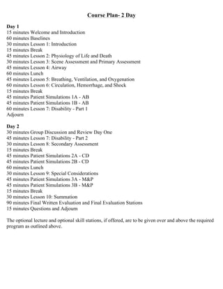
The document provides information on a 2-day PHTLS course, including the course plan, lesson topics, and assessment sequence. It also includes review materials and a pre-test for participants. The goal is to review trauma guidelines and management criteria prior to the course.
1) Pulmonary regurgitation is a common consequence of tetralogy of Fallot repair and can lead to right ventricular dilation and dysfunction over time.
2) Echocardiography and cardiac MRI are useful for evaluating the severity of pulmonary regurgitation and assessing the degree of right ventricular dilation and dysfunction.
3) Indications for pulmonary valve replacement include moderate or severe pulmonary regurgitation with signs of right heart failure or dilation out of proportion to age. It aims to prevent irreversible right ventricular damage.
This document provides an overview of adult bradycardia, including its definition, algorithms for assessing stable vs unstable bradycardia, recommended drugs and their dosages, and how to perform transcutaneous pacing. It defines bradycardia as a heart rate below 60 bpm, outlines an approach of ABCs, monitoring, IV access and 12-lead ECG, and recommends atropine as first-line treatment for unstable bradycardia while preparing for potential pacing. Transcutaneous pacing is described as a method to electrically stimulate the heart if bradycardia does not respond to drugs, with the goal of temporarily improving heart rate until more permanent pacing solutions can be established.
This document provides an overview of CT chest imaging, including the different types of CT chest scans, chest anatomy visualized on CT, and common abnormalities seen on CT chest exams. It discusses standard CT chest, HRCT, low dose CT, CT angiography, and combined PET/CT scans. It details the mediastinal compartments and lung segments seen on CT. It also provides examples of abnormalities such as pulmonary nodules/masses, pulmonary embolism, interstitial lung disease patterns, emphysema, atelectasis, pneumothorax, pleural effusions, and cardiomegaly. Virtual bronchoscopy and CT-guided biopsy procedures are also summarized.
This presentation is a simplified version of the various types of cardiac arrythmias seen in pediatric age groups. We have discussed supraventricular tachycarsias and prolonged QT syndrome in details here. Hope everyone finds it useful.
A 60-year-old male with multiple comorbid issues presented to the emergency room with chest pain. An ECG showed tall T waves and right bundle branch block. A CT/PET stress test revealed a small inferolateral infarct and significant peri-infarct ischemia. The patient was referred to cardiology to rule out acute coronary syndrome.
This document provides an overview of nuclear cardiology techniques used in the assessment of coronary artery disease (CAD). It discusses the history and development of nuclear medicine imaging including planar imaging and SPECT/PET. Key aspects summarized are:
1. SPECT imaging involves injection of radiotracers like thallium-201 or technetium-99m which distribute to the myocardium proportional to blood flow. Gamma photons are detected and reconstructed to provide 3D images of radiotracer distribution.
2. PET imaging uses positron-emitting radiotracers like rubidium-82 or ammonia-13 for perfusion and fluorodeoxyglucose for metabolism. It provides higher resolution functional imaging
This document discusses wide complex tachycardia (WCT), which is ventricular in origin 80% of the time. In patients with structural heart disease, 95% of WCT is ventricular tachycardia (VT). VT can be life-threatening and cause sudden death or tachycardia-induced cardiomyopathy. The document describes types of VT based on morphology and duration, symptoms of VT, features that appear on ECGs during VT like abnormal wide QRS complexes and AV dissociation, and examples of patients presenting with potential VT.
This document discusses toxicology and ECG interpretation in poisoned patients. It provides an overview of the cardiac conduction system and how specific drugs can affect it. Drugs that block sodium channels can cause a wide QRS complex, while potassium channel blockers prolong the QT interval and increase risk of Torsades de Pointes. A systematic approach to ECG interpretation is outlined, examining rate, rhythms, intervals, and morphology. Management strategies are presented for various cardiotoxic drugs like beta blockers, calcium channel blockers, sodium channel blockers, and cardiac glycosides.
This document discusses the natural history of ventricular septal defects (VSDs). It covers the incidence, classification, factors influencing outcomes, and potential complications of VSDs over time, including:
1. Cardiac failure in large VSDs due to left-to-right shunting.
2. Spontaneous closure or diminution, which is more common in smaller defects and those under 10 years of age.
3. Complications such as right ventricular outflow tract obstruction, aortic valve prolapse, pulmonary vascular disease, infective endocarditis, and arrhythmias.
The classification, mechanisms of closure, and guidelines for antibiotic prophylaxis for infective endocard
This document provides guidance on the assessment and treatment of arrhythmias presenting in the emergency department. It outlines an approach of first determining hemodynamic stability, then distinguishing between narrow and wide complex tachycardias, and finally determining the specific arrhythmia and appropriate treatment. For unstable patients with any arrhythmia, synchronized direct current cardioversion is recommended. Further treatment is tailored based on whether the arrhythmia has narrow or wide QRS complexes and is regular or irregular.
Radiology in newborn collected by Dr. Saiful islam MDDr. Habibur Rahim
This document summarizes a presentation on radiology in newborns. It discusses:
1. Types of radiographic examinations performed in newborns including chest x-rays, abdominal x-rays, and contrast studies.
2. How to assess the quality of chest x-rays and what normal findings look like.
3. Common chest x-ray findings for conditions like respiratory distress syndrome, transient tachypnea of the newborn, and pneumonia.
4. Positioning of tubes and catheters visible on chest x-rays.
5. Common abdominal x-ray findings including those for intestinal obstruction and duodenal atresia.
This document presents the case of a 4-year-old boy with tetralogy of Fallot. The key details are:
- He has had cyanosis, breathlessness and fainting episodes since infancy.
- Examination found cyanosis, clubbing and a grade IV/VI systolic murmur. Echocardiogram showed severe pulmonary stenosis, VSD, and RV hypertrophy consistent with tetralogy of Fallot.
- If left untreated, tetralogy of Fallot can cause serious complications like strokes from anoxic spells or cerebral thrombosis. Surgical repair is usually recommended in childhood.
1. The key points of an exercise testing manual include that systolic blood pressure measurement is most important for safety and should be adjusted based on the patient's history, age-predicted heart rate targets should not be used, the Borg scale of perceived exertion is better than heart rate for evaluating effort, and protocols should be tailored to each patient.
2. Factors that indicate when to stop a symptom-limited exercise test include dyspnea, fatigue, chest pain, a drop in systolic blood pressure, and ECG changes such as ST segment changes or arrhythmias.
3. The Borg scale estimate of perceived exertion is the most appropriate indicator of a maximal effort during exercise testing.
Dr Chong Shu Ling - Paediatric head injuryRahul Goswami
The document discusses the management of a 7-year-old girl who presented to the emergency department after being hit by a taxi while crossing the road. On examination, she was crying and oriented but became agitated and drowsy. Her GCS score was assessed and she displayed abnormal flexion in response to pain. The document discusses various clinical decision rules for determining which pediatric patients with head injuries require CT imaging, including the CHALICE, PECARN, and CATCH rules. It considers the risks of radiation exposure from CT scans for children and how to balance these risks with clinical need.
This document discusses atrioventricular septal defects (AVSDs), including their embryogenesis, classification, clinical features, imaging, and management. It describes the spectrum of AVSDs from partial to complete. Partial AVSDs involve a primum atrial septal defect with a cleft in the mitral valve. Complete AVSDs have a large ventricular septal defect with a common atrioventricular valve. Imaging like echocardiography is important for evaluating the anatomy and determining appropriate treatment, which ranges from observation to surgical repair.
This document provides an overview of a chest x-ray report. It discusses technical aspects like patient positioning and film quality. It describes the major chest x-ray views including posterior-anterior, anterior-posterior, lateral, and decubitus. Key anatomical structures are identified like the lungs, heart, diaphragm and bones. Common abnormalities seen on chest x-rays are outlined such as opacities, nodules, cavities and effusions. Examples of normal and abnormal x-rays are shown illustrating conditions like pneumonia, collapse, mass and pneumothorax.
Tetralogy of Fallot postoperative complications & management document discusses:
1. Common complications after tetralogy of Fallot repair include pulmonary regurgitation, residual RVOT obstruction, and arrhythmias.
2. Evaluation of postoperative patients includes annual echocardiograms and cardiac MRI to assess the severity of complications.
3. Interventions like pulmonary valve replacement may be needed to treat severe pulmonary regurgitation and RV dysfunction.
This document provides guidelines for pediatric advanced life support (PALS). It outlines the systematic approach algorithm which begins with checking responsiveness, calling for help, and checking for a pulse. The BLS assessment evaluates consciousness, breathing, and skin color to determine if the child is unresponsive with no breathing. For infants and children under 8, CPR should be provided first before calling for help, while those over 8 receive phone assistance first before CPR. The guidelines describe performing CPR, providing oxygen, inserting airways, monitoring the child, establishing IV/IO access, administering adrenaline, and considering reversible causes and an advanced airway. The primary and secondary assessment evaluates the child's airway, breathing, circulation, and
The majority of right ventricular myocardial infarctions (RVMI) result from occlusion of the proximal right coronary artery. RVMI commonly accompanies acute infarction of the inferior wall of the left ventricle, occurring in more than one third of cases. Poor outcomes from RVMI can include hemodynamic and electrical complications, but long-term prognosis is generally good for those who survive the initial events. Treatment of RVMI focuses on optimization of preload, heart rate, AV synchronization, and afterload as needed through careful use of fluids, pacing, inotropes, and vasopressors. Coronary reperfusion through fibrinolysis or primary PCI is also important.
Paediatric Congenital Heart Defects Case PresentationSCGH ED CME
A 12 week old girl presented with increased work of breathing and poor feeding. On examination, she was tachycardic, hypoxic, and floppy with crackles and a murmur.
Initial investigations showed severe metabolic acidosis and hyperkalemia. Echocardiogram revealed congenital mitral regurgitation, severe mitral regurgitation, and multiorgan failure.
She was diagnosed with congenital mitral regurgitation and shock from cardiac decompensation, precipitated by rhinovirus infection. She required intensive care management including ventilation, fluid resuscitation, and inotropic support.
Ventricular septal rupture (VSR) is a devastating complication following myocardial infarction. It occurs in 0.17-0.31% of cases post-primary PCI, usually 2-8 days post-MI. Risk factors include older age, female sex, late presentation, extensive MI, hypertension, and lytic therapy. Diagnosis involves acute deterioration, auscultation findings, echocardiogram, and CT scan. Management uses a hybrid approach of primary PCI of the culprit vessel followed by transient mechanical support like IABP, then definitive surgical or percutaneous repair. Without treatment, mortality is over 90% at 1 year. Challenges include friable tissue that cannot hold sutures early on and
This document discusses the evolution and advances in coronary CT angiography (CCTA) technology and its role in the assessment of coronary artery disease (CAD). Key points include:
- CCTA has advanced from early CT scanners with 4-minute scan times to modern multi-detector scanners that can image the entire heart in a single heartbeat.
- CCTA provides information on coronary artery anatomy, plaque characteristics, and has prognostic value when assessing coronary artery calcium scoring.
- CCTA has good accuracy for detecting CAD compared to invasive coronary angiography, especially for ruling out disease, though its role in asymptomatic patients is still unclear.
- CCTA is useful for evaluating coronary anomalies, bypass grafts,
1) The document defines wide complex tachycardia as a rhythm with a QRS duration ≥120ms and heart rate >100 bpm.
2) The main causes listed are ventricular tachycardia (80% of cases) and supraventricular tachycardia with aberrancy.
3) Key features that can help differentiate the underlying rhythm include QRS duration, axis, morphology, and the presence or absence of AV dissociation on electrocardiogram.
This document outlines the general approach and concepts for treating traumatic patients according to Advanced Trauma Life Support (ATLS) guidelines. It describes treating the greatest threats to life first using the ABCDE approach to assess the airway, breathing, circulation, disability, and exposure. The primary survey involves rapid assessment and interventions to stabilize the patient, while the secondary survey entails a full physical exam and diagnostic testing. Key interventions discussed include intubation, chest tube insertion, hemorrhage control, and use of the Focused Assessment with Sonography for Trauma (FAST) exam to evaluate for internal bleeding. Definitive care may involve transfer to the operating room or intensive care unit based on specialty consultations.
- Baby V is an 11-year-old female with tricuspid atresia, restrictive VSD, and PS who underwent a right-sided BDG at 10 months of age for cyanosis. She now presents with exertional dyspnea and increasing cyanosis.
- Examination found cyanosis, clubbing, and oxygen saturation of 50%. Echo showed patent glenn shunt and small restrictive VSD with moderate ASD, no forward flow across PA, and good ventricular function.
- Cardiac cath was performed to assess PA pressures, collaterals, and suitability for Fontan. It found elevated PA pressures but improved Qp/Qs ratio, making her a candidate for Font
The document discusses shock, including the types (hypovolemic, distributive, cardiogenic, obstructive), stages, pathophysiology, and case study of a 72-year-old male found lethargic in bed who is assessed using the AMLS pathway and diagnosed with sepsis from an untreated foot infection.
This document discusses the classification, causes, symptoms, and treatment of bradycardia. It defines different types of bradycardia based on rhythm and heart block. Common causes include medications, cardiac disease, metabolic abnormalities, and neurological or infectious etiologies. Symptoms range from dizziness to hypotension and shock. Treatment follows ACLS algorithms and may include atropine, transcutaneous pacing, or addressing underlying causes. Case examples demonstrate ECG findings and management of hyperkalemia-induced complete heart block, athlete's heart, and inferior STEMI with complete heart block.
A 6-year-old child was injured while sledding using a car hood pulled by an ATV. The child collided with a tree. Upon arrival, responders found the child with a positive level of consciousness but increased work of breathing. The remote winter location and snowing conditions complicated the response. The child was packaged for transport back to the vehicles, as the terrain prevented vehicle access to the scene.
The document provides guidance on assessing and managing patients at an emergency scene. It outlines establishing scene safety and patient priorities, performing a primary survey to assess airway, breathing, circulation, disability and environment, identifying critical patients needing rapid transport, and ongoing reassessment during transport. It emphasizes treating critical injuries, rapidly packaging and transporting critical patients to the closest appropriate facility like a trauma center.
The document provides guidance on evaluating and treating polytrauma patients. It outlines the goals of trauma resuscitation which include identifying life-threatening injuries. The Advanced Trauma Life Support (ATLS) approach is recommended as a safe standardized method, beginning with the primary survey of ABCDE (airway, breathing, circulation, disability, exposure). Key assessments include mechanism of injury, vital signs, neurological status, and bleeding control. A thorough secondary survey then involves a full head-to-toe examination. Guidance is given on managing specific injuries such as abdominal trauma, with operative intervention prioritized for unstable patients or those with signs of internal bleeding.
WFPS Spring 2014 Medical ConEd Field SessionpnairnWFPS
The document provides information on a continuing medical education session for paramedics, including objectives, demonstrations of new equipment, and case reviews. It discusses new intravenous catheters and solution sets being used, and provides instructions on preparing and accessing them. Two medical cases are then reviewed involving a woman found unresponsive at home and a man who fell from a third floor window. For each case, the document outlines the assessment approach, initial findings, further history, detailed exam, treatment, and questions from participants.
“Trauma” = Injury of one or more systems,that results in excessive bleeding and mayaffect the normal body functioning.
Defined as cellular disruption caused by anexchange with environmental energy that isbeyond the body's resilience.
This document outlines the key components of preoperative assessment for anaesthesia, including:
1. Taking a thorough patient history to identify any medical conditions or risks that could impact anaesthesia or surgery.
2. Performing a physical exam, including a focused assessment of the airway.
3. Determining the urgency of the planned surgery and communicating with the surgical team.
4. Ordering relevant preoperative investigations or tests based on the patient's age, health status, and type of surgery. The goals are to minimize risk and optimize preparation for anaesthesia and surgery.
Multiple trauma and it’s definition , classificationShehinSalim3
This document discusses multiple trauma and its management according to ATLS (Advanced Trauma Life Support) guidelines. It defines multiple trauma as injuries to two or more body systems that endanger vital signs. The mechanisms of injury include penetrating, blunt, blast, thermal and chemical injuries. It describes the trimodal distribution of death in trauma patients and the goals of prehospital retrieval and management. The core steps of ATLS including primary survey, resuscitation, secondary survey and definitive care are outlined. Key elements like airway management, breathing, circulation, disability and exposure are explained in detail.
Polytrauma and multiple traumata are medical terms describing the condition of a person who has been subjected to multiple traumatic injuries. This will be more prevalent in our country
Trauma results from the release of energy that causes injury and damages body systems. Globally, over 50 million people are disabled or injured each year from trauma. In India, vehicular accidents account for a large number of trauma cases, with a reported accident every 3 minutes resulting in death. The assessment and management of trauma patients follows the ABCDE approach - Airway, Breathing, Circulation, Disability, and Exposure. The primary survey focuses on stabilization and identifying life-threatening injuries, while the secondary survey provides a full examination and workup. Definitive treatment is based on the specific injuries identified.
Structured Approach to Critically Ill and Injured Patientmetriccertain
CERTAIN (Checklist for Early Recognition and Treatment of Acute Illness and iNjury) is designed and developed to standardize the approach to the evaluation and treatment of acutely decompensating patients. The design and content was informed by the survey of clinicians from diverse international settings. Available in electronic (laptop/mobile) and paper formats, CERTAIN provides evidence based diagnostic checklists, clinical decision support, educational modules on performing critical procedures, and has the ability to time and document real-time interventions. CERTAIN prompting has been shown to improve performance of clinical providers faced with simulated emergencies.
The document discusses Advance Trauma Life Support (ATLS). It describes ATLS as a general guideline for managing trauma patients that focuses on urgent problems during the critical "Golden Hour" period. The key components of ATLS include triage, primary survey and resuscitation (addressing ABCDE - Airway, Breathing, Circulation, Disability, Exposure), secondary survey to identify all injuries, and definitive care involving specialist treatment of injuries identified. The goal of ATLS is to prioritize assessment and rapid intervention for life-threatening injuries during the initial stages after trauma occurs.
The document summarizes the key changes to first aid guidelines published jointly by the American Red Cross and American Heart Association in 2005 based on a review of scientific evidence and expert consensus. Some of the major changes included recommendations around controlling external bleeding, treating wounds, burns, and frostbite. The guidelines aimed to provide medically sound, evidence-based assessments and interventions for first aid while avoiding delays in activating emergency medical services when needed. The development process involved an international collaborative effort to achieve scientific consensus on first aid practices.
This orientation provides information on medical emergency management (MEM) for healthcare providers. It outlines common medical emergencies like hemorrhage, anaphylaxis, sepsis, seizures, fainting, hypoglycemia, and cardiac issues. It teaches the ABCDE approach for assessing and treating patients, including airway management, breathing support, circulation support, disability assessment, exposure and environmental checks. It details basic life support skills like chest compressions and providing rescue breaths. The orientation emphasizes the importance of having emergency equipment and medicines accessible, trained staff, and arrangements for emergency transport and referral. It presents MEM as a team effort requiring everyone to understand their roles.
Initial management of polytrauma patients requires a systematic approach with airway, breathing, and circulation as top priorities. The primary survey assesses these areas to identify life-threatening injuries, while the secondary survey provides a full head-to-toe examination to identify all injuries and guide further treatment. Trauma mortality follows a trimodal distribution with immediate deaths from major vascular or brain injuries within an hour, early deaths from hemorrhage or respiratory failure within hours, and late deaths after 3 days often from sepsis or organ failure.
3. initial assessment and triage in er pptGirish Kumar
The document discusses the initial assessment and triage of pediatric patients in the emergency room. It outlines the goals of a triage system to rapidly assess patients and prioritize care based on acuity and severity of illness. The pediatric triage assessment involves a rapid 3-5 minute evaluation using the Pediatric Assessment Triangle (PAT) and ABCDE approach to primary assessment. The PAT evaluates appearance, breathing, and circulation within 30-40 seconds to identify life-threatening issues. Patients are then classified into 5 levels of triage acuity from resuscitation to non-urgent to prioritize treatment.
1) Trauma deaths follow a trimodal distribution, with peaks occurring within seconds-minutes of injury due to severe brain or spinal cord injury, within minutes-hours due to hemorrhage, and days-weeks later due to sepsis or multiple organ failure.
2) The initial assessment and management of polytrauma patients follows the ABCDE approach, with simultaneous attention to the airway, breathing, circulation, disability, and exposure while preparing for further care and monitoring.
3) Secondary surveys involve a full physical exam, history taking, and diagnostic tests to identify all injuries and guide definitive care, which may involve patient transfer to a higher level trauma center if needed.
1. Initial assessment and management of the trauma patient.pptxWalterBenites2
La primera etapa del curso ATLS (Advanced Trauma Life Support) se conoce como "Evaluación Inicial". En esta etapa, los estudiantes de medicina aprenden un enfoque sistemático y estructurado para evaluar a un paciente traumatizado de manera rápida y eficaz.
La Evaluación Inicial se centra en identificar y abordar de inmediato las lesiones que amenazan la vida del paciente.
- The document provides guidance on initial assessment and management of trauma patients, emphasizing the importance of quickly identifying and correcting life threats during the primary survey.
- The primary survey focuses on the ABCs - Airway, Breathing, Circulation. Oxygen should be given immediately if needed and breathing/ventilation issues addressed. Serious bleeding must be controlled.
- Only after life threats are stabilized should a more detailed exam and history be performed, and the patient transported without delay to definitive care. Rapid assessment and treatment is critical for trauma patients.
These lecture slides, by Dr Sidra Arshad, offer a simplified look into the mechanisms involved in the regulation of respiration:
Learning objectives:
1. Describe the organisation of respiratory center
2. Describe the nervous control of inspiration and respiratory rhythm
3. Describe the functions of the dorsal and respiratory groups of neurons
4. Describe the influences of the Pneumotaxic and Apneustic centers
5. Explain the role of Hering-Breur inflation reflex in regulation of inspiration
6. Explain the role of central chemoreceptors in regulation of respiration
7. Explain the role of peripheral chemoreceptors in regulation of respiration
8. Explain the regulation of respiration during exercise
9. Integrate the respiratory regulatory mechanisms
10. Describe the Cheyne-Stokes breathing
Study Resources:
1. Chapter 42, Guyton and Hall Textbook of Medical Physiology, 14th edition
2. Chapter 36, Ganong’s Review of Medical Physiology, 26th edition
3. Chapter 13, Human Physiology by Lauralee Sherwood, 9th edition
Integrating Ayurveda into Parkinson’s Management: A Holistic ApproachAyurveda ForAll
Explore the benefits of combining Ayurveda with conventional Parkinson's treatments. Learn how a holistic approach can manage symptoms, enhance well-being, and balance body energies. Discover the steps to safely integrate Ayurvedic practices into your Parkinson’s care plan, including expert guidance on diet, herbal remedies, and lifestyle modifications.
Clinic ^%[+27633867063*Abortion Pills For Sale In Tembisa Central19various
Clinic ^%[+27633867063*Abortion Pills For Sale In Tembisa Central Clinic ^%[+27633867063*Abortion Pills For Sale In Tembisa CentralClinic ^%[+27633867063*Abortion Pills For Sale In Tembisa CentralClinic ^%[+27633867063*Abortion Pills For Sale In Tembisa CentralClinic ^%[+27633867063*Abortion Pills For Sale In Tembisa Central
Osteoporosis - Definition , Evaluation and Management .pdfJim Jacob Roy
Osteoporosis is an increasing cause of morbidity among the elderly.
In this document , a brief outline of osteoporosis is given , including the risk factors of osteoporosis fractures , the indications for testing bone mineral density and the management of osteoporosis
Histololgy of Female Reproductive System.pptxAyeshaZaid1
Dive into an in-depth exploration of the histological structure of female reproductive system with this comprehensive lecture. Presented by Dr. Ayesha Irfan, Assistant Professor of Anatomy, this presentation covers the Gross anatomy and functional histology of the female reproductive organs. Ideal for students, educators, and anyone interested in medical science, this lecture provides clear explanations, detailed diagrams, and valuable insights into female reproductive system. Enhance your knowledge and understanding of this essential aspect of human biology.
These lecture slides, by Dr Sidra Arshad, offer a quick overview of the physiological basis of a normal electrocardiogram.
Learning objectives:
1. Define an electrocardiogram (ECG) and electrocardiography
2. Describe how dipoles generated by the heart produce the waveforms of the ECG
3. Describe the components of a normal electrocardiogram of a typical bipolar lead (limb II)
4. Differentiate between intervals and segments
5. Enlist some common indications for obtaining an ECG
6. Describe the flow of current around the heart during the cardiac cycle
7. Discuss the placement and polarity of the leads of electrocardiograph
8. Describe the normal electrocardiograms recorded from the limb leads and explain the physiological basis of the different records that are obtained
9. Define mean electrical vector (axis) of the heart and give the normal range
10. Define the mean QRS vector
11. Describe the axes of leads (hexagonal reference system)
12. Comprehend the vectorial analysis of the normal ECG
13. Determine the mean electrical axis of the ventricular QRS and appreciate the mean axis deviation
14. Explain the concepts of current of injury, J point, and their significance
Study Resources:
1. Chapter 11, Guyton and Hall Textbook of Medical Physiology, 14th edition
2. Chapter 9, Human Physiology - From Cells to Systems, Lauralee Sherwood, 9th edition
3. Chapter 29, Ganong’s Review of Medical Physiology, 26th edition
4. Electrocardiogram, StatPearls - https://www.ncbi.nlm.nih.gov/books/NBK549803/
5. ECG in Medical Practice by ABM Abdullah, 4th edition
6. Chapter 3, Cardiology Explained, https://www.ncbi.nlm.nih.gov/books/NBK2214/
7. ECG Basics, http://www.nataliescasebook.com/tag/e-c-g-basics
Does Over-Masturbation Contribute to Chronic Prostatitis.pptxwalterHu5
In some case, your chronic prostatitis may be related to over-masturbation. Generally, natural medicine Diuretic and Anti-inflammatory Pill can help mee get a cure.
REGULATION FOR COMBINATION PRODUCTS AND MEDICAL DEVICES.pptx
Phtls prep-packet-2-day
1. EMERGENCY MEDICAL CONSULTANTS INC.
Florida’s Premier Provider Of Quality Medical Training Programs
Nationally Accredited and OSHA Programs
CEU Provider
Since 1988
PHTLS
PREPARATION
PACKET
8th
Edition
Note: This Packet contains the latest Trauma guidelines, review information and pre-test. It is important
that participants review the textbook, complete the pre-test and be familiar with the PHTLS assessment
and management criteria prior to the course.
The pre-test will be collected at the beginning of the class.
Feel free to contact our office should you have any questions
(772) 878-3085 * Fax: (772) 878-7909 * Email: info@medicaltraining.cc
597 SE Port Saint Lucie Blvd * Port Saint Lucie, Florida 34984
Visit Our Website… EMCmedicaltraining.com
2.
3. Course Plan- 2 Day
Day 1
15 minutes Welcome and Introduction
60 minutes Baselines
30 minutes Lesson 1: Introduction
15 minutes Break
45 minutes Lesson 2: Physiology of Life and Death
30 minutes Lesson 3: Scene Assessment and Primary Assessment
45 minutes Lesson 4: Airway
60 minutes Lunch
45 minutes Lesson 5: Breathing, Ventilation, and Oxygenation
60 minutes Lesson 6: Circulation, Hemorrhage, and Shock
15 minutes Break
45 minutes Patient Simulations 1A - AB
45 minutes Patient Simulations 1B - AB
60 minutes Lesson 7: Disability - Part 1
Adjourn
Day 2
30 minutes Group Discussion and Review Day One
45 minutes Lesson 7: Disability - Part 2
30 minutes Lesson 8: Secondary Assessment
15 minutes Break
45 minutes Patient Simulations 2A - CD
45 minutes Patient Simulations 2B - CD
60 minutes Lunch
30 minutes Lesson 9: Special Considerations
45 minutes Patient Simulations 3A - M&P
45 minutes Patient Simulations 3B - M&P
15 minutes Break
30 minutes Lesson 10: Summation
90 minutes Final Written Evaluation and Final Evaluation Stations
15 minutes Questions and Adjourn
The optional lecture and optional skill stations, if offered, are to be given over and above the required
program as outlined above.
4. PHTLS Assessment Sequence
Assessment
Resources needed? YES Notify appropriate agencies
NO Proceed when safe
Standard Precautions
Airway
NO Patent?
YES
Breathing?
VR < 10 VR 12-20 VR >20
Assist ventilation Auscultate breath sounds1
Auscultate breath sounds Consider assisting
Ventilation ( Vt)
External hemorrhage? YES
NO
Circulation
Expose/ environment4
Life threats present?
YES NO
Spinal immobilization Assess vital signs
as indicated AMPLE history
Secondary survey
Consider PASG5
Reassess primary survey
Transport
Reassess primary survey YES Life threats?
IV fluid therapy6
NO
Trauma center Definitive field care7
If available
Spinal immobilization as indicated
Transport to appropriate facility
Assess Scene
• Safety
• Situation
Assess Patient
Secure airway as
needed
O2 to maintain
SpO2 >95%
Control as
appropriate2
Assess for shock3
Notes for Assessment Algorithm
1. Consider pleural decompression only if ALL are
present:
• Diminished or absent breath sounds
• Increased work of breathing or difficulty
ventilating with bag-valve-mask
• Decompensated shock/hypotension (SBP <90
mm Hg)
**Perform bilateral pleural decompression
only if patient is receiving positive
pressure ventilation
2. External hemorrhage control:
• Direct pressure/pressure dressing
• Tourniquet
*Consider topical hemostatic agent for prolonged
transport
3. Shock: tachycardia; cool, diaphoretic, pallorous
skin; anxiety; diminished or absent peripheral
pulses
4. Quick check for other life-threatening conditions;
cover patient to preserve body heat
5. PASG should be considered for suspected
unstable pelvic fracture with hypotension
6. Transport should not be delayed to initiate IV
fluid therapy. Initiate two large-bore IV lines:
uncontrolled bleed SBP 80-90, controlled bleed 1-
2 liters titrate SBP 80-90
7. Splint fractures and dress wounds as needed
5. PHTLS Shock
Classification of Hemorrhagic Shock
Class I Class II Class III Class IV
Blood loss (mL) Up to 750 750-1500 1500-2000 >2000
Blood loss (% vol) Up to 15% 15%-30% 30%-40% >40%
Pulse rate <100 100-120 120-140 >140
Blood pressure Normal Normal Decreased Decreased
Pulse pressure Normal or increased Decreased Decreased Decreased
Respiratory rate 14-20 20-30 30-40 >35
Urine output (mL/hr) >30 20-30 5-15 Negligible
CNS/ mental status Slightly anxious Mildly anxious Anxious, confused Confused, lethargic
Fluid replacement Crystalloid Crystalloid Crystalloid and blood Crystalloid and blood
Signs Associated with Types of Shock
Vital Sign Hypovolemic Neurogenic Septic Cardiogenic
Skin temperature Cool, clammy Warm, dry Cool, clammy Cool, clammy
Skin color Pale, cyanotic Pink Pale, mottled Pale, cyanotic
Blood pressure Drops Drops Drops Drops
Level of
consciousness
Altered Lucid Altered Altered
Capillary refilling
time
Slowed Normal Slowed Slowed
Management
-Ensure oxygenation and ventilation
-Control hemorrhage (external or internal)
-External- direct pressure or tourniquet or homeostatic agent
-Internal-direct pressure (extremity immobilization/ PASG for pelvis/ low abd.)* Consider Tranexamic Acid (TXA) for uncontrollable
bleeds.
-Move toward a definitive facility
-Control body temp (lower the pt)
-Fluid replacement for Class II, III, or IV shock
-Ideally blood or packed RBC’s (though not available prehospital)
-Isotonic crystalloids (Preferable no LR), replace at 3mL’s per mL’s blood loss (ideally warm)
-Controllable bleeds- 1-2 liters (adult) (20 mL/kg peds) – Titrated to SBP 80-90 mmHg
-Uncontrolled (internal) bleeds- the least amount of fluid required to maintain SBP 80-90 mmHg
Type of Fracture Blood Loss Potential
Rib 125 mL
Radius or ulna 250-500 mL
Humerus 500-750 mL
Tibia or fibula 500-1000 mL
Femur 1000-2000 mL
Pelvis 1000-unlimited mL
Shock Assessment
Vital Sign Compensated Decompensated
Pulse Increased; tachycardia Greatly increased;
marked tachycardia
that can progress to
bradycardia
Skin White, cool, moist White, cold waxy
Blood pressure range Normal Decreased
Level of consciousness Unaltered Altered, ranging from
disoriented to coma
Types of Shock There are three types of shock:
• Hypovolemic shock
• Vascular volume smaller than normal vascular size
• Loss of blood and fluid
• Hemorrhagic shock
• Loss of fluid and electrolytes
• Dehydration
• Distributive shock
• Vascular space is larger than normal
• Neurogenic “shock” (hypotension)
• Psychogenic shock
• Septic shock
• Anaphylactic shock
• Cardiogenic shock
• Pump failure
6. Spine Board Debate- Pg. 305
It is agreed that the long board is an appropriate device for extrication and patient movement on scene and to a
stretcher, but 2015 brought about documented controversy as to its effectiveness at truly immobilizing the spine
and its benefits; Key Issues:
- There are no documented studies to support that straight rigid board immobilization with a collar is
beneficial.
- Some patients’ anatomy actually flexes the head forward while others hyperextend the head when placed on
a board.
- Patients will all begin to complain of neck and back pain if left on a hard board.
- Skin breakdown can occur at points that contact the board.
- Obese patients are at risk for positional asphyxia
- Emergency airway procedures are more difficult to perform on immobilized patients
The lack of supporting benefit and the growing potential for detrimental side effects has led many areas to decrease
or completely remove the use of spine boards for anything more than extrication or movement. Instead opting for
placing a collar on the patient and lying the spine on the stretcher.
At the publishing of the 8th
Edition text (2016) this remains a controversial change and may be different by region.
7. PHTLS 8th
Ed. FINAL EVALUATION STATION FLOW SHEET
Student (Leader):____________________________________________________
Evaluator: _____________________________ Scenario Number: _____________
Beginning Time:________________________ Ending Time: _________________
Successful Station Completion: YES _______ NO ______
Completed
Yes No Assessment & Treatment
Identify Safe Scene
Proper Standard Precautions
Perform Primary Survey
Level of Consciousness/Response
Airway
Breathing
Ventilation/Air Exchange
Circulation/Perfusion
External Hemorrhage Control
Pulse
Skin Condition
Disability
Exposure of All Critical Body Areas for Assessment
Properly Identify Critical and Non-Critical Trauma Patients
Use of Appropriate Spinal Immobilization Technique(s)
Proper Use of Padding/Buttress Material
Identification of All Life-Threatening Injuries
Proper Treatment of All Life-Threatening Injuries
Performed Only Lifesaving Treatment(s) While On-Scene
Timely Transported When Indicated
Appropriate Level Trauma Facility When Indicated
Identification of All Non-Critical Injuries
Proper Treatment Performed En-Route
Completed Secondary Survey When Indicated
Completed Scenario Within 10 Minutes On-Scene Time
Reassessment of Patient's Conditions
Safety Observed Throughout Scenarios
Worked Together as A Team
Any mark(s) within the critical criteria area would indicate
the need for the group to repeat the station. Only mark those
comments that apply to the scenario. Please document
rationale for any checked critical criteria in the notation area.
Critical Criteria
_____Failure to utilize proper standard precaution techniques
_____Failure to identify safe scene
_____Failure to perform adequate/complete primary safety
_____Failure to identify all life-threatening
injuries/conditions
_____Failure to immediately treat life-threatening injuries/
conditions
_____Failure to identify critical patient based on assessment
_____Performed unnecessary treatment on-scene
_____Performed secondary survey before primary survey
_____Failure to assess and treat noncritical injuries
_____Failure to provide timely transport to an appropriate
level trauma facility
_____Failure to reassess the patient’s condition
_____Failure to perform scenario in a safe manner
_____Failure to perform in a team fashion
_____Failure to complete scenario within 10 minutes of on
scene time
NOTES: ______________________________
______________________________________
______________________________________
______________________________________
8. PHTLS 8th
EDITION PRETEST
Please respond to each question with the most correct answer from the given choices. There is only one answer
for each question.
1. You arrive at the scene of a motor vehicle collision in which a vehicle struck a tree. Which is the best indicator of
potential injury?
A) Circumference of the vehicle
B) Diameter of the tree
C) Mass of the vehicle
D) Speed of the vehicle
2. The potential for death or serious injury is greatest in which of the following motor vehicle collisions?
A) Down and under
B) Ejection from vehicle
C) Lateral compression
D) Up and over
3. Bilateral femur fractures are most often associated with which type of motorcycle crash?
A) Angular impact
B) Bike-road impact
C) Head-on impact
D) Rear impact
4. Which is the preferred fluid for resuscitation of hemorrhagic shock in the prehospital setting?
A) 5% dextrose in water
B) 7.5% hypertonic saline
C) Hetastarch
D) Lactated Ringer’s
5. Which is the most common cause of upper airway obstruction in the trauma patient?
A) Blood
B) Teeth
C) Tongue
D) Vomitus
6. Which is the preferred adjunct device for verifying placement of an endotracheal tube in a patient with a perfusing
rhythm?
A) End-tidal CO2
monitoring (capnography)
B) Esophageal detector device
C) Pulse oximeter
D) Stethoscope
7. Which is the most important reason to maintain an open airway in the trauma patient?
A) Prevents aspiration and pneumonia
B) Prevents hypoxemia and hypercarbia
C) Prevents snoring respirations
D) Prevents the tongue from blocking the pharynx
9. 8. Essential airway skills include manual clearing of the airway, manual maneuvers, suctioning and which of the
following?
A) Dual lumen airway
B) Endotracheal intubation
C) Laryngeal mask airway
D) Oropharyngeal airway
9. Your patient is a middle aged male who crashed his motorcycle. He is unresponsive. After opening the airway using
a modified jaw thrust, you note the patient has respirations at a rate of 6. Auscultation reveals breath sounds are absent
on the left side. Which of the following is the most appropriate next intervention?
A) Apply a non-rebreather mask
B) Begin ventilation with a BVM
C) Insert an endotracheal tube
D) Perform a needle decompression
10. Which best describes shock?
A) Decreased Glasgow Coma Scale (GCS)
B) Flushed, dry, hot skin combined with bradycardia
C) Generalized inadequate tissue perfusion
D) Low blood pressure combined with tachycardia
11. Your patient has a deep laceration to his antecubital fossa with significant bleeding. What is the most appropriate
initial action?
A) Apply a tourniquet
B) Apply direct pressure
C) Initiate rapid transport
D) Restore blood volume
12. Hypotension of unknown etiology in a trauma patient should be assumed to result from which of the following?
A) Blood loss
B) Cardiac tamponade
C) Spinal injury
D) Tension pneumothorax
13. Which assessment is most beneficial in differentiating hemorrhagic shock from neurogenic shock in the prehospital
setting?
A) Abdomen
B) Blood pressure
C) Neurologic status
D) Skin
14. The body initially compensates for blood loss through activation of which of the following?
A) Parasympathetic nervous system
B) Reticular activating system
C) Spinal reflex arcs
D) Sympathetic nervous system
15. Medication used by trauma patients for pre-existing conditions may cause which of the following?
A) Herbal preparations may enhance blood clotting
B) Anti-inflammatory agents may enhance blood clotting
C) Beta blockers may prevent tachycardia with blood loss
D) Calcium channel blockers may slow the onset of shock
10. 16. The target blood pressure for a trauma patient with suspected intraabdominal hemorrhage is which of the
following?
A) 60 – 70mm Hg
B) 80 – 90 mm Hg
C) 100 – 110 mm Hg
D) 120 – 130 mm Hg
17. Which best explains the mechanism by which gas exchange is impaired in pulmonary contusion?
A) Blood in the alveoli
B) Collapse of the alveoli
C) Compression of the lung tissue
D) Partial occlusion of the bronchi
18. Which of the following is a key finding that differentiates cardiac tamponade from tension pneumothorax?
A) Distended jugular veins
B) Equal breath sounds
C) Hypotension
D) Tachycardia
19. Your patient is a 20 year old male who struck his head on a teammate’s knee while diving to catch a football. He
was not wearing a helmet. He demonstrates decerebrate posturing and has a GCS score of 4. His heart rate is 58, blood
pressure 180/102 and his left pupil is dilated. What is the best ventilation rate to use when managing this patient?
A) 10 breaths per minute.
B) 20 breaths per minute.
C) 30 breaths per minute.
D) 35 breaths per minute.
20. A 20 year old female was ejected from her vehicle during a high speed roll-over motor vehicle collision. She has
significant bleeding from a large laceration. Your initial assessment reveals a GCS score of 7, systolic blood pressure of
70 mm Hg and pupils that are equal but respond sluggishly to light. After establishing two large bore IV lines, you
should titrate the infusion rate to achieve a target blood pressure of at least
A) 60 mm Hg.
B) 70 mm Hg.
C) 80 mm Hg.
D) 90 mm Hg.
21. Which of the following is the preferred prehospital wound management for a patient with a 36% body surface area
flame burn?
A) Cool moist dressings
B) Dry sterile dressings
C) Elastic bandages
D) Topical ointments
22. The most immediate life threatening condition resulting from injury to solid abdominal organs is which of the
following?
A) Acute respiratory failure
B) Hemorrhage.
C) Multiple organ failure.
D) Peritonitis
11. 23. An adult male sustained a deep laceration to his distal thigh. Bright red blood is spurting from the wound. Direct
pressure is not controlling the bleeding. What is the most appropriate next step?
A) Apply a topical hemostatic agent and transport
B) Apply a tourniquet and tighten it until bleeding stops
C) Elevate the leg and apply pressure to the femoral artery
D) Maintain direct pressure and transport immediately
24. An 18-year-old female was struck by a car and has sustained an apparent left femur fracture. Communication with
her is hampered because she only speaks a foreign language. Which finding, by itself, does not mandate immobilization
of the cervical spine?
A) Fracture of the femur
B) Inability to communicate
C) Mechanism of injury
D) Tenderness over the cervical spine
25. During the primary survey of a trauma patient, you note that the patient is agitated and confused, and has multiple
injuries from an altercation. Which of the following choices is the most appropriate first treatment priority?
A) Blood glucose determination
B) Correction of possible hypoxia
C) Full immobilization to a backboard
D) Obtain intravenous access