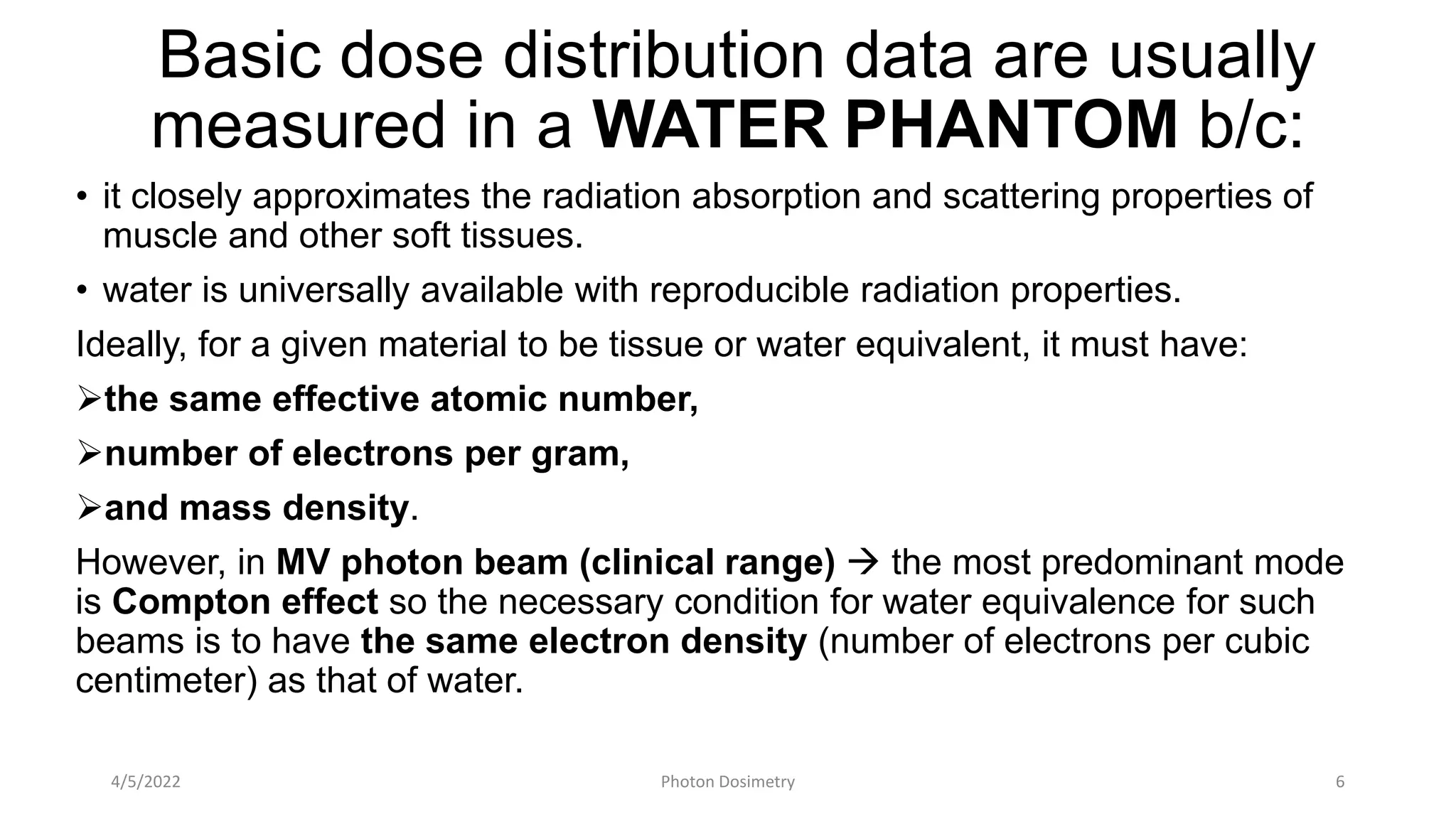
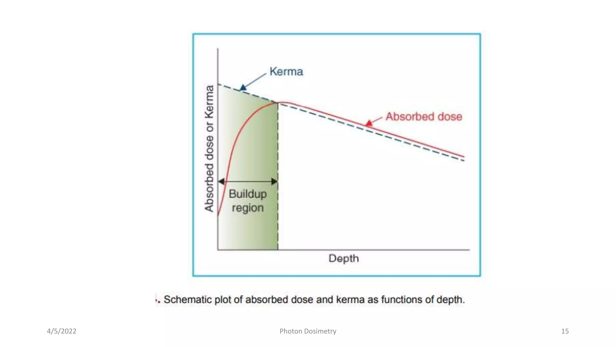
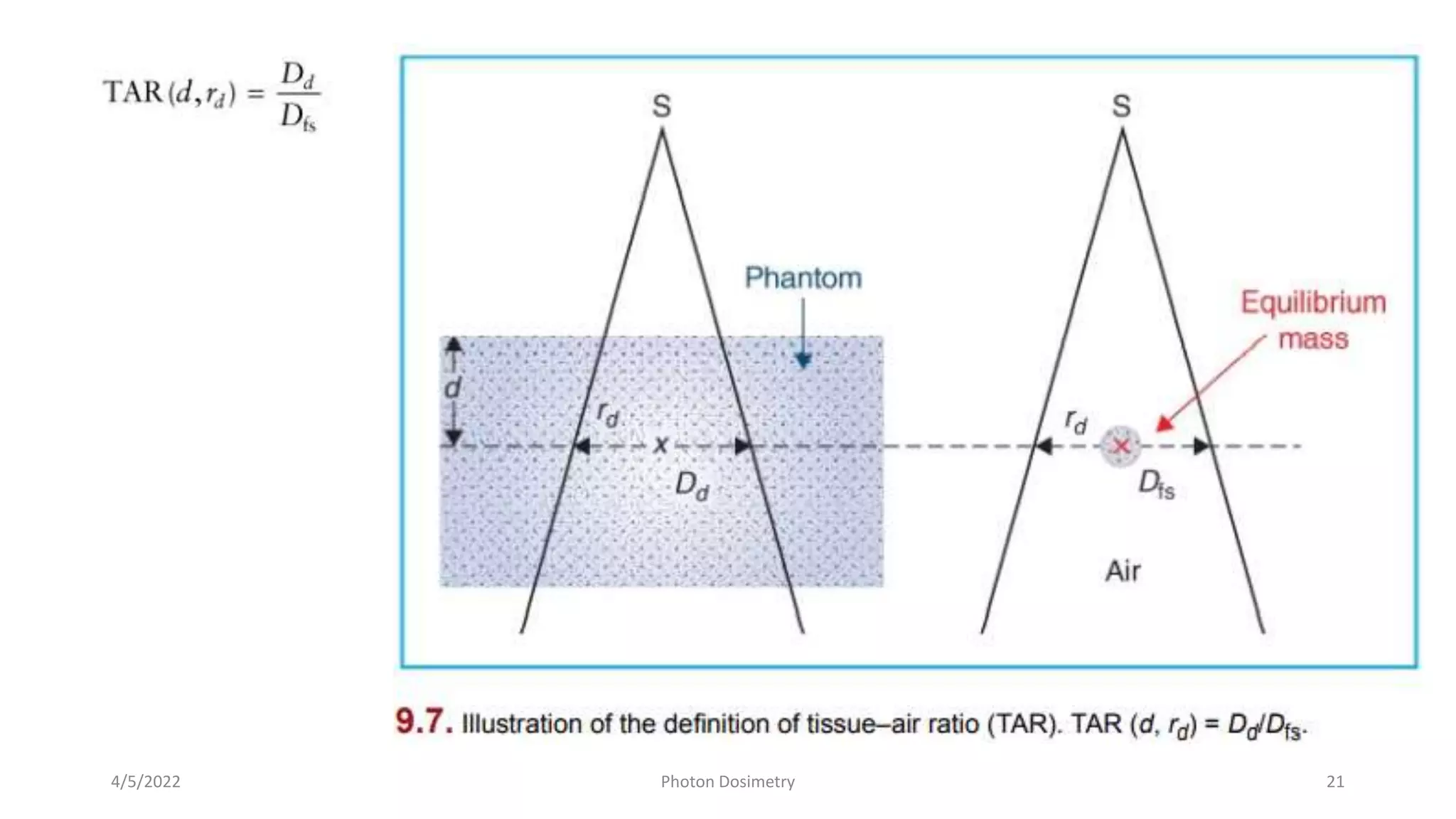
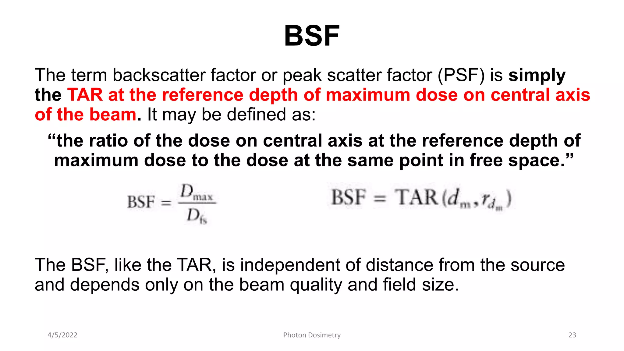
Dose Measurement of Photon
Beam
Dr. Saira Siddique
MSc. RMO fellow
4/5/2022 Photon Dosimetry 1
Presentation Layout
• Introduction
• Phantom
• Depth Dose Distribution
• PDD
• Definition
• It’s Dependence
• TAR & BSF
• TMR & TPR
4/5/2022 Photon Dosimetry 2
Introduction
It is seldom possible to measure dose distribution directly in patients
treated with radiation.
Data on dose distribution are almost entirely derived from measurements
in phantoms—tissue-equivalent materials, usually large enough in
volume to provide fullscatter conditions for the given beam.
These basic data are used in a dose calculation system devised to
predict dose distribution in an actual patient. Various dosimetric quantities
and methodologies have been devised to facilitate dose calculation in
clinical situations.
4/5/2022 Photon Dosimetry 3
The methods based on quantities such as percent depth
doses (PDDs), tissue–air ratios (TARs), and scatter–air
ratios (SARs) have been used traditionally for dose
calculation involving low energy beams (up to 60Co),
which were usually calibrated in terms of exposure rate in
air or dose rate in free space.
Current methods of dose calculation use tissue–phantom
ratios (TPRs) or tissue–maximum ratios (TMRs), which
are better suited for higher energy beams and involve
measurements in phantom rather than in air.
4/5/2022 Photon Dosimetry 4
Low energy – Exposure Rate/ Dose rate in FREE AIR  PDD, TAR, SAR
Higher energy – in phantom  TPR, TMR
TMT Various Phantom materials
4/5/2022 Photon Dosimetry 5
Basic dose distribution data are usually
measured in a WATER PHANTOM b/c:
• it closely approximates the radiation absorption and scattering properties of
muscle and other soft tissues.
• water is universally available with reproducible radiation properties.
Ideally, for a given material to be tissue or water equivalent, it must have:
the same effective atomic number,
number of electrons per gram,
and mass density.
However, in MV photon beam (clinical range)  the most predominant mode
is Compton effect so the necessary condition for water equivalence for such
beams is to have the same electron density (number of electrons per cubic
centimeter) as that of water.
4/5/2022 Photon Dosimetry 6
For clinical dosimetry 
anthropomorphic phantoms are
frequently used.
One such commercially available
system, known as Alderson Radiation
Therapy Phantom (formerly the
Alderson Rando Phantom),
it incorporates materials to simulate
various body tissues—muscle, bone,
lung, and air cavities.
It is shaped into a human torso and is
sectioned transversely into slices for
inserting film or other dosimeters
4/5/2022 Photon Dosimetry 7
DEPTH DOSE DISTRIBUTION
As the beam is incident on a patient (or a phantom), the absorbed dose in the
patient varies with depth.
This variation depends on many conditions: beam energy, depth, field size,
distance from source, and beam collimation system.
Thus, the calculation of dose in a patient involves considerations in regard to these
parameters and others as they affect depth dose distribution.
An essential step in the dose calculation system is to establish depth dose variation
along the central axis of the beam.
A number of quantities, are defined for this purpose, major among these being
PDDs, TARs, TPRs, and TMRs.
These quantities are usually derived from measurements made in water phantoms
using small ionization chambers. Although other dosimetry systems such as
thermoluminescent dosimeters (TLDs), diodes, and film are occasionally used, ion
chambers are preferred because of their better precision and smaller energy
dependence.
4/5/2022 Photon Dosimetry 8
PERCENTAGE DEPTH DOSE
One way of characterizing the central axis dose distribution is to
normalize dose at depth with respect to dose at a reference
depth.
The quantity percentage depth dose is defined as
“the quotient, expressed as a percentage, of the absorbed
dose at any depth d to the absorbed dose at a reference
depth d0 , along the central axis of the beam.”
• For orthovoltage (up to 400 kV) & lower-energy x-rays, the
reference depth is usually the surface (d0 = 0).
• For higher energies, the reference depth is usually taken at the
position of the peak absorbed dose (d0 = dmax)
4/5/2022 Photon Dosimetry 9
PDD
4/5/2022 Photon Dosimetry 10
PDD depends on:
• BEAM QUALITY AND DEPTH
• FIELD SIZE AND SHAPE
• SSD
4/5/2022 Photon Dosimetry 11
PDD _ Dependence on Beam QUALITY & DEPTH
PDD (beyond the depth of maximum dose) decreases with
depth and increases with beam energy.
Higher-energy beams have greater penetrating power and
thus deliver a higher PDD at a given depth
the beam quality affects the PDD by virtue of the average
attenuation coefficient . As it decreases, the beam becomes
more penetrating, resulting in a higher PDD at any given
depth beyond the buildup region.
4/5/2022 Photon Dosimetry 12
4/5/2022 Photon Dosimetry 13
Initial Dose Buildup
PDD decreases with depth beyond the depth of maximum dose. However, there
is an initial buildup of dose that becomes more and more pronounced as the
energy is increased. In the case of the orthovoltage or lower-energy x-rays, the
dose builds up to a maximum on or very close to the surface. But for higher-
energy beams, the point of maximum dose lies deeper into the tissue or
phantom.
“The region between the surface and the point of maximum dose is called
the dose buildup region.”
dose buildup effect (higher-energy)  the skin-sparing effect.
For MV beams (cobalt-60 & higher energies), the surface dose is much smaller
than the Dmax . This offers a distinct advantage over the lower-energy beams
for which the Dmax occurs at or very close to the skin surface.
Thus, in MV beams, higher doses can be delivered to deep-seated tumors
without exceeding the tolerance of the skin. This, of course, is possible because
of both the higher PDD at the tumor and the lower surface dose at the skin.
4/5/2022 Photon Dosimetry 14
4/5/2022 Photon Dosimetry 15
PDD _ EFFECT OF FIELD SIZE AND SHAPE
Field size may be specified either
geometrically
dosimetrically.
The geometric field size: “the projection, on a plane
perpendicular to the beam axis, of the distal end of the
collimator as seen from the front center of the source”
The dosimetric, or the physical, field size is the distance
intercepted by a given isodose curve (usually 50% isodose)
on a plane perpendicular to the beam axis at a stated
distance from the source.
4/5/2022 Photon Dosimetry 16
PDD _ EFFECT OF FIELD SIZE AND SHAPE
For a sufficiently small field, the depth dose at a point is primarily the result
of the primary radiation (the photons that have traversed the overlying
medium without interacting).
The contribution of the scattered photons to the depth dose in this case is
negligibly small and may be assumed to be 0.
But as the field size is increased, the contribution of the scattered
photons to the absorbed dose increases because the volume that can
scatter radiation gets larger with field size. Also, this increase in scattered
dose will be greater at greater depths.
The increase in PDD caused by increase in field size depends on beam
quality. Because the scattering probability or scatter cross section decreases
with increase in energy and the higher-energy photons are scattered more
predominantly in the forward direction, the field size dependence of PDD is
less pronounced for the higher-energy than for the lower-energy beams.
4/5/2022 Photon Dosimetry 17
PDD _ DEPENDENCE ON SSD
Photon fluence emitted by a point source of radiation varies inversely as a
square of the distance from the source. The source is considered as a point
source (at large SSDs – 80cm). Thus, the exposure rate or “dose rate in free
space” from such a source varies inversely as the square of the distance
(assuming it as a Primary beam – no scatter).
PDD increases with SSD as a result of the inverse square law.
Although the actual dose rate at a point decreases with increase in distance
from the source, the PDD, which is a relative dose with respect to a
reference point, increases with SSD.
4/5/2022 Photon Dosimetry 18
Relative dose rate from a point
source is plotted as a function of
distance from the source, following
the inverse square law.
The plot shows that:
the drop in dose rate
between two points is much
greater at smaller distances
from the source than at large
distances.
This means that the PDD, which
represents depth dose relative to a
reference point, decreases more
rapidly near the source than far away
from the source.
4/5/2022 Photon Dosimetry 19
TISSUE–AIR RATIO
TAR may be defined as:
“the ratio of the dose at a given point in the phantom (Dd) to the
dose in free space at the same point (Dfs).”
For a given quality beam, TAR depends on
 depth d
 field size at that depth.
Because the PDD depends on the SSD, the SSD correction to the
PDD will have to be applied to correct for the varying SSD—a
cumbersome procedure to apply routinely in clinical practice. TAR—
has been defined to remove the SSD dependence.
concept of TAR has been refined to facilitate calculations not only for
rotation therapy but also for stationary isocentric techniques as well
as irregular fields. (specifically for rotation therapy calculation in past)
4/5/2022 Photon Dosimetry 20
4/5/2022 Photon Dosimetry 21
TAR
EFFECT OF DISTANCE:
The most important properties attributed to TAR is that it is
independent of the distance from the source. TAR for the primary
beam is only a function of depth, not of SSD.
VARIATION WITH ENERGY, DEPTH, AND FIELD SIZE
TAR varies with energy, depth, and field size very much like the PDD.
For the megavoltage beams, the TAR builds up to a maximum at the
depth of maximum dose (dm ) and then decreases with depth.
4/5/2022 Photon Dosimetry 22
BSF
The term backscatter factor or peak scatter factor (PSF) is simply
the TAR at the reference depth of maximum dose on central axis
of the beam. It may be defined as:
“the ratio of the dose on central axis at the reference depth of
maximum dose to the dose at the same point in free space.”
The BSF, like the TAR, is independent of distance from the source
and depends only on the beam quality and field size.
4/5/2022 Photon Dosimetry 23
TMR & TPR
4/5/2022 Photon Dosimetry 24
TMR & TPR
TPR (Tissue Phantom Ratio) is defined as the ratio of the dose
rate at a given depth in phantom to the dose rate at the
same source-point distance, but at a reference depth.
If dmax is adopted as a fixed reference depth, the quantity TPR
gives rise to the TMR.
Thus, TMR is a special case of TPR
TMR (Tissue Maximum Ratio) is defined as the ratio of the
dose rate at a given point in phantom to the dose rate at the
same source-point distance and at the reference depth of
maximum dose.
4/5/2022 Photon Dosimetry 25
THANK YOU!
4/5/2022 Photon Dosimetry 26

Photon dosimetry 31 01-2022

  • 1.
    Dose Measurement ofPhoton Beam Dr. Saira Siddique MSc. RMO fellow 4/5/2022 Photon Dosimetry 1
  • 2.
    Presentation Layout • Introduction •Phantom • Depth Dose Distribution • PDD • Definition • It’s Dependence • TAR & BSF • TMR & TPR 4/5/2022 Photon Dosimetry 2
  • 3.
    Introduction It is seldompossible to measure dose distribution directly in patients treated with radiation. Data on dose distribution are almost entirely derived from measurements in phantoms—tissue-equivalent materials, usually large enough in volume to provide fullscatter conditions for the given beam. These basic data are used in a dose calculation system devised to predict dose distribution in an actual patient. Various dosimetric quantities and methodologies have been devised to facilitate dose calculation in clinical situations. 4/5/2022 Photon Dosimetry 3
  • 4.
    The methods basedon quantities such as percent depth doses (PDDs), tissue–air ratios (TARs), and scatter–air ratios (SARs) have been used traditionally for dose calculation involving low energy beams (up to 60Co), which were usually calibrated in terms of exposure rate in air or dose rate in free space. Current methods of dose calculation use tissue–phantom ratios (TPRs) or tissue–maximum ratios (TMRs), which are better suited for higher energy beams and involve measurements in phantom rather than in air. 4/5/2022 Photon Dosimetry 4 Low energy – Exposure Rate/ Dose rate in FREE AIR  PDD, TAR, SAR Higher energy – in phantom  TPR, TMR
  • 5.
    TMT Various Phantommaterials 4/5/2022 Photon Dosimetry 5
  • 6.
    Basic dose distributiondata are usually measured in a WATER PHANTOM b/c: • it closely approximates the radiation absorption and scattering properties of muscle and other soft tissues. • water is universally available with reproducible radiation properties. Ideally, for a given material to be tissue or water equivalent, it must have: the same effective atomic number, number of electrons per gram, and mass density. However, in MV photon beam (clinical range)  the most predominant mode is Compton effect so the necessary condition for water equivalence for such beams is to have the same electron density (number of electrons per cubic centimeter) as that of water. 4/5/2022 Photon Dosimetry 6
  • 7.
    For clinical dosimetry anthropomorphic phantoms are frequently used. One such commercially available system, known as Alderson Radiation Therapy Phantom (formerly the Alderson Rando Phantom), it incorporates materials to simulate various body tissues—muscle, bone, lung, and air cavities. It is shaped into a human torso and is sectioned transversely into slices for inserting film or other dosimeters 4/5/2022 Photon Dosimetry 7
  • 8.
    DEPTH DOSE DISTRIBUTION Asthe beam is incident on a patient (or a phantom), the absorbed dose in the patient varies with depth. This variation depends on many conditions: beam energy, depth, field size, distance from source, and beam collimation system. Thus, the calculation of dose in a patient involves considerations in regard to these parameters and others as they affect depth dose distribution. An essential step in the dose calculation system is to establish depth dose variation along the central axis of the beam. A number of quantities, are defined for this purpose, major among these being PDDs, TARs, TPRs, and TMRs. These quantities are usually derived from measurements made in water phantoms using small ionization chambers. Although other dosimetry systems such as thermoluminescent dosimeters (TLDs), diodes, and film are occasionally used, ion chambers are preferred because of their better precision and smaller energy dependence. 4/5/2022 Photon Dosimetry 8
  • 9.
    PERCENTAGE DEPTH DOSE Oneway of characterizing the central axis dose distribution is to normalize dose at depth with respect to dose at a reference depth. The quantity percentage depth dose is defined as “the quotient, expressed as a percentage, of the absorbed dose at any depth d to the absorbed dose at a reference depth d0 , along the central axis of the beam.” • For orthovoltage (up to 400 kV) & lower-energy x-rays, the reference depth is usually the surface (d0 = 0). • For higher energies, the reference depth is usually taken at the position of the peak absorbed dose (d0 = dmax) 4/5/2022 Photon Dosimetry 9
  • 10.
  • 11.
    PDD depends on: •BEAM QUALITY AND DEPTH • FIELD SIZE AND SHAPE • SSD 4/5/2022 Photon Dosimetry 11
  • 12.
    PDD _ Dependenceon Beam QUALITY & DEPTH PDD (beyond the depth of maximum dose) decreases with depth and increases with beam energy. Higher-energy beams have greater penetrating power and thus deliver a higher PDD at a given depth the beam quality affects the PDD by virtue of the average attenuation coefficient . As it decreases, the beam becomes more penetrating, resulting in a higher PDD at any given depth beyond the buildup region. 4/5/2022 Photon Dosimetry 12
  • 13.
  • 14.
    Initial Dose Buildup PDDdecreases with depth beyond the depth of maximum dose. However, there is an initial buildup of dose that becomes more and more pronounced as the energy is increased. In the case of the orthovoltage or lower-energy x-rays, the dose builds up to a maximum on or very close to the surface. But for higher- energy beams, the point of maximum dose lies deeper into the tissue or phantom. “The region between the surface and the point of maximum dose is called the dose buildup region.” dose buildup effect (higher-energy)  the skin-sparing effect. For MV beams (cobalt-60 & higher energies), the surface dose is much smaller than the Dmax . This offers a distinct advantage over the lower-energy beams for which the Dmax occurs at or very close to the skin surface. Thus, in MV beams, higher doses can be delivered to deep-seated tumors without exceeding the tolerance of the skin. This, of course, is possible because of both the higher PDD at the tumor and the lower surface dose at the skin. 4/5/2022 Photon Dosimetry 14
  • 15.
  • 16.
    PDD _ EFFECTOF FIELD SIZE AND SHAPE Field size may be specified either geometrically dosimetrically. The geometric field size: “the projection, on a plane perpendicular to the beam axis, of the distal end of the collimator as seen from the front center of the source” The dosimetric, or the physical, field size is the distance intercepted by a given isodose curve (usually 50% isodose) on a plane perpendicular to the beam axis at a stated distance from the source. 4/5/2022 Photon Dosimetry 16
  • 17.
    PDD _ EFFECTOF FIELD SIZE AND SHAPE For a sufficiently small field, the depth dose at a point is primarily the result of the primary radiation (the photons that have traversed the overlying medium without interacting). The contribution of the scattered photons to the depth dose in this case is negligibly small and may be assumed to be 0. But as the field size is increased, the contribution of the scattered photons to the absorbed dose increases because the volume that can scatter radiation gets larger with field size. Also, this increase in scattered dose will be greater at greater depths. The increase in PDD caused by increase in field size depends on beam quality. Because the scattering probability or scatter cross section decreases with increase in energy and the higher-energy photons are scattered more predominantly in the forward direction, the field size dependence of PDD is less pronounced for the higher-energy than for the lower-energy beams. 4/5/2022 Photon Dosimetry 17
  • 18.
    PDD _ DEPENDENCEON SSD Photon fluence emitted by a point source of radiation varies inversely as a square of the distance from the source. The source is considered as a point source (at large SSDs – 80cm). Thus, the exposure rate or “dose rate in free space” from such a source varies inversely as the square of the distance (assuming it as a Primary beam – no scatter). PDD increases with SSD as a result of the inverse square law. Although the actual dose rate at a point decreases with increase in distance from the source, the PDD, which is a relative dose with respect to a reference point, increases with SSD. 4/5/2022 Photon Dosimetry 18
  • 19.
    Relative dose ratefrom a point source is plotted as a function of distance from the source, following the inverse square law. The plot shows that: the drop in dose rate between two points is much greater at smaller distances from the source than at large distances. This means that the PDD, which represents depth dose relative to a reference point, decreases more rapidly near the source than far away from the source. 4/5/2022 Photon Dosimetry 19
  • 20.
    TISSUE–AIR RATIO TAR maybe defined as: “the ratio of the dose at a given point in the phantom (Dd) to the dose in free space at the same point (Dfs).” For a given quality beam, TAR depends on  depth d  field size at that depth. Because the PDD depends on the SSD, the SSD correction to the PDD will have to be applied to correct for the varying SSD—a cumbersome procedure to apply routinely in clinical practice. TAR— has been defined to remove the SSD dependence. concept of TAR has been refined to facilitate calculations not only for rotation therapy but also for stationary isocentric techniques as well as irregular fields. (specifically for rotation therapy calculation in past) 4/5/2022 Photon Dosimetry 20
  • 21.
  • 22.
    TAR EFFECT OF DISTANCE: Themost important properties attributed to TAR is that it is independent of the distance from the source. TAR for the primary beam is only a function of depth, not of SSD. VARIATION WITH ENERGY, DEPTH, AND FIELD SIZE TAR varies with energy, depth, and field size very much like the PDD. For the megavoltage beams, the TAR builds up to a maximum at the depth of maximum dose (dm ) and then decreases with depth. 4/5/2022 Photon Dosimetry 22
  • 23.
    BSF The term backscatterfactor or peak scatter factor (PSF) is simply the TAR at the reference depth of maximum dose on central axis of the beam. It may be defined as: “the ratio of the dose on central axis at the reference depth of maximum dose to the dose at the same point in free space.” The BSF, like the TAR, is independent of distance from the source and depends only on the beam quality and field size. 4/5/2022 Photon Dosimetry 23
  • 24.
    TMR & TPR 4/5/2022Photon Dosimetry 24
  • 25.
    TMR & TPR TPR(Tissue Phantom Ratio) is defined as the ratio of the dose rate at a given depth in phantom to the dose rate at the same source-point distance, but at a reference depth. If dmax is adopted as a fixed reference depth, the quantity TPR gives rise to the TMR. Thus, TMR is a special case of TPR TMR (Tissue Maximum Ratio) is defined as the ratio of the dose rate at a given point in phantom to the dose rate at the same source-point distance and at the reference depth of maximum dose. 4/5/2022 Photon Dosimetry 25
  • 26.