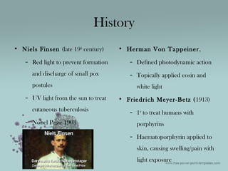
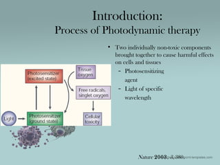
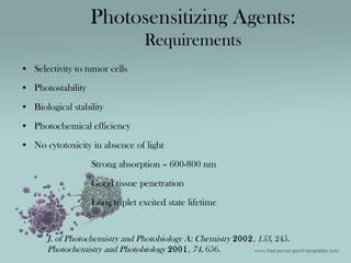
Photodynamic therapy involves using a photosensitizing agent and light of a specific wavelength to generate reactive oxygen species that are toxic to cancer cells. The history of photodynamic therapy began in the late 19th century with researchers using light therapy to treat various skin conditions. Key developments included the use of hematoporphyrin derivatives and the first human trials in the 1970s-1980s. Effective photosensitizers require properties like selectivity for tumor cells and absorption of light in the 600-800 nm range for good tissue penetration. The mechanisms of photodynamic therapy cytotoxicity can be direct tumor cell killing or indirect effects on the tumor vasculature or immune response.
Photodynamic therapy (PDT) is a two-stage treatment that combines light energy with a drug (photosensitizer) designed to destroy cancerous and precancerous cells after light activation. Photosensitizers are activated by a specific wavelength of light energy, usually from a laser.
The combined use of radiation therapy and chemotherapy in cancer treatment is a logical and reasonable approach that has already proven beneficial for several malignancies.
Particle beam – proton,neutron & heavy ion therapyAswathi c p
particle therapy is advanced external beam therapy used to treat cancer , which uses beams of protons or other charged particles such as helium, carbon or other ions instead of photons. charged particles have different depth-dose distributions compared to photons. They deposit most of their energy in the last final millimeters of their trajectory (when their speed slows). This results in a sharp and localized peak of dose, known as the Bragg peak.
Photodynamic therapy in treatment of oral lichen planus: Dr AparnaAparna Srivastava
PHOTODYNAMIC THERAPY is also known as Photoradiation therapy,
Phototherapy,
Photochemotherapy.
Photodynamic therapy (PDT) is a treatment that uses a drug, called a photosensitizer or photosensitizing agent.
Photosensitizers are exposed to a specific wavelength of light, photoactivation causes the formation of singlet oxygen, which produces peroxidative reactions that can cause cell damage and death.
Photodynamic Therapy using the Hydrosun 750thombody
Photodynamic therapy (PDT) involves the use of photochemical reactions mediated through the interaction of photosensitizing agents, light, and oxygen for the treatment of malignant or benign diseases. Photodynamic therapy is a two-step procedure. In the first step, the photosensitizer is administered to the patient by topical application, and it is allowed to be taken up by the target cells. The second step involves the activation of the photosensitizer in the presence of oxygen with a specific wavelength of light directed toward the target tissue. Because the photosensitizer is preferentially absorbed by hyperproliferative tissue and the light source is directly targeted on the lesional tissue, photodynamic therapy achieves dual selectivity, minimizing damage to adjacent healthy structures.
Cheratosi Attinica: lesioni cliniche e campo di cancerizzazioneMyskin
Definizione e classificazione della Cheratosi Attinica, presentazione del modello di progressione in Carcinoma Squamocellulare e spiegazione del concetto di campo di cancerizzazione
Early Stage Breast Cancer and Radiation TherapyMatthew Katz
These slides are intended as an educational overview for newly diagnosed early stage breast cancer patients. My hope is that it can complement and enhance the doctor-patient relationship and shared decision making. I welcome any feedback on how to improve it.
Introduction to Targeted Therapies in OncologyMohamed Abdulla
Describes the molecular background which represents the core for developing targeted therapies against specific biological events in malignant cellular clones.
Natural allergens,photosensitizing agents and fungal toxinPratik Rahate
It is very helpful ppt for B.pharma students.
In this contain information about natural allergens,photosensitizing agents and fungal toxins which are useful in pharmacognosy.
Fluoroscopy ,Radiation safety and contrast agents including adverse effect an...Dr Ravi Shankar Sharma
IT includes everything related to fluoroscopy, radiation exposure, it,s effects, contrast agents , and it,s newer variants including gadolinium, anaphylaxis reactions and it,s management, images for epidural,intrathecal,subdural, intrarterial and intravenous contrast picture.
Those who administer ionizing radiation must become familiar with the magnitude of exposure encountered in medicine, dentistry and every day life; the possible risks associated with such exposure; and the methods used to affect exposure.
Practitioners should remain informed about safety updates to further improve diagnostic quality of radiographs and decrease radiation exposure.
Light is an integral part of our life. Advances in technology are increasing and changing the ways that the patient experience dental treatment. One of the milestones in technological advancements in dentistry is the use of lasers The early 20th century saw one of the greatest inventions in science & technology, in that LASERS which later went on to became a gift to health sciences. Albert Einstein is usually credited for the development of the laser theory. He was the first one to coin the term “Stimulated Emission” in his publication “Zur Quantentheorie der Strahlung”, published in 1917 in the “Physikalische Zeitschrift”
Lasers are devices that produce beams of coherent and very high intensity light. The word LASER is an acronym for “Light Amplification by Stimulated\Emission of Radiation”. A crystal or gas is excited to emit light photons of a characteristic wavelength that are amplified and filtered to make a coherent light beam. The effect of the laser depends upon the power of the beam and the extent to which the beam absorbed. Several types of lasers are available based on the wavelengths. These range from long wavelengths (infrared), to visible wavelengths, to short wavelengths (ultraviolet), to special ultraviolet lasers called excimers. Lasers are used nowadays in many areas in the field of dentistry It is of the most captivating technologies in dental practice. Even though, introduced as an alternative to the traditional halogen curing light, laser now has become the instrument of choice, in many dental applications. Its advancements in the field of dentistry are playing a major role in patient care and well being.
Paper presentation on World Oral Cancer Day.
World Cancer Day aims to save millions of preventable deaths each year by raising awareness and education about cancer.
Protocol of Dental Treatment in Radiotherapy Indicated Patients.pptxHoor-E-Jannath Prity
The dental management of patients who are to or have received radiotherapy pose a great challenge for general dentists. It is very important that we adhere to the established treatment regime to avoid any complications that may occur because of unplanned dental treatments.
CONTENTS
Introduction
Brief overview of the history of phototherapy
Types of phototherapy
Mechanism
Phototherapy protocols
Indications for phototherapy in dermatology
Side effects and complications
Contraindications
Laser science is principally concerned with quantum electronics, laser construction, optical cavity design, the physics of producing a population inversion in laser media, and the temporal evolution of the light field in the laser. It is also concerned with the physics of laser beam propagation, particularly the physics of Gaussian beams, with laser applications, and with associated fields such as non-linear optics and quantum optics.
Flu Vaccine Alert in Bangalore Karnatakaaddon Scans
As flu season approaches, health officials in Bangalore, Karnataka, are urging residents to get their flu vaccinations. The seasonal flu, while common, can lead to severe health complications, particularly for vulnerable populations such as young children, the elderly, and those with underlying health conditions.
Dr. Vidisha Kumari, a leading epidemiologist in Bangalore, emphasizes the importance of getting vaccinated. "The flu vaccine is our best defense against the influenza virus. It not only protects individuals but also helps prevent the spread of the virus in our communities," he says.
This year, the flu season is expected to coincide with a potential increase in other respiratory illnesses. The Karnataka Health Department has launched an awareness campaign highlighting the significance of flu vaccinations. They have set up multiple vaccination centers across Bangalore, making it convenient for residents to receive their shots.
To encourage widespread vaccination, the government is also collaborating with local schools, workplaces, and community centers to facilitate vaccination drives. Special attention is being given to ensuring that the vaccine is accessible to all, including marginalized communities who may have limited access to healthcare.
Residents are reminded that the flu vaccine is safe and effective. Common side effects are mild and may include soreness at the injection site, mild fever, or muscle aches. These side effects are generally short-lived and far less severe than the flu itself.
Healthcare providers are also stressing the importance of continuing COVID-19 precautions. Wearing masks, practicing good hand hygiene, and maintaining social distancing are still crucial, especially in crowded places.
Protect yourself and your loved ones by getting vaccinated. Together, we can help keep Bangalore healthy and safe this flu season. For more information on vaccination centers and schedules, residents can visit the Karnataka Health Department’s official website or follow their social media pages.
Stay informed, stay safe, and get your flu shot today!
Pulmonary Thromboembolism - etilogy, types, medical- Surgical and nursing man...VarunMahajani
Disruption of blood supply to lung alveoli due to blockage of one or more pulmonary blood vessels is called as Pulmonary thromboembolism. In this presentation we will discuss its causes, types and its management in depth.
TEST BANK for Operations Management, 14th Edition by William J. Stevenson, Ve...kevinkariuki227
TEST BANK for Operations Management, 14th Edition by William J. Stevenson, Verified Chapters 1 - 19, Complete Newest Version.pdf
New Drug Discovery and Development .....NEHA GUPTA
The "New Drug Discovery and Development" process involves the identification, design, testing, and manufacturing of novel pharmaceutical compounds with the aim of introducing new and improved treatments for various medical conditions. This comprehensive endeavor encompasses various stages, including target identification, preclinical studies, clinical trials, regulatory approval, and post-market surveillance. It involves multidisciplinary collaboration among scientists, researchers, clinicians, regulatory experts, and pharmaceutical companies to bring innovative therapies to market and address unmet medical needs.
263778731218 Abortion Clinic /Pills In Harare ,sisternakatoto
263778731218 Abortion Clinic /Pills In Harare ,ABORTION WOMEN’S CLINIC +27730423979 IN women clinic we believe that every woman should be able to make choices in her pregnancy. Our job is to provide compassionate care, safety,affordable and confidential services. That’s why we have won the trust from all generations of women all over the world. we use non surgical method(Abortion pills) to terminate…Dr.LISA +27730423979women Clinic is committed to providing the highest quality of obstetrical and gynecological care to women of all ages. Our dedicated staff aim to treat each patient and her health concerns with compassion and respect.Our dedicated group ABORTION WOMEN’S CLINIC +27730423979 IN women clinic we believe that every woman should be able to make choices in her pregnancy. Our job is to provide compassionate care, safety,affordable and confidential services. That’s why we have won the trust from all generations of women all over the world. we use non surgical method(Abortion pills) to terminate…Dr.LISA +27730423979women Clinic is committed to providing the highest quality of obstetrical and gynecological care to women of all ages. Our dedicated staff aim to treat each patient and her health concerns with compassion and respect.Our dedicated group of receptionists, nurses, and physicians have worked together as a teamof receptionists, nurses, and physicians have worked together as a team wwww.lisywomensclinic.co.za/
Lung Cancer: Artificial Intelligence, Synergetics, Complex System Analysis, S...Oleg Kshivets
RESULTS: Overall life span (LS) was 2252.1±1742.5 days and cumulative 5-year survival (5YS) reached 73.2%, 10 years – 64.8%, 20 years – 42.5%. 513 LCP lived more than 5 years (LS=3124.6±1525.6 days), 148 LCP – more than 10 years (LS=5054.4±1504.1 days).199 LCP died because of LC (LS=562.7±374.5 days). 5YS of LCP after bi/lobectomies was significantly superior in comparison with LCP after pneumonectomies (78.1% vs.63.7%, P=0.00001 by log-rank test). AT significantly improved 5YS (66.3% vs. 34.8%) (P=0.00000 by log-rank test) only for LCP with N1-2. Cox modeling displayed that 5YS of LCP significantly depended on: phase transition (PT) early-invasive LC in terms of synergetics, PT N0—N12, cell ratio factors (ratio between cancer cells- CC and blood cells subpopulations), G1-3, histology, glucose, AT, blood cell circuit, prothrombin index, heparin tolerance, recalcification time (P=0.000-0.038). Neural networks, genetic algorithm selection and bootstrap simulation revealed relationships between 5YS and PT early-invasive LC (rank=1), PT N0—N12 (rank=2), thrombocytes/CC (3), erythrocytes/CC (4), eosinophils/CC (5), healthy cells/CC (6), lymphocytes/CC (7), segmented neutrophils/CC (8), stick neutrophils/CC (9), monocytes/CC (10); leucocytes/CC (11). Correct prediction of 5YS was 100% by neural networks computing (area under ROC curve=1.0; error=0.0).
CONCLUSIONS: 5YS of LCP after radical procedures significantly depended on: 1) PT early-invasive cancer; 2) PT N0--N12; 3) cell ratio factors; 4) blood cell circuit; 5) biochemical factors; 6) hemostasis system; 7) AT; 8) LC characteristics; 9) LC cell dynamics; 10) surgery type: lobectomy/pneumonectomy; 11) anthropometric data. Optimal diagnosis and treatment strategies for LC are: 1) screening and early detection of LC; 2) availability of experienced thoracic surgeons because of complexity of radical procedures; 3) aggressive en block surgery and adequate lymph node dissection for completeness; 4) precise prediction; 5) adjuvant chemoimmunoradiotherapy for LCP with unfavorable prognosis.
HOT NEW PRODUCT! BIG SALES FAST SHIPPING NOW FROM CHINA!! EU KU DB BK substit...GL Anaacs
Contact us if you are interested:
Email / Skype : kefaya1771@gmail.com
Threema: PXHY5PDH
New BATCH Ku !!! MUCH IN DEMAND FAST SALE EVERY BATCH HAPPY GOOD EFFECT BIG BATCH !
Contact me on Threema or skype to start big business!!
Hot-sale products:
NEW HOT EUTYLONE WHITE CRYSTAL!!
5cl-adba precursor (semi finished )
5cl-adba raw materials
ADBB precursor (semi finished )
ADBB raw materials
APVP powder
5fadb/4f-adb
Jwh018 / Jwh210
Eutylone crystal
Protonitazene (hydrochloride) CAS: 119276-01-6
Flubrotizolam CAS: 57801-95-3
Metonitazene CAS: 14680-51-4
Payment terms: Western Union,MoneyGram,Bitcoin or USDT.
Deliver Time: Usually 7-15days
Shipping method: FedEx, TNT, DHL,UPS etc.Our deliveries are 100% safe, fast, reliable and discreet.
Samples will be sent for your evaluation!If you are interested in, please contact me, let's talk details.
We specializes in exporting high quality Research chemical, medical intermediate, Pharmaceutical chemicals and so on. Products are exported to USA, Canada, France, Korea, Japan,Russia, Southeast Asia and other countries.
These lecture slides, by Dr Sidra Arshad, offer a quick overview of physiological basis of a normal electrocardiogram.
Learning objectives:
1. Define an electrocardiogram (ECG) and electrocardiography
2. Describe how dipoles generated by the heart produce the waveforms of the ECG
3. Describe the components of a normal electrocardiogram of a typical bipolar leads (limb II)
4. Differentiate between intervals and segments
5. Enlist some common indications for obtaining an ECG
Study Resources:
1. Chapter 11, Guyton and Hall Textbook of Medical Physiology, 14th edition
2. Chapter 9, Human Physiology - From Cells to Systems, Lauralee Sherwood, 9th edition
3. Chapter 29, Ganong’s Review of Medical Physiology, 26th edition
4. Electrocardiogram, StatPearls - https://www.ncbi.nlm.nih.gov/books/NBK549803/
5. ECG in Medical Practice by ABM Abdullah, 4th edition
6. ECG Basics, http://www.nataliescasebook.com/tag/e-c-g-basics
Ethanol (CH3CH2OH), or beverage alcohol, is a two-carbon alcohol
that is rapidly distributed in the body and brain. Ethanol alters many
neurochemical systems and has rewarding and addictive properties. It
is the oldest recreational drug and likely contributes to more morbidity,
mortality, and public health costs than all illicit drugs combined. The
5th edition of the Diagnostic and Statistical Manual of Mental Disorders
(DSM-5) integrates alcohol abuse and alcohol dependence into a single
disorder called alcohol use disorder (AUD), with mild, moderate,
and severe subclassifications (American Psychiatric Association, 2013).
In the DSM-5, all types of substance abuse and dependence have been
combined into a single substance use disorder (SUD) on a continuum
from mild to severe. A diagnosis of AUD requires that at least two of
the 11 DSM-5 behaviors be present within a 12-month period (mild
AUD: 2–3 criteria; moderate AUD: 4–5 criteria; severe AUD: 6–11 criteria).
The four main behavioral effects of AUD are impaired control over
drinking, negative social consequences, risky use, and altered physiological
effects (tolerance, withdrawal). This chapter presents an overview
of the prevalence and harmful consequences of AUD in the U.S.,
the systemic nature of the disease, neurocircuitry and stages of AUD,
comorbidities, fetal alcohol spectrum disorders, genetic risk factors, and
pharmacotherapies for AUD.
The prostate is an exocrine gland of the male mammalian reproductive system
It is a walnut-sized gland that forms part of the male reproductive system and is located in front of the rectum and just below the urinary bladder
Function is to store and secrete a clear, slightly alkaline fluid that constitutes 10-30% of the volume of the seminal fluid that along with the spermatozoa, constitutes semen
A healthy human prostate measures (4cm-vertical, by 3cm-horizontal, 2cm ant-post ).
It surrounds the urethra just below the urinary bladder. It has anterior, median, posterior and two lateral lobes
It’s work is regulated by androgens which are responsible for male sex characteristics
Generalised disease of the prostate due to hormonal derangement which leads to non malignant enlargement of the gland (increase in the number of epithelial cells and stromal tissue)to cause compression of the urethra leading to symptoms (LUTS
Title: Sense of Smell
Presenter: Dr. Faiza, Assistant Professor of Physiology
Qualifications:
MBBS (Best Graduate, AIMC Lahore)
FCPS Physiology
ICMT, CHPE, DHPE (STMU)
MPH (GC University, Faisalabad)
MBA (Virtual University of Pakistan)
Learning Objectives:
Describe the primary categories of smells and the concept of odor blindness.
Explain the structure and location of the olfactory membrane and mucosa, including the types and roles of cells involved in olfaction.
Describe the pathway and mechanisms of olfactory signal transmission from the olfactory receptors to the brain.
Illustrate the biochemical cascade triggered by odorant binding to olfactory receptors, including the role of G-proteins and second messengers in generating an action potential.
Identify different types of olfactory disorders such as anosmia, hyposmia, hyperosmia, and dysosmia, including their potential causes.
Key Topics:
Olfactory Genes:
3% of the human genome accounts for olfactory genes.
400 genes for odorant receptors.
Olfactory Membrane:
Located in the superior part of the nasal cavity.
Medially: Folds downward along the superior septum.
Laterally: Folds over the superior turbinate and upper surface of the middle turbinate.
Total surface area: 5-10 square centimeters.
Olfactory Mucosa:
Olfactory Cells: Bipolar nerve cells derived from the CNS (100 million), with 4-25 olfactory cilia per cell.
Sustentacular Cells: Produce mucus and maintain ionic and molecular environment.
Basal Cells: Replace worn-out olfactory cells with an average lifespan of 1-2 months.
Bowman’s Gland: Secretes mucus.
Stimulation of Olfactory Cells:
Odorant dissolves in mucus and attaches to receptors on olfactory cilia.
Involves a cascade effect through G-proteins and second messengers, leading to depolarization and action potential generation in the olfactory nerve.
Quality of a Good Odorant:
Small (3-20 Carbon atoms), volatile, water-soluble, and lipid-soluble.
Facilitated by odorant-binding proteins in mucus.
Membrane Potential and Action Potential:
Resting membrane potential: -55mV.
Action potential frequency in the olfactory nerve increases with odorant strength.
Adaptation Towards the Sense of Smell:
Rapid adaptation within the first second, with further slow adaptation.
Psychological adaptation greater than receptor adaptation, involving feedback inhibition from the central nervous system.
Primary Sensations of Smell:
Camphoraceous, Musky, Floral, Pepperminty, Ethereal, Pungent, Putrid.
Odor Detection Threshold:
Examples: Hydrogen sulfide (0.0005 ppm), Methyl-mercaptan (0.002 ppm).
Some toxic substances are odorless at lethal concentrations.
Characteristics of Smell:
Odor blindness for single substances due to lack of appropriate receptor protein.
Behavioral and emotional influences of smell.
Transmission of Olfactory Signals:
From olfactory cells to glomeruli in the olfactory bulb, involving lateral inhibition.
Primitive, less old, and new olfactory systems with different path
2. History
• Niels Finsen (late 19th
century)
– Red light to prevent formation
and discharge of small pox
postules
– UV light from the sun to treat
cutaneous tuberculosis
– Nobel Prize 1903
• Herman Von Tappeiner,
– Defined photodynamic action
– Topically applied eosin and
white light
• Friedrich Meyer-Betz (1913)
– 1st
to treat humans with
porphyrins
– Haematoporphyrin applied to
skin, causing swelling/pain with
light exposure
3. History
• Samuel Schwartz (1960’s)
Developed haematoporphyrin
derivative (HpD)
Haematoporphyrin treated with
acetic and sulfuric acids,
neutralized with sodium acetate
• I. Diamond (1972) Use PDT to
treat cancer
• Thomas Dougherty (1975)
– HpD and red light
– Eradicated mammary tumor
growth in mice
• J.F. Kelly (1976)
– 1st
human trials using HpD
– Bladder cancer
• Canada (1999)
– 1st
PDT drug approved
4. Photodynamic therapy is based on the concept
(1) certain photosensitizers can be localized (somewhat preferentially) in
neoplastic tissue, and
(2) subsequently, these photosensitizers can be activated with the appropriate
wavelength (energy) of light to generate active molecular species, such as free
radicals and singlet oxygen (1O2) that are toxic to cells and tissues
5. • Two individually non-toxic components
brought together to cause harmful effects
on cells and tissues
– Photosensitizing
agent
– Light of specific
wavelength
Introduction:
Process of Photodynamic therapy
Nature 2003, 3, 380.
6. • Type 1:
– Direct reaction with substrate (cell membrane or molecule)
– Transfer of H atom to form radicals
– Radicals react with O2 to form oxygenated products
• Type 2: Transfer of energy to O2 to form 1
O2
Ratio of Type 1/Type 2 depends on:
Photosensitizing agent, concentration of substrate and O2,
binding affinity of photosensitizing agent to substrate
Reactive oxygenated species (ROS)
Free radicals or 1
O2
Half-life of 1
O2 < 0.04 µs
Radius affected < 0.02 µm
Introduction:
Reaction Mechanisms
7.
8. • Selectivity to tumor cells
• Photostability
• Biological stability
• Photochemical efficiency
• No cytotoxicity in absence of light
Strong absorption – 600-800 nm
Good tissue penetration
Long triplet excited state lifetime
Photosensitizing Agents:
Requirements
J. of Photochemistry and Photobiology A: Chemistry 2002, 153, 245.
Photochemistry and Photobiology 2001, 74, 656.
9. MECHANISMS OF PDT
CYTOTOXICITY
• INDIRECT–
changes in tumor
microenvironment
- anti-vascular effects
- anti-tumor immune response
• DIRECT-
direct tumor cell killing due to
macromolecule damage
- apoptosis
- necrosis/ by-stander effect
10. INDIRECT CYTOTOXICITY
ANTI-VASCULAR
EFFECTS
- vessel leakage
- vasocontriction
- thrombosis
strongly dependent on—
photosensitizer used & time interval
between the administration of
photosensitizer & light
ANTI-TUMOR
IMMUNE RESPONSE
- release of pro-inflammatory
cytokines
- fixation of complement
- release of tumor associated
antigens
11. • The lifetime of singlet oxygen is 0.03 to 0.18 mcs, &
corresponds to a diffusion distance of less than 0.2
mcm, or about 1/50th of a cell diameter.
• Thus, the macromolecular damage inside the cell occurs very
close to the location of photosensitizer activation/singlet oxygen
production.
• Different photosensitizers are known to localize to - plasma
membrane, lysosome, mitochondria, Golgi apparatus,
endoplasmic reticulum, or nuclear membrane.
DIRECT CYTOTOXICITY
12. • Apoptotic cell death tends to predominate in the most PDT-sensitive
cell lines at lower light/photosensitizer doses
• necrotic/ nonapoptotic mechanisms tend to predominate at higher
light/photosensitizer doses.
The percentage apoptosis achieved, as well as the
mechanism of apoptosis (extrinsic vs. intrinsic) is
dependent upon-
1. Tumor cell line
2. Photosensitizer
DIRECT CYTOTOXICITY
15. • Limitations:
– Contains 60 compounds
– Difficult to reproduce composition
– At 630 nm, molar absorption coefficient is low (1,170 M-1
cm-1
)
– Main absorption at 400 nm
– High concentrations of drug and light needed
– Not very selective toward tumor cells
– Absorption by skin cells causes long-lasting photosensitivity (½ life = 452
hr)
Photosensitizing Agents:
Photofrin
Nature 2003, 3, 380. J. of Photochemistry and Photobiology A: Chemistry 2002, 153,
245.
16. Photosensitizing Agents:
Foscan
•Chlorin photosensitizing agent
•Approved for treatment of head and
neck cancer
•Low drug dose (0.1 mg/kg body
weight)
5-Aminolevulinic acid (5-
ALA)
•Approved for treatment of actinic
keratosis and BCC of skin
•Topical application most frequently
used
•Endogenous photosensitizing agent
– 5-ALA not directly
photosensitizing
– Creates porphyria-like
syndrome
Nature 2003, 3, 380.
17. Photosensitizing Agents:
Mono-L-aspartyl chlorin e6 (NPe6)
•Derived from chlorophyll a
•Chemically pure
•Absorption at 664 nm
•Localizes in lysosomes (instead of
mitochondria)
•Reduced limitations compared to
Photofrin
•Decreased sensitivity to sunlight (1
week)
– ½ life = 105.9 hr
Phthalocyanines
•Ring of 4 isoindole units linked by
N-atoms
•Stable chelates with metal cations
•Sulfonate groups increase water
solubility
•Examples (AlPcS4, ZnPcS2)
• More prolonged
photosensitization than
HpD
• Less skin sensitivity in
sunlight
18. Photochemistry and Photobiology 2001, 74, 656. Int. J. Cancer 2001, 93, 720.
• 2nd
generation
• Improved red light absorption
• 25-30 times more potent than HpD
• More selective toward tumor cells
• Most active photosensitizer with low drug and light doses
• Not granted approval
Photosensitizing Agents:
Meta-tetra(hydroxyphenyl)porphyrins (mTHPP)
19. Photosensitizer
Excitation
Wavelength
Clinical Uses
Porfimer sodium
(Photofrin)
630 nm Barrett's esophagus+*
, endobroncheal cancer*+
,
esophageal+
, serosal cancers (pleural peritoneal), bladder
cancer, skin cancer Bowen's disease or AK), breast
cancer metastases, head and neck cancer, brain
ALA (Levulan),
mALA (Metvixv)
400-450 nm
635 nm
AK*+
, BCC+
, Bowen's disease, bladder cancer, vulvar
cancer
BPD (Visudyne) 690 nm Macular degeneration+*
, BCC
mTHCP (Foscan) 652 nm Head and neck+
, pancreatic cancer, cancer, pleural
cancers, brain
HPPH
(Photochlor)
665 nm BCC, pleural cancers
Silicon
pthalocyanine-4
(Pc-4)
672 nm Cutaneous and subcutaneous metastases malignancies
PHOTOSENSITIZERS
21. difficult to couple them to light delivery fibers
without reducing their optical power.
difficult to calculate the effective delivered light
dose
power output is limited to a maximum of 1 W.
Filters are also required to cut off UV radiation
and infrared emission
LIGHT APPLICATION
22. • LASERS -- emit light of precise wavelengths in
easily focused beams.
Early lasers were expensive, large, immobile
machines that required a level of technical
support.
LIGHT APPLICATION
23. • SEMICONDUCTOR DIODE TECHNOLOGY resulted in cheaper
systems, which are compact and portable while still retaining high power
output.
• However, diode lasers offer only a single output wavelength, limiting their
versatility.
LIGHT APPLICATION
24. • LIGHT EMITTING DIODES (LEDs) are less
expensive than otherlight sources, are small, and
can provide a power output up to 150 mW/cm2
at wavelengths in the rangeof 350–1,100 nm
LIGHT APPLICATION
25. • OPTICAL FIBER TECHNOLOGY
meet the demands of illuminationat
different localizations.
• For superficial illumination of, for example, oral
mucosa, optic fibers with a lens tip are used to
spread the light over the target area.
LIGHT APPLICATION
26. • OPTICAL FIBER TECHNOLOGY
In hollow organs ---- endobronchial, esophagus, and bladder,
illumination is often performed with cylindrical diffusers
combined withinflated balloons for uniform light distribution.
Black coating of one side of the balloon is sometimes used to
shield adjacentnormal tissue areas for protection.
LIGHT APPLICATION
27. OPTICAL FIBER TECHNOLOGY
In hollow organs ---- endobronchial, esophagus, and bladder, illumination is
often performed with cylindrical diffusers combined withinflated balloons for
uniform light distribution.
Black coatingof one side of the balloon is sometimes used to shield adjacent
normal tissue areas for protection.
LIGHT APPLICATION
28. • Experiments on oxic and hypoxic cells and
tissues show that pretreatment tumor hypoxia
significantly decreases the efficacy of
PDT.
• Limited studies of PDT and tumor hypoxia in
clinical samples confirm this relationship
between hypoxia and decreased PDT efficacy
OXYGEN EFFECTS
29. • ADVANTAGES OF PDT
single injection of drug followed after a certain time interval by single
illumination
local, rather than systemic, treatment
limited light penetration protects normal tissue from phototoxicity
functional recovery withoutscarring
can be repeated
CLINICAL APPLICATION
30. • Most promising treatment using PDT
– Skin highly accessible to light exposure
• Most common method
– Topical administration of 5-ALA
– Non-invasive, short photosensitization period, treat multiple lesions,
good cosmetic results, well accepted by patients, no side effects
PDT Trials on Tumor Cells:
Skin Cancer
Pharmaceutical Research 2000, 17, 1447.
31. PDT Trials on Tumor Cells:PDT Trials on Tumor Cells:
Skin CancerSkin Cancer
• Clinical Studies performed on superficial skin cancer types:
– Actinic keratosis (AK)
– Basal cell carcinoma (BCC)
– Squamous cell carcinoma (SCC)
– Bowen’s disease (BD)
• Complete response (CR) – no clinical or histopathologic signs after follow-up
• Minimal side effects
Pharmaceutical Research 2000, 17, 1447.
32. PDT Trials on Tumor Cells:
Skin Cancer
Pharmaceutical Research 2000, 17, 1447.
34. • Clinical trials (continued)
– 5 different intravenous doses of NPe6 over 30 minutes (0.5 mg/kg – 3.5
mg/kg)
• 4-8 hr prior to light administration (due to number of lesions)
– Light dose – 25-200 J/cm2
• Argon-pumped tunable dye laser set at 664 nm
• Dose dependent on tumor size/shape
PDT Trials on Tumor Cells:
Skin Cancer
Photodermatol Photoimmunol Photomed 2005, 21, 72.
35. PDT Trials on Tumor Cells:
Skin Cancer
Photodermatol Photoimmunol Photomed 2005, 21, 72.
36. • Results:
– 4 weeks later: 20 of 22 BCC – CR, 18 of 27 other – CR
• CR – no evidence of tumor in treatment field
• PR – >50% reduction in tumor size
– Photosensitivity gone within 1 week (12 of 14)
• 3 patients – mild to moderate pruritis, facial edema or blistering,
erythema, tingling
• 1 patient – severe intermittent burning pain
• 1 patient – erythema, edema, moderate pain (gone within 2 weeks)
PDT Trials on Tumor Cells:
Skin Cancer
Photodermatol Photoimmunol Photomed 2005, 21, 72.
37. • EARLY STAGE, ENDOBRONCHIAL LUNG CANCER
In a phase II trial, porfimer sodium (2 mg/kg) was administered to 51 patients
with 61 total carcinoma lesions, and PDT was performed 48 hours later using
150 to 200 J/cm2
630 nm light.
complete response rate was 85% no grade 3 or 4 toxicities were reported.
PDT for Early Stage Cancers
38. • BARETT’S ESOPHAGUS
At 18 months of follow-up, 75% of patients treated with PDT-PPI showed
ablation of HGD versus 36% of patients treated with PPI alone (P <.0001).
BARETT’S ESOPHAGUS
52% of patients treated with PDT-PPI showed complete return to normal
squamous epithelium versus 7% of patients treated with PPI (P <.0001).
Finally, with an average follow-up of nearly a year, 13% of the patients in the
PDT-PPI arm showed progression to cancer versus 28% of patients on the
PPI arm (P <.006).
PDT for Early Stage Cancers
39. HEAD AND NECK CANCER patients used HpD or porfimer sodium but
nowadays mTHPC is more often used in combination with 10–20J/cm2
.
For early-stage primary tumors of the oral cavity or oropharynx, a CR rate of
85% at 1 year, decreasing to 77% at 2 years, is reported with an even higher
CR rate of96% for lip carcinoma
PDT for Early Stage Cancers
40. • Dosage:
– Diode laser used to generate λ = 652 nm
• 3 patients
– 0.10 mg/kg total body weight
– 48 hr under 5 J/cm2
• 4 patients
– 0.15 mg/kg total body weight
– 96 hr under 10 J/cm2
PDT Trials on Tumor Cells:
Breast Cancer
Int. J. Cancer 2001, 93, 720.
41. • Chest wall recurrences – problem with mastectomy treatment (5-19%)
• Study:
– 7 patients, 57.6 years old (12.6)
– 89 metastatic nodes treated
– 11 PDT sessions
– Photosensitizing agent: (m-THPC)
meta-tetra(hydroxyphenyl)chlorin
• 2nd
generation photosensitizing agent
PDT Trials on Tumor Cells:
Breast Cancer
Int. J. Cancer 2001, 93, 720.
42. • Results:
– Complete response in all 7 patients
– Pain – 10 days, Healing – 8-10 weeks
– Patients advised to use sun block or clothing to protect skin from light
for 2 weeks
• 4 days after treatment – 1 patient with skin erythema and edema
from reading light
– 6 of 7 patients given medication for pain
• Mostly based on size, not lightdose
– Recurrences in 2 patients (2 months)
PDT Trials on Tumor Cells:
Breast Cancer
Int. J. Cancer 2001, 93, 720.
43. • INTRAPERITONEAL PHOTODYNAMIC THERAPY FOR
CARCINOMATOSIS OR SARCOMATOSIS
intraoperative PDT following maximal surgical debulking resulted in a 76%
complete cytologic response rate with tolerable toxicity
ADVANCED & PALLIATIVE
SETTINGS
44. • INTRAPERITONEAL PHOTODYNAMIC THERAPY FOR
CARCINOMATOSIS OR SARCOMATOSIS
associated with a postoperative capillary leak syndrome that necessitated
massive fluid resuscitation in the immediate postoperative period that was in
excess of the typical fluid needs of patients who receive surgery alone
ADVANCED & PALLIATIVE
SETTINGS
45. • Postoperative Photodynamic Therapy for Pleural-Based Spread of Non
Small-Cell Lung Cancer and Mesothelioma
• Palliation of Obstructing Lesions
• Prostate and Bladder Cancers
• Brain Tumors
ADVANCED & PALLIATIVE
SETTINGS
46. • PDT of cancer regulated by:
– Type of photosensitizing agent
– Type of administration
– Dose of photosensitizer
– Light dose
– Fluence rate
– O2 availability
– Time between administration of photosensitizer and
light
Conclusions
47. • Tumor cells show some selectivity for photosensitizing agent uptake
• Limited damage to surrounding tissues
• Less invasive approach
• Outpatient procedure
• Various application types
• Well accepted cosmetic results
Conclusions
49. • Mechanism by which HpD selectively accumulates in tumor cells – not well
understood
– High vascular permeability of agents?
• Testing photosensitizing agents:
– Porphyrins, haematoporphyrins, HpD, ALA-D
– Administer photosensitizer and monitor fluorescence with endoscope
– SCC shows increased fluorescence
– More invasive tumors show even greater fluorescence
Future Applications:
Tumor Detection Using Fluorescence
Nature 2003, 3, 380.
50. • a: Green vascular endothelial cells of a tumor
• b: Red photosensitizing agent localizes to vascular
endothelial cells after intravenous injection
Future Applications:
Tumor Detection Using Fluorescence
Nature 2003, 3, 380.
51. • Improved Specificity and Potency
– Better photosensitizers developed and under investigation in
clinical trials
– Use of carriers – conjugated antibodies directed to tumor-
associated antigens
– New compounds that absorb light of longer wavelength –
better tissue penetration
– New compounds with less skin photosensitivity
• Improved Efficacy
– Creating a preferred treatment of cancer
Future Applications:
Photosensitizing Drugs
Nature 2003, 3, 380.