
Recommended
More Related Content
What's hot
What's hot (20)
Viewers also liked
Viewers also liked (20)
Similar to Blood Banking.. Experiment # 2
Similar to Blood Banking.. Experiment # 2 (20)
Recently uploaded
Recently uploaded (20)
Blood Banking.. Experiment # 2
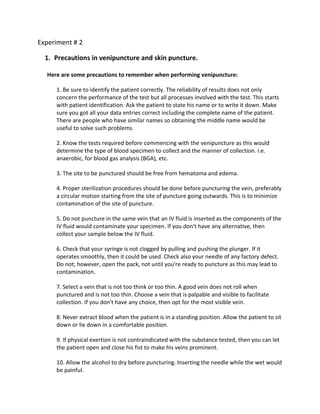
- 1. Experiment # 2 1. Precautions in venipuncture and skin puncture. Here are some precautions to remember when performing venipuncture: 1. Be sure to identify the patient correctly. The reliability of results does not only concern the performance of the test but all processes involved with the test. This starts with patient identification. Ask the patient to state his name or to write it down. Make sure you got all your data entries correct including the complete name of the patient. There are people who have similar names so obtaining the middle name would be useful to solve such problems. 2. Know the tests required before commencing with the venipuncture as this would determine the type of blood specimen to collect and the manner of collection. i.e. anaerobic, for blood gas analysis (BGA), etc. 3. The site to be punctured should be free from hematoma and edema. 4. Proper sterilization procedures should be done before puncturing the vein, preferably a circular motion starting from the site of puncture going outwards. This is to minimize contamination of the site of puncture. 5. Do not puncture in the same vein that an IV fluid is inserted as the components of the IV fluid would contaminate your specimen. If you don't have any alternative, then collect your sample below the IV fluid. 6. Check that your syringe is not clogged by pulling and pushing the plunger. If it operates smoothly, then it could be used. Check also your needle of any factory defect. Do not; however, open the pack, not until you're ready to puncture as this may lead to contamination. 7. Select a vein that is not too think or too thin. A good vein does not roll when punctured and is not too thin. Choose a vein that is palpable and visible to facilitate collection. If you don't have any choice, then opt for the most visible vein. 8. Never extract blood when the patient is in a standing position. Allow the patient to sit down or lie down in a comfortable position. 9. If physical exertion is not contraindicated with the substance tested, then you can let the patient open and close his fist to make his veins prominent. 10. Allow the alcohol to dry before puncturing. Inserting the needle while the wet would be painful.
- 2. 11. The tourniquet should be released first before the needle. This is to avoid bleeding of patients who have clotting disorders. 12. The bevel should be facing upwards to allow the smooth flow of blood into the barrel of the syringe. 13. When blood starts to flow into the barrel, the plunger should not be pulled too fast to avoid collapse of the veins. It should also not be too slow to avoid clotting before you could transfer the blood to an appropriate container. 14. Transfer the blood immediately after extraction to an anti-coagulated tube if you want plasma and to a plain tube if you want serum. You could also make use of vacutainer tubes to facilitate extraction and preservation of blood. Being aware of these precautions would make you more skilled in venipuncture procedures. Do not attempt to extract if you don't have any formal or hands-on training because venipuncture is a skill that should be perfected. Precautions When Selecting a Puncture Site on the Heel. The puncture should not be done in a previous puncture site because of the possibility of infection. Do not do punctures in the central arch area of the foot. Puncture in this area may result in damage to nerves, tendons, and cartilage and offers no advantage over a heel puncture. 1. Do not puncture deeper that 2.4 mm. 2. Do not puncture through previous puncture sites. 3. Do not puncture the area between the imaginary boundaries. 4. Do not puncture the posterior curvature of the heel. 5. Do not puncture in the area of the arch. 6. Do not puncture areas of the foot other than the heel.2 Precautions Collecting the Blood Sample Several precautions must be observed to produce the most accurate specimen. Hemolysis is the greatest concern with microcollection samples. Hemolysis may occur because of the following situations: 1. The alcohol used to clean the skin was not allowed to dry.
- 3. 2. The heel was squeezed too hard to produce a greater blood flow. 3. Newborn infants have increased red blood cell fragility and a high red blood cell volume. These factors result in a higher amount of hemolysis. 4. Instead of allowing the blood to flow into the microcollection container, the blood was scraped off the skin surface. 5. Collect samples for hematology first (purple micro-container). Additional Precautions to Protect Well-Being of Infant: 1. The baby's heel may be punctured a maximum of two times. Do not stick a baby more than twice to obtain a specimen at any given time. 2. Do not puncture a foot if there are bruises, abrasions, or sloughing skin present. Notify the baby's nurse. 3. To help obtain a free-flowing puncture wound from a baby who doesn't bleed freely, wrap the baby's heel in a warm towel for 10 to 15 minutes before puncture is made. This is mandatory for collection of blood gases. 4. Never repuncture old puncture wounds. 5. Never remove a baby from its bassinet or change its position in any way without the approval of a nurse. 6. Use only gentle massage when obtaining blood. It is sufficient to massage with your thumb and forefinger or gentle squeezing. 2. Discuss briefly the vacutainer method of collecting blood. The venipuncture procedure is complex, requiring both knowledge and skill to perform. Each phlebotomist generally establishes a routine that is comfortable for her or him. Several essential steps are required for every successful collection procedure: 1. Identify the patient. 2. Assess the patient's physical disposition (i.e. diet, exercise, stress, basal state). 3. Check the requisition form for requested tests, patient information, and any special requirements. 4. Select a suitable site for venipuncture. 5. Prepare the equipment, the patient and the puncture site. 6. Perform the venipuncture. 7. Collect the sample in the appropriate container. 8. Recognize complications associated with the phlebotomy procedure.
- 4. 9. Assess the need for sample recollection and/or rejection. 10. Label the collection tubes at the bedside or drawing area. 11. Promptly send the specimens with the requisition to the laboratory. ORDER OF DRAW: Blood collection tubes must be drawn in a specific order to avoid cross-contamination of additives between tubes. The recommended order of draw for plastic vacutainer tubes is: 1. First - blood culture bottle or tube (yellow or yellow-black top) 2. Second - coagulation tube (light blue top). If just a routine coagulation assay is the only test ordered, then a single light blue top tube may be drawn. If there is a concern regarding contamination by tissue fluids or thromboplastins, then one may draw a non-additive tube first, and then the light blue top tube. 3. Third - non-additive tube (red top) 4. Last draw - additive tubes in this order: 1. SST (red-gray or gold top). Contains a gel separator and clot activator. 2. Sodium heparin (dark green top) 3. PST (light green top). Contains lithium heparin anticoagulant and a gel separator. 4. EDTA (lavender top) 5. ACDA or ACDB (pale yellow top). Contains acid citrate dextrose. 6. Oxalate/fluoride (light gray top) NOTE:Tubes with additives must be thoroughly mixed. Erroneous test results may be obtained when the blood is not thoroughly mixed with the additive. 3. What precautions should phlebotomist observe when performing venipuncture to avoid exposure of blood? As per the guidelines from The Centers for Disease Control and Prevention (CDC) several disease-specific precautionary policies for patients known to be or suspected of being infected with certain pathogens are recommended to be followed by phlebotomists. Universal standard precautions assume that all specimens are potentially infectious and should be handled accordingly. It is assumed that every direct contact with body fluids is potentially infectious as per protocols for infection control in Standard precautions. Cross-transmission and exposure of the skin and mucous membranes to infectious microbes can be prevented by avoiding direct contact with patient specimens and every possible precaution for barrier protection are taken when contact cannot be avoided.
- 5. It should be routine for phlebotomist to exercise certain consistent precautions. The infectious potential of any patient specimen is recognized by standard precautions and cross- transmission of infectious disease to patients is prevented and the laboratory personnel are protected from infected patients. To protect against potential exposure to HBV, a licensed inactivated vaccine (HB) is recommended. The CDCs advisory committee on Immunization practices recommends the use of his vaccine as a precautionary step for persons who are at a greater risk for Hepatitis B infection ” clinical laboratory workers, phlebotomists, and pathologists. Phlebotomists working with blood specimens should follow safe work practices to eliminate the risk of transmitting infectious pathogens like: - To disrupt transmission of infectious pathogens frequent hand washing according to the procedure – Eating, drinking, smoking should be prohibited in lab area. – Gloves, gowns, lab coats, masks, eye protectors, etc. Personal Protective Equipment used as required – Clean up any infectious fluids/blood spills immediately and minimize aerosolization – Waste disposal measures as recommended by OSHA and NCCLS are followed. Puncture”resistant sharps containers are used to dispose disposable syringes and needles, scalpel blades, and other sharp items. – Needles should not be recapped by hand, purposely bent or broken by hand, removed from disposable syringes, or otherwise manipulated by hand to prevent needle stick injuries. – Prophylactic measures for pre-exposure and post-exposure for handling potential occupational transmission of certain pathogens should be known by phlebotomist. A professional phlebotomist should be knowledgeable in general safety regulations governing the clinical laboratory, OSHA (Occupational Safety and Health Administration) mandated plans for chemical hygiene and for occupational exposure to blood-borne pathogens, the importance of safety manual, and general emergency procedures. A hazardous procedure for all health care staff is the disposal of sharps. The risks involved in venipuncture should be known by the phlebotomist. All healthcare professionals who carry out venipuncture should be properly training in closed systems for blood collecting like vacutainers to minimize the needle stick injuries. Case of a needle stick injury should be handled by following: - Bleeding should be encouraged from the injury – Injured site should be washed with water and disinfected. – Suitable dressing should cover the site – A note of patients name, identification should be made – Incident reporting procedure should be followed – Injuries should be always reported to lab manager – Needle stick should be reported to Occupational Health, GP or Accident and Emergency Department as appropriate.
- 6. Accredited phlebotomist technician programs teach all of the necessary techniques and procedures needed to be a competent and skilled phlebotomist. Some of the subjects in phlebotomy training courses are: Anatomy and physiology, Blood and cell composition, Blood sampling procedures, Laboratory safety, and CPR. Other phlebotomy courses that might be included in training program are: Professional behavior, Quality control, Legal issues, Computer training. Each phlebotomy training school will offer something a little different depending upon the regulations and training for work settings. 4. Definition of Terms. Arteries- are blood vessels that carry blood away from the heart. This blood is normally oxygenated, exceptions made for the pulmonary and umbilical arteries. Vein- In the circulatory system, veins are blood vessels that carry blood towards the heart. Most veins carry deoxygenated blood from the tissues back to the heart; exceptions are the pulmonary and umbilical veins, both of which carry oxygenated blood to the heart. Veins differ from arteries in structure and function; for example, arteries are more muscular than veins, veins contain valves, and they carry blood away from the heart. Capillaries- are the smallest of a body's blood vessels and are parts of the microcirculation. They are only 1 cell thick. These microvessels, measuring 5-10 μm in diameter, connect arterioles and venules, and enable the exchange of water, oxygen, carbon dioxide, and many other nutrient and waste chemical substances between blood and surrounding tissues. Cephalic Vein- In human anatomy, the cephalic vein (or antecubital vein) is a superficial vein of the upper limb. Ante Cubital Fossa- It communicates with the basilic vein via the median cubital vein at the elbow and is located in the superficial fascia along the anterolateral surface of the biceps brachii muscle. Anatomically the antecubital fossa the fold of the elbow, where it is common for a stethoscope to be placed during blood pressure measurement at the forearm. The antecubital fossa provides the safest route for central venous access, and blood is usually taken from any palpable vein passing through this region. These veins are the basilic vein, cephalic vein and the median cubital vein. hematoma- or haematoma, is an extravasation of blood outside the blood vessels,[1] generally the result of hemorrhage. A hematoma is a pocket or localized collection of blood usually in liquid form within the tissue. This distinguishes it from an ecchymosis, which is the spread of blood under the skin in a thin layer, commonly called a bruise. Internal bleeding is generally considered to be a spreading of blood within the abdomen or skull, not within muscle. Phlebotomy- Obtaining blood from a vein. In the old days, this was done by incising (cutting) a vein and just letting the blood flow into a container. Today phlebotomy is done more neatly by puncturing a vein with a needle.
- 7. Allen test- A test for occlusion of the radial or ulnar artery, in which one of these arteries is compressed after blood has been forced out of the hand by clenching it into a fist; failure of the blood to diffuse into the hand when opened indicates that the artery not compressed is occluded. Oxygenated blood- is blood in which oxygen is attached to the haemoglobin molecules in the red blood cells. It is bright red because the attached oxygen makes the normally blue haemoglobin molecules turn red. Deoxygenated Blood- When blood has delivered oxygen to the cells, it is described as deoxygenated. It now looks a very dark red because so many of the haemoglobin molecules have turned blue again. The mixture of blue and red molecules looks dark red to our eyes. 5. Why is the first blood wipe away in skin puncture? -Blood should wipe away after the first drop of blood appears for it contains tissue juices and dead epidermal cells. 6. What are the test done in capillary and venous blood in the field of blood banking? In a transfusion service there are a number procedures routinely done. The ones noted in red are those done even in small hospitals whereas the rest are more likely done at larger hospitals and reference laboratories: • ABO/Rh(D) typing • Antigen typing from other blood group systems such as Rh antigens other than D, Kell, Kidd, and Duffy • Antibody screening for antibodies form to blood group antigens other than A and B • Antibody identification to determine the specific antibodies detected in the antibody screening • Crossmatch, or compatibility testing, which determines whether donor blood can probably be safely transfused to the recipient 7. Specify the ideal site for skin puncture. The best site for skin puncture are the lateral or medial plantar surface of the heel which is generally performed on infants <1 year of age. The palmar surface of the distal phalanx of a finger which is generally performed in children or adults. Skin-puncture of the earlobe is not recommended.
- 8. 8. State the advantages and disadvantages of skin puncture. Advantages of skin puncture using the finger: 1. It is easily accessible to the operator. 2. It is easy to manipulate. 3. It is ideal for peripheral blood smears 4. It is less intimidating. Advantages of skin puncture using the Earlobe: 1. It is less painful (due to lesser nerve endings.) 2. There is more free flow of blood (due to thinner skin.) 3. There is less tissue juice contamination of blood (due to lesser tissue and muscles in the earlobe.) 4. It is ideal when searching for abnormal cells. Disadvantages of skin puncture: 1. Less amount of blood can be obtained. 2. Additional and repeated tests cannot be done. 3. Blood obtained by skin puncture hemolyses easily. 9. Site situation or cases in the application of skin puncture. A good situation or cases that best apply the skin puncture procedure are to the newly born babies in which blood is drawn using capillary method by puncturing their heels in order for the doctors to determine or to detect possible diseases or illnesses that the babies acquired or developed during their fetal life’s. this will also help the doctors to treat a certain disease of the babies and to prolong their life. 10. List down hematologic examinations which can be done in capillary blood collected through skin puncture. The laboratory study of blood is divided into 2 groups: 1. Tests for the detection of diseases of the blood. 2. Tests for the detection of diseases of other organs. The laboratory tests for the detection of diseases of blood: 1. Chemical tests (like hemoglobin determination and its abnormalities. 2. Morphologic tests.
- 9. a. Qualitative test- determination of the appearance of the cells as analyzed and classified. b. Quantitative test- determination of the number of cells such as RBC count, WBC count, Platelet count, etc. The complete Blood Count (CBC): 1. Red Blood Cell Count 2. White Blood Cell Count 3. Hemoglobin Determination 4. Hematocrit Determination 5. Differential Count 6. Stained Red Cell Examination
- 10. CAPILLARY PUNCTURE NOTE: To be a valid report, work done on capillary blood must be from a free-flowing puncture wound. Procedure. (1) The puncture site should be warm to assure good circulation of blood. If it is cold, apply warm water (38º-40ºC) for a few minutes. If blood is to be drawn from the ear, the edge of the lobe, not the flat side, should be punctured. (2) The site to be punctured is first rubbed with alcohol prep pads to remove dirt and epithelial debris, increase circulation, and render the area reasonably disinfected (see figure 3-4 capillary puncture procedure, a through d). Figure 3-4a. Capillary puncture procedure: Clean the puncture site. (3) Allow sufficient time for the circulation to equalize. (4) While making a finger puncture, apply gentle pressure to the finger to hold the skin taut. Hold the finger in one hand and the lancet in the other. The puncture is made perpendicular to the lines of the fingerprints, which results in a more free-flowing wound (see figure 3-4b). Figure 3-4b. Capillary puncture procedure: Puncture the finger.
- 11. (5) The first drop of blood that appears is wiped away before specimens are taken (see figure 3-4c). Figure 3-4c. Capillary puncture procedure: Wipe away first drop of blood. (6) The blood must not be squeezed out since this dilutes it with fluid from the tissues, thus altering the ratio of cellular elements to fluid, as well as the ratio of cellular elements to each other. (7) After the desired specimens have been collected, have the patient hold a sterile dry gauze pad over the wound until bleeding stops (see figure 3-4d). Figure 3-4d. Capillary puncture procedure: Apply pressure to the site.
- 12. VENIPUNCTURE Figure 3-1. Site of venipuncture. CAUTION: Do not allow the tourniquet to remain in place for more than 2 minutes. Check the pulse at the wrist to make sure that arterial circulation is not cut off. Figure 3-2a. Venipuncture procedure: Locate the vein.
- 13. Figure 3-2b. Venipuncture procedure: Clean the puncture site. 3-2c. Venipuncture procedure: Guide needle toward the vein.
- 14. Figure 2-3d. Venipuncture procedure: Insert needle into the vein. Figure 3-2e. Venipuncture procedure: Aspirate the blood. Figure 3-2f. Venipuncture procedure: Remove the tourniquet.
- 15. CAUTION: Do not remove the needle now. If the needle were remove prior to the Tourniquet being removed, blood would be forced out of the venipuncture site, resulting in blood loss and/or hematoma formation (tumor-like cluster of blood forming under the skin). Figure 3-2g. Venipuncture procedure: Place a sterile pad over the site and withdraw the needle. Figure 3-2h. Venipuncture procedure: Have the patient extend the arm and maintain light pressure on the site.