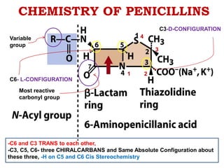
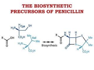
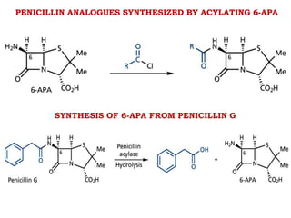
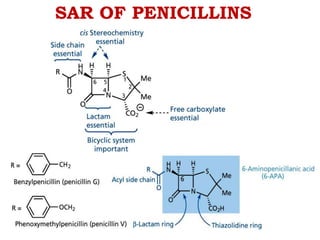
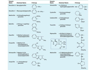
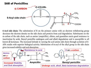
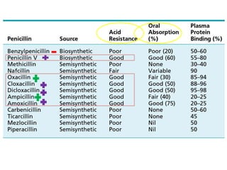
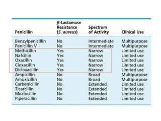
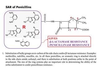
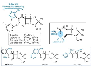
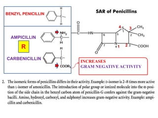
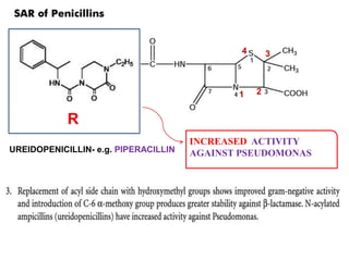
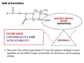
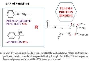
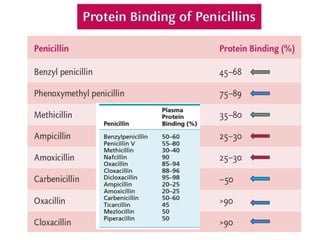
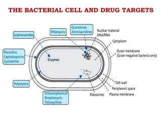
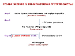
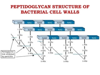
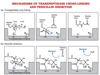
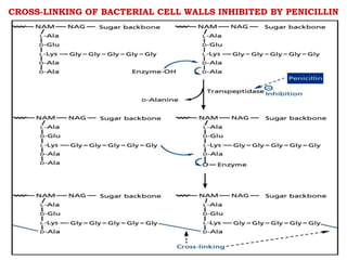
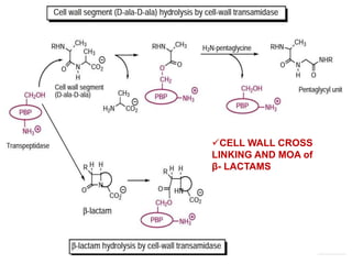
CHEMISTRY, SAR AND MOA
OF PENICILLINS
Dr. Vishal S. More,
Assistant Professor,
Dept. of Pharmaceutical Chemistry,
Amrutvahini College of Pharmacy, Sangamner.
CHEMISTRY OF PENICILLINS
 
(-LACTAM RING/
2- AZETIDINONE RING)
1
3
2
4
THIAZOLIDINE RING
5
3
2
7
6
1
4
Ring and numbering
systems of clinically
available -Lactam
antibiotics type
CHEMISTRY OF PENICILLINS
C6- L-CONFIGURATION
C3-D-CONFIGURATION
3
2
7
1
1 4
-C6 and C3 TRANS to each other,
-C3, C5, C6- three CHIRALCARBANS and Same Absolute Configuration about
these three, -H on C5 and C6 Cis Stereochemistry
Variable
group
Most reactive
carbonyl group
THE BIOSYNTHETIC
PRECURSORS OF PENICILLIN
PENICILLIN ANALOGUES SYNTHESIZED BY ACYLATING 6-APA
SYNTHESIS OF 6-APA FROM PENICILLIN G
SAR OF PENICILLINS
-C-CO-HN-
 CARBON
6-Acyl side chain
Amino
on -
carbon
Ampicillin Benzyl penicillin
3
2
1
4
H on -carbon
Resistance to acid
Increased Stability
and Oral absorption Undergo acid, alkali, Beta lactamase degradation
R
GIVES
--LACTAMASE RESISTANCE
-PENICILLINASE RESISTANCE
3
2
1
4
BENZYL PENICILLIN
AMPICILLIN
CARBENICILLIN
INCREASES
GRAM NEGATIVE ACTIVITY
R
3
2
1
4
UREIDOPENICILLIN- e.g. PIPERACILLIN
INCREASED ACTIVITY
AGAINST PSEUDOMONAS
R
3
2
1
4
ACETOXY METHYL
ESTER
DERIVATIVES
INCREASED
LIPOPHILICITY AND
ACID STABILITY PRODRUG
3
2
1
4
3
2
1
4
O, -CH2-, CH-β-CH3
=Broad Spectrum
antibacterial activity Characteristic
of penicillins
Ester or salt/ derivative prepared only with the
possibility of generation of free penicillin
carboxylic acid in vivo
(COOR/ COO-Na+/K+)
PLASMA
PROTEIN
BINDING
PHENOXY METHYL
PENICILLIN-75%
AMPICILLIN-25%
R
3
2
1
4
THE BACTERIAL CELL AND DRUG TARGETS
MODE OF ACTION OF β-LACTAM ANTIBIOTICS
The cell wall of bacteria is essential for the normal growth
and development. Peptidoglycan is a heteropolymeric
component of the cell wall that provides rigid mechanism for
stability by virtue of its highly cross-linked lattice-wise structure.
The peptidoglycan is composed of glycan chains, which are linear
strands of two alternating amino sugars (N-acetyl glucosamine
and N-acetylmuramic acid) that are cross-linked by peptide chains
of an enzyme, transpeptidase.
Penicillins inhibit the transpeptidase activity to the synthesis
of cell walls. They also block cleavage of terminal D-alanine
during the cell wall synthesis.
The biosynthesis of peptidoglycan involves three stages. Β-lactam
antibiotics inhibit the last step in peptidoglycan synthesis. The
transpeptidase enzyme that contains serine is probably acylated
by β-lactam antibiotics with the cleavage of -CO-N-bond of the β-
lactam ring. This renders the enzyme inoperative and inhibits
peptidoglycan synthesis.
Step I Uridine diphosphate (UDP) acetyl muramyl pentapeptide
(Precursor formation)
Step II
+UDP-acetyl glucosamine
Glu NAC-mur NAC-pentapeptide
(Long polymer)
Step III Transpeptidone-Ser-OH
Peptidoglycan
(Cross-linked polymer)
STAGES INVOLVED IN THE BIOSYNTHESIS OF PEPTIDOGLYCAN
β-Lactam antibiotics Inhibit
PEPTIDOGLYCAN STRUCTURE OF
BACTERIAL CELL WALLS
MECHANISMS OF TRANSPEPTIDASE CROSS-LINKING
AND PENICILLIN INHIBITION
CROSS-LINKING OF BACTERIAL CELL WALLS INHIBITED BY PENICILLIN
CELL WALL CROSS
LINKING AND MOA of
β- LACTAMS
ΒETA-LACTAMASE DEACTIVATION
OF PENICILLIN
MECHANISM OF ACTION
The molecular mode of action of the β-lactam antibiotics
is a selective and irreversible inhibition of the enzymes
processing the developing peptidoglycan layer. Just
before cross-linking occurs, the peptide pendant from the
lactate carboxyl of a muramic acid unit terminates in a d-
alanyl-d-alanine unit.
The terminal d-alanine unit is exchanged for a glycine unit
on an adjacent strand in a reaction catalyzed by a cell wall
transamidase.
This enzyme is one of the PBPs (carboxypeptidases,
endopeptidases, and transpeptidases) that normally reside
in the bacterial inner membrane and perform construction,
repair, and housekeeping functions, maintaining cell wall
integrity and playing a vital role in cell growth and division.
They differ significantly from bacterium to bacterium, and
this is used to rationalize different potency and morphologic
outcomes following β-lactam attack on the different
bacteria.
The cell wall transamidase uses a serine hydroxyl group to
attack the penultimate d-alanyl unit forming a covalent
ester bond, and the terminal d-alanine, which is released by
this action, diffuses away.
The enzyme peptidoglycan ester bond is attacked by the free
amino end of a pentaglycyl unit of an adjacent strand,
regenerating the transpeptidase’s active site for further
catalytic action and producing a new amide bond, which
connects two adjacent strands together.
The three-dimensional geometry of the active site of the enzyme
perfectly accommodates to the shape and separation of the
amino acids of its substrate. Because the substrate has
unnatural stereochemistry at the critical residues, this enzyme
is not expected to attack host peptides or even other bacterial
peptides composed of natural amino acids. The penicillins and
the other β-lactam antibiotics have a structure that closely
resembles that of acylated d-alanyl-d-alanine. The enzyme
mistakenly accepts the penicillin as though it were its normal
substrate.
The highly strained β-lactam ring is much more reactive than a
normal amide moiety, particularly when fused into the
appropriate bicyclic system. The intermediate acyl-enzyme
complex, however, is rather different structurally from the
normal intermediate in that the hydrolysis does not break
penicillin into two pieces as it does with its normal substrate. In
the penicillins, a heterocyclic residue is still covalently bonded
and cannot diffuse away as the natural terminal d-alanine unit
does.
This presents a steric barrier to approach by the nearby
pentaglycyl unit and thus keeps the enzyme’s active site from
being regenerated and the cell wall precursors from being
cross-linked. The result is a defective cell wall and an
inactivated enzyme. The relief of strain that is obtained on
enzymatic β-lactam bond cleavage is so pronounced that
there is virtually no tendency for the reaction to reverse.
Water is also an insufficiently effective nucleophile and
cannot hydrolyze the complex either.
Thus, the cell wall transamidase is stoichiometrically
inactivated. The gaps in the cell wall produced by this
covalent interruption are not filled in because the enzyme is
now inactivated. The resulting cell wall is structurally weak
and subject to osmotic stress. Cell lysis can result, and the
cell rapidly dies assisted by another class of bacterial
enzymes, the autolysins.
THANK YOU

Penicillins

  • 1.
    CHEMISTRY, SAR ANDMOA OF PENICILLINS Dr. Vishal S. More, Assistant Professor, Dept. of Pharmaceutical Chemistry, Amrutvahini College of Pharmacy, Sangamner.
  • 2.
    CHEMISTRY OF PENICILLINS  (-LACTAM RING/ 2- AZETIDINONE RING) 1 3 2 4 THIAZOLIDINE RING 5 3 2 7 6 1 4
  • 3.
    Ring and numbering systemsof clinically available -Lactam antibiotics type
  • 4.
    CHEMISTRY OF PENICILLINS C6-L-CONFIGURATION C3-D-CONFIGURATION 3 2 7 1 1 4 -C6 and C3 TRANS to each other, -C3, C5, C6- three CHIRALCARBANS and Same Absolute Configuration about these three, -H on C5 and C6 Cis Stereochemistry Variable group Most reactive carbonyl group
  • 5.
  • 6.
    PENICILLIN ANALOGUES SYNTHESIZEDBY ACYLATING 6-APA SYNTHESIS OF 6-APA FROM PENICILLIN G
  • 7.
  • 9.
    -C-CO-HN-  CARBON 6-Acyl sidechain Amino on - carbon Ampicillin Benzyl penicillin 3 2 1 4 H on -carbon Resistance to acid Increased Stability and Oral absorption Undergo acid, alkali, Beta lactamase degradation
  • 12.
  • 14.
  • 15.
    UREIDOPENICILLIN- e.g. PIPERACILLIN INCREASEDACTIVITY AGAINST PSEUDOMONAS R 3 2 1 4
  • 16.
  • 17.
    3 2 1 4 O, -CH2-, CH-β-CH3 =BroadSpectrum antibacterial activity Characteristic of penicillins Ester or salt/ derivative prepared only with the possibility of generation of free penicillin carboxylic acid in vivo (COOR/ COO-Na+/K+)
  • 18.
  • 20.
    THE BACTERIAL CELLAND DRUG TARGETS
  • 21.
    MODE OF ACTIONOF β-LACTAM ANTIBIOTICS The cell wall of bacteria is essential for the normal growth and development. Peptidoglycan is a heteropolymeric component of the cell wall that provides rigid mechanism for stability by virtue of its highly cross-linked lattice-wise structure. The peptidoglycan is composed of glycan chains, which are linear strands of two alternating amino sugars (N-acetyl glucosamine and N-acetylmuramic acid) that are cross-linked by peptide chains of an enzyme, transpeptidase. Penicillins inhibit the transpeptidase activity to the synthesis of cell walls. They also block cleavage of terminal D-alanine during the cell wall synthesis. The biosynthesis of peptidoglycan involves three stages. Β-lactam antibiotics inhibit the last step in peptidoglycan synthesis. The transpeptidase enzyme that contains serine is probably acylated by β-lactam antibiotics with the cleavage of -CO-N-bond of the β- lactam ring. This renders the enzyme inoperative and inhibits peptidoglycan synthesis.
  • 22.
    Step I Uridinediphosphate (UDP) acetyl muramyl pentapeptide (Precursor formation) Step II +UDP-acetyl glucosamine Glu NAC-mur NAC-pentapeptide (Long polymer) Step III Transpeptidone-Ser-OH Peptidoglycan (Cross-linked polymer) STAGES INVOLVED IN THE BIOSYNTHESIS OF PEPTIDOGLYCAN β-Lactam antibiotics Inhibit
  • 23.
  • 24.
    MECHANISMS OF TRANSPEPTIDASECROSS-LINKING AND PENICILLIN INHIBITION
  • 25.
    CROSS-LINKING OF BACTERIALCELL WALLS INHIBITED BY PENICILLIN
  • 26.
    CELL WALL CROSS LINKINGAND MOA of β- LACTAMS
  • 27.
  • 28.
    MECHANISM OF ACTION Themolecular mode of action of the β-lactam antibiotics is a selective and irreversible inhibition of the enzymes processing the developing peptidoglycan layer. Just before cross-linking occurs, the peptide pendant from the lactate carboxyl of a muramic acid unit terminates in a d- alanyl-d-alanine unit. The terminal d-alanine unit is exchanged for a glycine unit on an adjacent strand in a reaction catalyzed by a cell wall transamidase. This enzyme is one of the PBPs (carboxypeptidases, endopeptidases, and transpeptidases) that normally reside in the bacterial inner membrane and perform construction, repair, and housekeeping functions, maintaining cell wall integrity and playing a vital role in cell growth and division.
  • 29.
    They differ significantlyfrom bacterium to bacterium, and this is used to rationalize different potency and morphologic outcomes following β-lactam attack on the different bacteria. The cell wall transamidase uses a serine hydroxyl group to attack the penultimate d-alanyl unit forming a covalent ester bond, and the terminal d-alanine, which is released by this action, diffuses away. The enzyme peptidoglycan ester bond is attacked by the free amino end of a pentaglycyl unit of an adjacent strand, regenerating the transpeptidase’s active site for further catalytic action and producing a new amide bond, which connects two adjacent strands together.
  • 30.
    The three-dimensional geometryof the active site of the enzyme perfectly accommodates to the shape and separation of the amino acids of its substrate. Because the substrate has unnatural stereochemistry at the critical residues, this enzyme is not expected to attack host peptides or even other bacterial peptides composed of natural amino acids. The penicillins and the other β-lactam antibiotics have a structure that closely resembles that of acylated d-alanyl-d-alanine. The enzyme mistakenly accepts the penicillin as though it were its normal substrate. The highly strained β-lactam ring is much more reactive than a normal amide moiety, particularly when fused into the appropriate bicyclic system. The intermediate acyl-enzyme complex, however, is rather different structurally from the normal intermediate in that the hydrolysis does not break penicillin into two pieces as it does with its normal substrate. In the penicillins, a heterocyclic residue is still covalently bonded and cannot diffuse away as the natural terminal d-alanine unit does.
  • 31.
    This presents asteric barrier to approach by the nearby pentaglycyl unit and thus keeps the enzyme’s active site from being regenerated and the cell wall precursors from being cross-linked. The result is a defective cell wall and an inactivated enzyme. The relief of strain that is obtained on enzymatic β-lactam bond cleavage is so pronounced that there is virtually no tendency for the reaction to reverse. Water is also an insufficiently effective nucleophile and cannot hydrolyze the complex either. Thus, the cell wall transamidase is stoichiometrically inactivated. The gaps in the cell wall produced by this covalent interruption are not filled in because the enzyme is now inactivated. The resulting cell wall is structurally weak and subject to osmotic stress. Cell lysis can result, and the cell rapidly dies assisted by another class of bacterial enzymes, the autolysins.
  • 32.