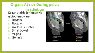
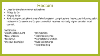
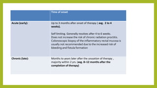
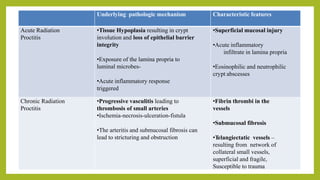
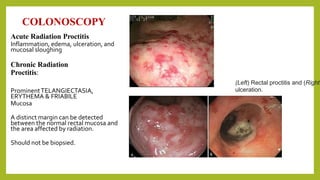
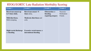
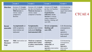
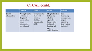
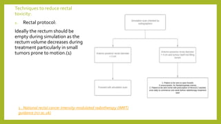
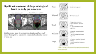
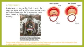
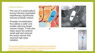
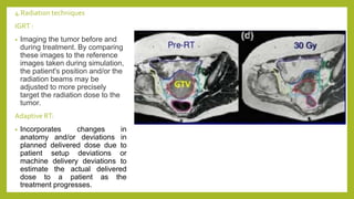
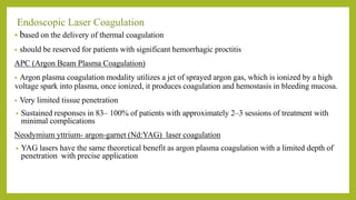
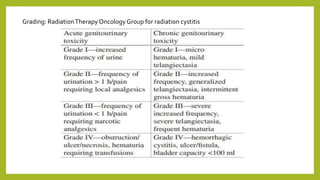
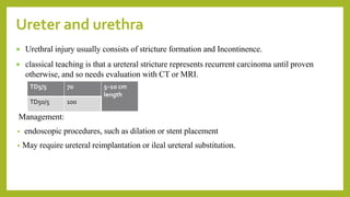
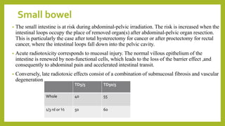
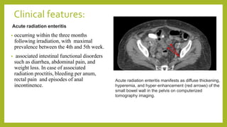
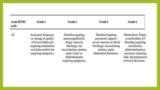
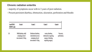
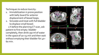
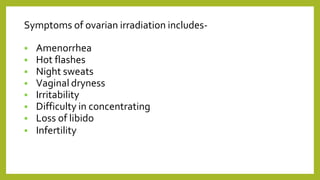
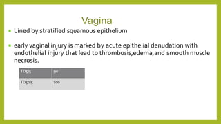
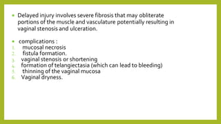
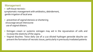
The document discusses classifications of mammalian cell sensitivity to radiation and the radiobiological basis of radiation-induced toxicities. It then focuses on the rectum as an organ at risk for pelvic irradiation, describing the acute and chronic effects of radiation proctitis. Techniques to reduce rectal toxicity during radiotherapy planning and treatment are also outlined, including emptying the rectum during simulation and using rectal spacers or balloons.
This document discusses the treatment of lung cancer with radiation. Stage I-II lung cancers are typically treated with surgery and sometimes post-operative chemotherapy or radiation. Stage III cancers usually receive chemotherapy and radiation, sometimes followed by surgery. Stage IV cancers are treated with chemotherapy or radiation. Advanced techniques like CT-guided planning, adaptive radiotherapy using daily CT images, and stereotactic body radiation therapy can help target radiation doses precisely to tumors while minimizing exposure to healthy lung tissue. Radiation is generally well-tolerated but can cause short-term effects like cough and long-term effects like fibrosis. Careful treatment planning aims to limit radiation doses to normal lungs.
SBRT is a precise form of radiation therapy that delivers very high ablative doses of radiation to tumors in a small number of fractions. It has become the standard of care for early stage non-small cell lung cancer (NSC LC) that is not surgically resectable. Key aspects of SBRT planning and delivery include delineating targets and organs at risk on imaging, determining appropriate dose and fractionation based on tumor location, using motion management strategies to account for tumor motion, precise daily image guidance, and ensuring dose constraints are met to minimize risks to critical structures like the spinal cord. SBRT provides superior local tumor control compared to conventional fractionation for early stage NSCLC with a favorable toxicity profile.
Principles of radiotherapy in gastric carcinomaAnil Gupta
This document discusses principles of radiotherapy for gastric carcinoma. It summarizes that post-operative radiotherapy can reduce local recurrence rates after surgery for gastric cancer, although no survival benefit has been proven. Newer radiation techniques like IMRT and VMAT may further reduce doses to organs-at-risk compared to 3D conformal radiotherapy. Pre-operative radiotherapy can also improve resectability in some inoperable cases.
This document summarizes the results of updated meta-analyses on the use of altered fractionated radiotherapy in head and neck squamous cell carcinoma (HNSCC) patients. The March 2006 meta-analysis found improved overall and progression-free survival with altered fractionation compared to conventional radiotherapy, with hyperfractionation showing the greatest benefit. The updated 2017 meta-analysis confirmed these findings based on 34 trials and over 11,000 patients, with longer follow-up. It found hyperfractionated radiotherapy continued to have a significant survival benefit over moderately or very accelerated regimens.
1) The document discusses various radiation techniques for treating cancer of the esophagus including 2D, 3D conformal radiation therapy, IMRT, and IGRT.
2) It covers topics like target volume delineation, field design considerations for different esophageal subsites, and evolution from 2D to 3D treatment planning.
3) While there is no consensus, most contemporary trials use margins of 3-5cm cranially and caudally on the gross tumor with approximately a 2cm radial margin.
Stomach cancer accounts for 1.3% of new cancers and 1.9% of cancer deaths in the US. Risk factors include H. pylori infection, smoking, and diet low in fruits and vegetables. Symptoms often present at advanced stages and include weight loss, abdominal pain, and nausea. Treatment involves surgery, with chemotherapy and radiation sometimes used adjuvantly or palliatively. Radiation improves local control after surgery and survival outcomes compared to surgery alone. It is also effective for palliation of symptoms from advanced disease.
This document outlines the key aspects of radiotherapy treatment planning for rectal cancer, including:
1) The epidemiology of rectal cancer, stages of disease, and patient positioning and immobilization techniques.
2) How to define the target volumes including the gross tumor, clinical target volume, and planning target volume based on disease stage and risk of lymph node involvement.
3) Typical three-field beam arrangements and doses of 45-50.4 Gy given in 1.8 Gy fractions for preoperative or postoperative radiotherapy, with additional boost doses sometimes used.
4) The acute and chronic complications of radiotherapy and dose constraints for organs at risk like the small bowel and bladder.
This document discusses radiation therapy options for prostate cancer. It notes that treatment depends on risk level: low risk may receive external beam radiation or seeds alone, intermediate risk should receive some external beam, and high risk should receive hormone therapy plus radiation. Newer techniques like IMRT and IGRT reduce side effects by more precisely targeting the prostate. Side effects of radiation include short term issues like urinary frequency and diarrhea as well as long term risks like radiation cystitis and impotence in some cases.
This document discusses reirradiation in recurrent head and neck cancer. It notes that radiation therapy plays a central role in head and neck cancer treatment but recurrence still occurs in 20-35% of patients. Reirradiation presents challenges due to prior radiation exposure and damage to normal tissues. The document discusses treatment options, appropriate patient selection, techniques like IMRT to minimize dose to organs at risk, optimal timing and dosing of reirradiation, and management of toxicities.
Radiotherapy plays an important role in the management of urinary bladder cancers. It can be used as part of bladder-preserving protocols for muscle-invasive bladder cancer or as palliative treatment in elderly patients. Combined modality treatment with transurethral resection and concurrent chemoradiotherapy provides 5-year overall survival of 50-65% and bladder preservation in 38-43% of patients. External beam radiotherapy is typically delivered with a 4-field box technique to the whole pelvis at 45-50 Gy followed by a bladder boost to 60-65 Gy.
Ewing sarcoma is the second most common bone tumor in children. Radiotherapy plays an important role in the treatment of both localized and metastatic Ewing sarcoma. For localized disease, radiotherapy is recommended for patients who cannot undergo surgery or have unresectable tumors. It is also used post-operatively if there is residual disease. For metastatic disease, radiotherapy can help control the primary tumor and reduce pulmonary metastases when combined with chemotherapy. Advances in radiotherapy planning and techniques have improved outcomes while reducing long-term side effects.
Induction chemotherapy followed by concurrent ct rt versus ct-rt in advanced ...Santam Chakraborty
Induction chemotherapy followed by concurrent chemoradiation (CT-RT) has been studied as an alternative to primary CT-RT for locally advanced head and neck cancers. Meta-analyses have found induction chemotherapy provides no survival benefit compared to primary CT-RT and is associated with increased toxicity. Recent large randomized trials could not demonstrate an improvement with induction chemotherapy due to inadequate accrual and poor compliance with subsequent CT-RT. While induction chemotherapy may improve organ preservation or outcomes for select subgroups like HPV-negative cancers, current evidence indicates primary CT-RT remains the standard of care for most patients.
The combined use of radiation therapy and chemotherapy in cancer treatment is a logical and reasonable approach that has already proven beneficial for several malignancies.
RADIOTHERAPY IN CARCINOMA BREAST (EARLY AND LOCALLY ADVANCED)DrAnkitaPatel
This document discusses radiation therapy for breast cancer. It begins by outlining the important role of radiation therapy at various stages of breast cancer, including as part of breast conservation and after mastectomy. It then discusses indications for adjuvant radiation therapy based on factors like tumor size and lymph node involvement. The document reviews evidence from clinical trials demonstrating the benefits of radiation therapy after breast-conserving surgery in reducing recurrence rates and improving survival. It also discusses techniques, dosing, and toxicity considerations for radiation therapy delivery.
This document provides contouring and treatment planning guidelines for stereotactic body radiation therapy (SBRT). It discusses indications, contraindications, simulation, target volume delineation, organ at risk contouring, dose prescription, and plan evaluation for SBRT treatment of lung, spine, liver, and other cancers. Key considerations include ensuring accurate tumor targeting given organ motion, minimizing dose to nearby organs at risk, and prescribing ablative doses in a small number of fractions to achieve tumor control.
Radiotherapy is used as primary treatment for early-stage Hodgkin lymphoma or as part of combined modality treatment with chemotherapy. Historically, large mantle fields covering lymph node regions from the skull to the pelvis were used. More modern approaches use smaller involved field radiotherapy targeting only initially involved lymph node regions after chemotherapy based on imaging. Proper delineation of clinical target volumes requires pre-chemotherapy imaging ideally with PET/CT to define original disease extent.
Management of cacrinoma cervix: Techniques of radiotherapy (2D conventional, 3D Conformal radiotherapy (3DCRT) and IMRT with a review of various contouring guidelines.
Accelerated partial breast irradiation is an alternative to whole breast irradiation in carcinoma breast patients Post breast conserving surgery with equivalent outcome, less duration & less burden on the patient.
1. Radiation therapy plays an important role in the treatment of Wilms tumor, especially for advanced or high-risk cases.
2. It is used preoperatively, postoperatively, and for metastatic disease to reduce the risk of recurrence.
3. The indications and techniques for radiation therapy depend on factors like tumor stage, histology, response to chemotherapy, and whether metastases are present. Precise radiation treatment planning is required to effectively target tumors while sparing healthy tissues.
This document discusses the history and techniques of stereotactic radiosurgery (SRS) and stereotactic body radiation therapy (SBRT). It begins by outlining the early development of SRS by Lars Leksell in the 1950s. It then defines key terms like SRS, SBRT, and fractionated stereotactic radiosurgery. The document goes on to discuss the rationale and advantages of SRS/SBRT, including its ability to deliver high radiation doses with steep dose gradients using multiple beams and image guidance. It also covers topics like tumor oxygenation, cell kill mechanisms, and recent technological advances in the field like VMAT, flattening filter free beams, and 4D
This document provides an overview of interstitial brachytherapy principles and concepts. It discusses the history and evolution of brachytherapy sources from radium to modern radioactive sources like iridium-192. Key concepts covered include dose rate calculations, implant systems like the Paris system, and factors that influence dose distribution from a radioactive source like distance, absorption and scattering. The document also describes temporary and permanent brachytherapy sources and different methods of source application including preloading, afterloading and remote afterloading.
Radiation Therapy in the Management of Lung Cancerflasco_org
This document discusses modern radiation therapy techniques for lung cancer, focusing on non-small cell lung cancer (NSCLC). It summarizes that stereotactic ablative radiotherapy (SABR) is now the standard of care for inoperable stage I NSCLC, providing local control and survival rates comparable or superior to surgery with less toxicity. For stage III NSCLC, concurrent chemotherapy and radiation improves survival compared to sequential treatment, though local control remains challenging and toxicities can be significant. Ongoing studies are exploring dose escalation using intensity-modulated radiation therapy (IMRT) and proton therapy to improve outcomes while reducing normal tissue damage.
TARGET DELINEATION OF CANCER ESOPHAGUSKanhu Charan
1. The document discusses guidelines for delineating target volumes for radiation treatment planning in esophageal cancer.
2. It describes expanding the gross tumor volume (GTV) to create the clinical target volume (CTV) with margins of 4cm above and below the tumor and 1-1.5cm radially, plus inclusion of involved lymph nodes.
3. The planning target volume (PTV) is created by expanding the CTV by 0.5-1cm to account for setup variability and organ motion.
Smart radiotherapy aims to precisely target tumor cells while sparing healthy cells. New techniques described in the document include using hypoxic cell sensitizers to target hypoxic tumor regions, anti-angiogenic agents to inhibit tumor blood vessels, and nanoparticles to enhance radiation dose and selectively deliver drugs. Molecular imaging helps optimize treatment by identifying tumor characteristics. Combining radiotherapy with immunotherapy or targeted depletion of host cells may also improve outcomes. Overall, the document discusses developing more precise radiation approaches through better understanding of tumor biology and microenvironment.
Role of Post-op Radiotherapy in Head and Neck CancersAshutosh Mukherji
This document discusses the role of adjuvant radiation therapy in head and neck cancers. It begins by outlining the use of radical and palliative treatment for stage III and IV diseases. It then reviews several landmark studies that established the benefits of postoperative radiation therapy (PORT) over surgery alone in improving local control and survival. Key factors that determine the need for adjuvant therapy like extracapsular extension, positive margins, and T3/T4 stage are discussed. The document also addresses optimal radiation dose, timing, use of concurrent chemotherapy and altered fractionation schedules based on evidence from clinical trials. While targeted therapies in the adjuvant setting have not proven beneficial so far, ongoing studies are exploring their potential role.
Evaluation of radiotherapy treatment planningAmin Amin
This document discusses the evaluation of radiotherapy treatment planning through the use of various tools and indices. The goals of treatment planning are to ensure the prescription dose adequately covers and conforms to the target volume while minimizing doses to surrounding healthy tissues. Key evaluation tools discussed include isodose distributions, orthogonal planes, dose volume histograms, dose statistics, homogeneity indices, and conformity/coverage indices. These tools provide both qualitative and quantitative assessments of the dose distribution and how well it meets the goals of treatment planning.
Acute and Late Radiation Related Side Effects and their Management in Pelvic ...Dr Kartik Kadia
Acute and Late Radiation Related Side Effects and their Management in Pelvic Malignancies
Carcinoma Cervix - Radiation Related Toxicities and Management
This document discusses the use of radioactive iodine (131I) for diagnosis and treatment of thyroid cancer. Some key points:
- 131I localizes in thyroid tissue and can be used to ablate thyroid remnants after surgery or treat thyroid cancer metastases. It emits beta and gamma radiation.
- For remnant ablation, lower doses (30-100 mCi) are usually sufficient while higher doses (100-200 mCi) may be needed for more aggressive cancers. Success rates are similar between low vs high doses and thyroid hormone withdrawal vs rhTSH.
- Post-therapy scans 2-10 days after treatment can identify additional metastases not seen on diagnostic scans in 10-26% of
1. Small cell lung carcinoma is a highly aggressive malignancy associated with tobacco exposure. It is characterized pathologically by small, round, blue cells with scant cytoplasm and fine chromatin.
2. Prognostic factors include stage, performance status, gender, and normal LDH levels. Staging workup involves imaging of the chest, abdomen, brain and bone as well as biopsy of suspicious lesions.
3. Treatment depends on stage - limited stage receives chemotherapy with thoracic radiation while extensive stage receives chemotherapy alone with consideration of prophylactic cranial irradiation for those who respond to initial treatment. The standard chemotherapy regimen is etoposide and platinum.
Small cell lung cancer (SCLC) accounts for 13% of lung cancers. It is strongly associated with smoking and most patients have metastases at diagnosis. SCLC is classified as limited stage, confined to one hemithorax, or extensive stage with distant metastases. Treatment involves platinum-based chemotherapy with radiation for limited stage. Prophylactic cranial irradiation and chemotherapy are recommended after response to lower risk of brain metastases. Outcomes remain poor due to frequent relapse and development of resistance.
Gastric carcinoma is the second most common cancer-related death worldwide. It typically spreads through direct extension, lymphatics, or hematogenously to distant sites like the liver. Staging involves endoscopy, imaging, and biopsy to determine the depth of invasion and lymph node involvement. Surgery aims to perform a curative resection with negative margins and lymph node dissection, but outcomes remain poor with high rates of recurrence.
- Small cell lung cancer (SCLC) accounts for 10-20% of lung cancers and is strongly linked to smoking. It is an aggressive neuroendocrine tumor that typically recurs after initial treatment.
- SCLC is classified as limited stage (confined to one lung and regional lymph nodes) or extensive stage (has spread widely). Limited stage has a median survival of 16-24 months with chemotherapy and radiation, while extensive stage has a median survival of 6-12 months with chemotherapy alone.
- First-line treatment is platinum-based chemotherapy for both stages. Limited stage also receives chest radiation. Prophylactic cranial irradiation reduces the risk of brain metastases. However, recurrence is common due to S
1. Case 1 involves an 18-year old male presenting with right lower back pain and burning urination for 10 days. Imaging shows kinking of the right ureter at the PUJ and 4.14cm away.
2. Case 2 is a 72-year old male with a history of bladder cancer who now presents with hematuria and oliguria for 10 days. Imaging reveals a large lobulated bladder mass and lung cysts concerning for metastases. Laboratory findings include elevated creatinine and pus cells and red blood cells in the urine.
3. Both cases involve the urinary system, with Case 1 suggestive of possible ure
Ovarian cancer arises from the ovaries and is the 8th most common cancer in women in the US. Approximately 5,500 women in the UK and 21,000 women in the USA are diagnosed with ovarian cancer each year. Risk factors include age, nulliparity, family history, and genetic mutations. Diagnosis involves imaging tests and biopsy of suspicious tissue. Staging determines how far the cancer has spread. Treatment includes surgery to remove the ovaries and nearby tissue, followed by chemotherapy with drugs like paclitaxel and carboplatin to kill any remaining cancer cells. Chemotherapy can cause side effects by damaging rapidly dividing cells, but aims to cure the cancer or prolong life by controlling its growth.
This document summarizes the management of small cell lung cancer (SCLC). It discusses that SCLC typically presents as a large central hilar mass with mediastinal lymph node involvement. Standard treatment for limited stage SCLC is platinum-based chemotherapy with concurrent thoracic radiotherapy. The optimal radiotherapy dose and fractionation schedule remains to be determined. Multiple studies have shown improved survival when radiotherapy is given early with chemotherapy compared to late or delayed radiotherapy. Chemotherapy with etoposide and cisplatin is the standard first-line regimen for SCLC.
This document provides information about breast cancer including types, symptoms, screening recommendations, diagnosis, treatment options, and nursing considerations. It discusses ductal carcinoma, lobular carcinoma, and other less common types. Symptoms may include painless masses, skin changes, or lymph node enlargement. Screening includes monthly breast self-exams and annual mammograms starting at age 40. Treatment involves surgery such as lumpectomy or mastectomy as well as radiation and chemotherapy. Nurses should provide education on wound care, exercises, and lifestyle changes after treatment.
Nuclear medicine techniques such as radioactive iodine scans and therapy are important in evaluating and treating thyroid diseases. Radioactive iodine is selectively taken up and concentrated in the thyroid gland, allowing functional imaging and selective internal radiotherapy for hyperthyroidism and thyroid cancer. Radioactive iodine therapy is the primary treatment for Graves' disease and toxic multinodular goiter. It is also used to ablate residual thyroid tissue after surgery and treat thyroid cancer metastases. Precautions must be taken after radioactive iodine therapy to limit radiation exposure to others.
Nuclear medicine in systemic lymphomasGanesh Kumar
Nuclear medicine plays an important role in managing systemic lymphomas through techniques like FDG PET/CT, radioimmunotherapy (RIT), and assessing atypical presentations. PET/CT is useful for staging, prognostication via interim PET, and response evaluation using Deauville criteria. RIT with radiolabeled antibodies like Zevalin and Bexxar is an effective treatment for relapsed lymphomas. Unusual presentations can include neurolymphomatosis where PET/CT helps with diagnosis, and AIDS-related lymphomas where PET/CT aids in differentiation and treatment guidance.
This document outlines the anatomy, epidemiology, risk factors, diagnosis, and management of thyroid cancer. It discusses the different histological subtypes including papillary, follicular, hurthle cell, anaplastic, and medullary carcinomas. Total thyroidectomy is the primary treatment for localized disease. Radioactive iodine is commonly used as adjuvant therapy for differentiated cancers. External beam radiation therapy may be used for residual or unresectable neck disease. Long term monitoring with serum thyroglobulin is important for recurrence detection.
Radiation can cause both acute and chronic effects depending on the dose. High doses over a short period can cause immediate organ damage and cell death, while low doses over long periods increase cancer risks. Whole body irradiation can lead to acute radiation syndrome depending on dose, with hematopoietic, gastrointestinal and cardiovascular/CNS syndromes being characterized by declining blood counts, diarrhea/dehydration, and organ failure respectively. Radiation is carcinogenic by damaging DNA and causing mutations, increasing risks of cancers like leukemia, thyroid cancer and others. The biological effects of radiation include damage to molecules like DNA, cell death or abnormal function, and tissue/organ damage depending on cell type and proliferation rates.
Effects of radiation
Signs and symptoms of radiation
Infected period of radiation
Dosage
Calculation of dosage
Units and SI units used
Diseases caused by radiation
Radioresistant
Thyroid cancer & hashimotos disease.pptxRITIKARana18
Hashimoto's disease and thyroid cancer are conditions that affect the thyroid gland. Hashimoto's disease is an autoimmune disorder where the body's immune system attacks the thyroid, causing inflammation. It is the most common cause of hypothyroidism in the US. Thyroid cancer develops from the cells of the thyroid and there are several types including papillary, follicular, medullary, and anaplastic carcinomas. Diagnosis involves medical history, physical exam, blood tests and imaging. Treatment depends on the condition but may include hormone replacement therapy, surgery, radiation therapy and nursing management of symptoms.
This document provides an overview of oropharyngeal squamous cell carcinoma. It discusses the background, risk factors, diagnosis, staging, treatment options, and nutritional considerations for this type of head and neck cancer. A case study is presented of a 62-year-old male patient diagnosed with oropharyngeal cancer who underwent chemotherapy and radiation treatment. He experienced significant weight loss but was able to regain weight through enteral nutrition support via a PEG tube. His cancer is now in remission. The document also reviews several research studies on links between diet and cancer risk/outcomes.
The most common type of cancer arising in the kidney: Renal cell carcinoma(RCC)(also known as Hypernephroma or Grawitz tumor).
Renal cell carcinoma accounts for over 3% of all adult malignancies and has several histological subtypes.
Approximately 85% of kidney tumors are renal cell carcinoma, and approximately 70% of these have a Clear cell histology. Its diagnostic work-up, staging and management.
- Thyroid cancer originates from follicular or parafollicular thyroid cells and can present as papillary, follicular, or anaplastic thyroid cancer.
- Signs and symptoms may include a neck lump, difficulty swallowing or breathing, or hoarseness. Some cancers are found incidentally.
- The goal of treatment is to eliminate all cancer cells and may include surgery, radioactive iodine, thyroid hormone therapy, external radiation, or chemotherapy depending on cancer type and stage. Patients require lifelong thyroid hormone replacement and monitoring after treatment.
1. The patient presented with a swelling in his neck for 2 years that had been gradually increasing in size and causing pain for the past 2 months.
2. Examination and investigations including lymph node biopsy, ultrasound neck, and CT neck revealed metastatic papillary carcinoma of the thyroid.
3. The patient underwent a total thyroidectomy with modified radical neck dissection. Post-operative recovery was unremarkable.
Pictorial and detailed description of patellar instability with sign and symptoms and how to diagnose , what investigations you should go with and how to approach with treatment options . I have presented this slide in my 2nd year junior residency in orthopedics at LLRM medical college Meerut and got good reviews for it
After getting it read you will definitely understand the topic.
Nano-gold for Cancer Therapy chemistry investigatory projectSIVAVINAYAKPK
chemistry investigatory project
The development of nanogold-based cancer therapy could revolutionize oncology by providing a more targeted, less invasive treatment option. This project contributes to the growing body of research aimed at harnessing nanotechnology for medical applications, paving the way for future clinical trials and potential commercial applications.
Cancer remains one of the leading causes of death worldwide, prompting the need for innovative treatment methods. Nanotechnology offers promising new approaches, including the use of gold nanoparticles (nanogold) for targeted cancer therapy. Nanogold particles possess unique physical and chemical properties that make them suitable for drug delivery, imaging, and photothermal therapy.
The biomechanics of running involves the study of the mechanical principles underlying running movements. It includes the analysis of the running gait cycle, which consists of the stance phase (foot contact to push-off) and the swing phase (foot lift-off to next contact). Key aspects include kinematics (joint angles and movements, stride length and frequency) and kinetics (forces involved in running, including ground reaction and muscle forces). Understanding these factors helps in improving running performance, optimizing technique, and preventing injuries.
Summer is a time for fun in the sun, but the heat and humidity can also wreak havoc on your skin. From itchy rashes to unwanted pigmentation, several skin conditions become more prevalent during these warmer months.
STUDIES IN SUPPORT OF SPECIAL POPULATIONS: GERIATRICS E7shruti jagirdar
Unit 4: MRA 103T Regulatory affairs
This guideline is directed principally toward new Molecular Entities that are
likely to have significant use in the elderly, either because the disease intended
to be treated is characteristically a disease of aging ( e.g., Alzheimer's disease) or
because the population to be treated is known to include substantial numbers of
geriatric patients (e.g., hypertension).
- Video recording of this lecture in English language: https://youtu.be/Pt1nA32sdHQ
- Video recording of this lecture in Arabic language: https://youtu.be/uFdc9F0rlP0
- Link to download the book free: https://nephrotube.blogspot.com/p/nephrotube-nephrology-books.html
- Link to NephroTube website: www.NephroTube.com
- Link to NephroTube social media accounts: https://nephrotube.blogspot.com/p/join-nephrotube-on-social-media.html
Giloy in Ayurveda - Classical Categorization and SynonymsPlanet Ayurveda
Giloy, also known as Guduchi or Amrita in classical Ayurvedic texts, is a revered herb renowned for its myriad health benefits. It is categorized as a Rasayana, meaning it has rejuvenating properties that enhance vitality and longevity. Giloy is celebrated for its ability to boost the immune system, detoxify the body, and promote overall wellness. Its anti-inflammatory, antipyretic, and antioxidant properties make it a staple in managing conditions like fever, diabetes, and stress. The versatility and efficacy of Giloy in supporting health naturally highlight its importance in Ayurveda. At Planet Ayurveda, we provide a comprehensive range of health services and 100% herbal supplements that harness the power of natural ingredients like Giloy. Our products are globally available and affordable, ensuring that everyone can benefit from the ancient wisdom of Ayurveda. If you or your loved ones are dealing with health issues, contact Planet Ayurveda at 01725214040 to book an online video consultation with our professional doctors. Let us help you achieve optimal health and wellness naturally.
These lecture slides, by Dr Sidra Arshad, offer a simplified look into the mechanisms involved in the regulation of respiration:
Learning objectives:
1. Describe the organisation of respiratory center
2. Describe the nervous control of inspiration and respiratory rhythm
3. Describe the functions of the dorsal and respiratory groups of neurons
4. Describe the influences of the Pneumotaxic and Apneustic centers
5. Explain the role of Hering-Breur inflation reflex in regulation of inspiration
6. Explain the role of central chemoreceptors in regulation of respiration
7. Explain the role of peripheral chemoreceptors in regulation of respiration
8. Explain the regulation of respiration during exercise
9. Integrate the respiratory regulatory mechanisms
10. Describe the Cheyne-Stokes breathing
Study Resources:
1. Chapter 42, Guyton and Hall Textbook of Medical Physiology, 14th edition
2. Chapter 36, Ganong’s Review of Medical Physiology, 26th edition
3. Chapter 13, Human Physiology by Lauralee Sherwood, 9th edition
The Nervous and Chemical Regulation of Respiration
Pelvic irradiation toxicity
1. RADIATION INDUCED
TOXICITIES IN PELVIC
IRRADIATION
Dr KilitoliChophy
Junior Resident
Department of radiation oncology
16th March 2021
2. Casarett classification of mammalian cell
• Arranged from most sensitive (Group I) to least sensitive (Group IV).
• Group I:Vegetative Intermitotic Divides constantly with no differentiation.
Includes basal epithelial cells (skin, intestinal crypts, etc), undifferentiated hematopoietic stem cells and
germ cells.
• Group II: Differentiating Intermitotic Divides for a finite amount of cycles before differentiating into a non-
dividing cell.
• Includes all of the cells that are intermediate between stem cells and differentiated cells. For example,
myelocytes, spermatogonia, etc.
• Group III: Reverting Postmitotic : normally non-dividing cell that retains the potential to divide (“revert”).
• Includes liver, kidneys and glandular tissues such as pancreas, adrenal, thyroid and pituitary.
• Group IV: Fixed Postmitotic :A permanently non-dividing cell.
• Includes permanent cells such as nerves and muscles, as well as short-lived differentiated cells such as
neutrophils, red blood cells, and superficial epithelial cells.
3. • alpha by beta ratio :radiation dose at which the alpha
killing (lethal) is equal to beta killing (combinations of
sublethal killing leading to lethal killing)
• Early responding tissues:
α/β is large, as a consequence i.e. irreparable
damage dominates at low doses, and dose response
curve has marked initial slope & bends at higher doses.
Cells with poor repair capability (e.g. tumours) tend to
develop more lethal damage
• Late responding normal tissue:
α/β is small ,i.e. β term (repairable damage) has an
influence at low doses.
normal late responding tissue around the tumour with a
lower alpha by beta ratio tends to get damaged more.
Radiobiological basis:
4. • Therefore, adopting a dose per fraction more
than the alpha by beta ratio will kill more cells
than when dose per fraction is less than the
alpha by beta ratio for the same given total
dose.
5. • The therapeutic index (TI) defines how theTCP relates to NTCP for different doses of radiation .
• TCP and NTCP curves are sigmoid in shape
• TI = NTCP/TCP
• Usually radiosensitive tumours like seminoma have a wide therapeutic index, while those with
radioresistant tumors have a narrow therapeutic index.
• An ideal radiotherapy plan where there is 100% chance of tumor control and 0% chance of normal tissue
toxicity. Achieving an optimal balance betweenTCP and NTCP is a basic aim of any radiotherapy plan.
Therapeutic Index
6. ModifyingTherapeutic Index
1. Reducing the size of the target volume and the margins by using image guidance in radiotherapy
planning.
2. Hyperfractionation— small dose per fraction with two or three fraction delivered per day is used to
achieve a higher biologically effective dose to the tumor. Reduces the chances of long term normal
tissue complications
3. Concurrent Chemotherapy—The use of concurrent chemotherapy acts as a radiosensitizer and thereby
shifts theTCP to left.
4. RadiationSensitizers—The use of radiosensitizers helps in optimizing therapeutic index by overcoming
hypoxia
5. Extracorporeal radiotherapy
6. Radio Protectors—The radio protectors (e.g.,Amifostine) mainly act by neutralizing free radicals
generated by ionizing radiations in the normal tissue, thereby reducing normal tissue complication
rates
7. These are normal tissues whose radiation may significantly
influence the treatment planning or prescribed dose.
It is divided into three classes.
1. Class I-Radiation lesions are fatal or result in
severe morbidity.
2. Class II-Radiation lesion result in mild to moderate morbidity.
3. Class III-Radiation lesion are mild, transient and reversible or result
into no significant morbidity.
ICRU 62
8. CLASSIFICATION OF ORGANS AT
RISK
•Serial – whole organ is a continuous unit and damage at
one point will cause complete damage of the organ
(spinal cord, digestive system). So even point dose is
significant.
•Parallel – organ consists of several functional units and
if one part is damaged, the rest of the organ makes up
for the loss (lung, bladder). Dose delivered to a given
volume or average/mean dose is considered .
•Serial-parallel – kidney (glomerulus- parallel, tubules-
serial), heart (myocardium- parallel, coronary arteries-
serial). ICRU-62
9. Pelvic RT indications:
1. Ca cervix : Radical concurrent Chemoradiation, Adjuvant radiation
45-50 Gy+ brachytherapy
2. Ca Endometrium: adjuvant RT 45Gy/25# + brachytherapy
3. Ca bladder: Radical RT 64Gy/32#
4. Ca Prostate: Radical RT Radical 74Gy/37#, Hypofractionation 60Gy/20#, post op adjuvant RT
, salvage RT, (brachy alone or as boost in early prostate)
5. Ca vulva and Ca vagina : Concurrent Chemoradiation pelvis 45 to 60Gy +/- brachytherapy
6. Ca Penis : Adjuvant pelvic + inguinal RT , radical Concurrent Chemoradiation (penis +LNs)
7. Ca anal canal : Concurrent Chemoradiation 50-60 Gy
8. Ca rectum NACCRT 50.4Gy/28# , short course NART 25 Gy/5# hypofractionation.
10. Organ at risk during pelvic
radiotherapy are:
1. Bladder
2. Rectum
3. Urethra & Ureter
4. Small bowel
5. Vagina
6. Gonads
11. • Lined by simple columnar epithelium.
• TD5/5 60 Gy
• TD50/5 80 Gy
• Radiation proctitis (RP) is one of the long term complications that occurs following pelvic
radiation in Ca cervix and Ca prostate which requires relatively higher dose for local
control.
Symptoms:
•diarrhea (common)
•fecal urgency
•tenesmus
•mucous discharge
Rectum
•constipation
•fecal incontinence
•Anorectal dysfunction
•mucous discharge
•rectal bleeding
12. Time of onset
Acute (early): Up to 3 months after onset of therapy ( avg. 2 to 4
weeks).
Self limiting. Generally resolves after 4 to 6 weeks.
Does not increase the risk of chronic radiation proctitis.
Colonoscopic biopsy of the inflammatory rectal mucosa is
usually not recommended due to the increased risk of
bleeding and fistula formation
Chronic (late): Months to years later after the cessation of therapy ,
majority within 2 yrs. (avg. 8–12 months after the
completion of therapy)
13. Underlying pathologic mechanism Characteristic features
Acute Radiation
Proctitis
•Tissue Hypoplasia resulting in crypt
involution and loss of epithelial barrier
integrity
•Exposure of the lamina propria to
luminal microbes-
•Acute inflammatory response
triggered
•Superficial mucosal injury
•Acute inflammatory
infiltrate in lamina propria
•Eosinophilic and neutrophilic
crypt abscesses
Chronic Radiation
Proctitis
•Progressive vasculitis leading to
thrombosis of small arteries
•Ischemia-necrosis-ulceration-fistula
•The arteritis and submucosal fibrosis can
lead to stricturing and obstruction
•Fibrin thrombi in the
vessels
•Submucosal fibrosis
•Telangiectatic vessels –
resulting from network of
collateral small vessels,
superficial and fragile,
Susceptible to trauma
14. COLONOSCOPY
Acute Radiation Proctitis
Inflammation, edema, ulceration, and
mucosal sloughing
Chronic Radiation
Proctitis:
ProminentTELANGIECTASIA,
ERYTHEMA & FRIABILE
Mucosa
A distinct margin can be detected
between the normal rectal mucosa and
the area affected by radiation.
Should not be biopsied.
(Left) Rectal proctitis and (Right)
ulceration.
15. RTOG/EORTC Late Radiation Morbidity Scoring
Grade 1 Grade 2 Grade 3 Grade 4
Bowel movement up
to 5 times daily,
Mild diarrhoea,
Mild cramping
Slight rectal discharge
or bleeding
Bowel movement >5
times daily,
Moderate diarrhoea and
colic
Excessive rectal mucus or
intermittent bleeding
Obstruction or
bleeding
requiring surgery
Necrosis/
Perforation
Fistula
16. CTCAE 4
Grade 1 Grade 2 Grade 3 Grade 4
Diarrhea
Rectal
mucositis
Increase of <4 stools
per day over baseline;
Mild increase in
ostomy output
compared to
Baseline
Asymptomatic or
mild symptoms;
intervention not
indicated
Increase of 4 - 6 stools
per day over baseline;
Moderate increase in
ostomy output
compared to baseline
Symptomatic;
medical intervention
indicated; limiting
instrumentalADL
Increase of ≥7 stools
per day over baseline;
incontinence;
hospitalization
indicated; severe
increase in ostomy
output compared to
baseline; limiting self
care ADL
Severe symptoms;
limiting self
careADL
Life-threatening
consequences;
urgent intervention
indicated
Life-threatening
consequences;
urgent
operative
intervention
indicated
Rectal
Haemorr
hage
Mild; intervention not
indicated
Moderate symptoms;
medical intervention
or minor cauterization
indicated
Transfusion,
radiologic,
endoscopic, or
elective
operative intervention
indicated
Life-threatening
consequences;
urgent
Operative
intervention
indicated
17. CTCAE contd.
Grade 1 Grade 2 Grade 3 Grade 4
Rectal
obstruction
Asymptomatic
; clinical or
diagnostic
observations
only;
intervention
not indicated
Symptomatic;
altered GI
function;
limiting
instrumental
ADL
Hospitalizatio n
indicated;
elective
operative
intervention
indicated;
limiting self
care
ADL; disabling
Life-
threatening
consequences;
urgent
operative
intervention
indicated
18. Techniques to reduce rectal
toxicity:
1. Rectal protocol:
Ideally the rectum should be
empty during simulation as the
rectum volume decreases during
treatment particularly in small
tumors prone to motion.(1)
1 . National rectal cancer intensity-modulated radiotherapy (IMRT)
guidance (rcr.ac.uk)
20. 2. Rectal spacers:
Rectal spacers are used to limit dose to the
anterior rectal wall in high dose external beam
radiation therapy of the prostate and have
been shown to reduce radiation induced
toxicity.
Rectum-spacer related acute toxicity – endoscopy results of 403 prostate cancer patients after implantation of gel
or balloon spacers | Radiation Oncology | FullText (biomedcentral.com)
21. 3. Rectal balloon:
• The use of a rectal balloon
during intensity-modulated
radiotherapy significantly
reduces prostate motion.
• Prostate immobilization
thus allows a safer and
smaller planning target
volume margin. It has also
helps spare the anterior
rectal wall and reduced
the rectal volume that
received high-dose
radiation
The use of rectal balloon during the delivery of intensity modulated
radiotherapy (IMRT) for prostate cancer: more than just a prostate
gland immobilization device? - PubMed (nih.gov)
22. 4.Radiation techniques
IGRT :
• Imaging the tumor before and
during treatment. By comparing
these images to the reference
images taken during simulation,
the patient's position and/or the
radiation beams may be
adjusted to more precisely
target the radiation dose to the
tumor.
Adaptive RT:
• Incorporates changes in
anatomy and/or deviations in
planned delivered dose due to
patient setup deviations or
machine delivery deviations to
estimate the actual delivered
dose to a patient as the
treatment progresses.
23. 5. Radioprotectors
Amifostine rectal suspension
A thiol derivative, acts as a free radical scavenger and radioprotector
Simone et al. 2008
2 g vs. 1 g of amifostine given in a rectal suspension to patients receiving radiotherapy for Prostate cancer
placed 30–45 min before each radiation treatment.
Reduced RTOG ≥ Grade 2 toxicity & improved acute and late bowel quality of life scores compared to patients
who received 1 g of amifostine.
25. Acute Proctitis: Self-limiting
• Cessation of therapy is usually the definitive treatment and surgical
interventions are rarely needed.
• Sitz baths
• Stool softeners,
• Antimotility agents
• Ethamsylate/Tranexamic acid
• Adequate oral hydration
• Probiotics:
Modulate the pro-inflammatory pathways and prevent transcription of the
dependent pro-inflammatory genes by intestinal epithelial cells
26. • steroid or 5-aminosalicylate enemas
• Sucralfate:
It mechanically protects the gastrointestinal mucosa by forming a
protective coating on inner surface of the bowel and stimulate healing by
increasing angiogenesis.
Administration of sucralfate enema given as 2gm/day bd.
• Hyperbaric oxygen (HBO)—HBO is also an effective modality in
management of RP, especially in patients not responding to conservative
management. A
27. Formalin
• scleroses and seals fragile neovasculature in radiation damaged
tissues to prevent further bleeding through chemical
cauterization
• its success is entirely dependent on accurate localization.
• rectal instillation of 4% formalin solution or direct topical
application of a 10% formalin solution. Topical formalin is
generally applied through a rigid proctoscope, flexible
endoscope, or by direct application with formalin soaked gauze
Contact with formalin for 2-3 minutes (until slight blanching of
the mucosa is achieved) is allowed.
28. Endoscopic Laser Coagulation
• based on the delivery of thermal coagulation
• should be reserved for patients with significant hemorrhagic proctitis
APC (Argon Beam Plasma Coagulation)
• Argon plasma coagulation modality utilizes a jet of sprayed argon gas, which is ionized by a high
voltage spark into plasma, once ionized, it produces coagulation and hemostasis in bleeding mucosa.
• Very limited tissue penetration
• Sustained responses in 83– 100% of patients with approximately 2–3 sessions of treatment with
minimal complications
Neodymium yttrium- argon-garnet (Nd:YAG) laser coagulation
• YAG lasers have the same theoretical benefit as argon plasma coagulation with a limited depth of
penetration with precise application
29. Surgery
Reserved for severe, complicated cases refractory to other options.
• The most frequent indications for surgery are fistulas and strictures causing
obstruction
• Fistulas- often require a diverting colostomy before
definitive resection or in high risk surgical candidates.
• Rectal strictures may also be relieved by dilators and stent
• Other indications are perforation or refractory bleeding- may be treated by a variety
of surgical techniques including combinations of excision, diversion, and
reconstruction
30. The urinary bladder and ureters are covered by urothelium—
transitional epithelium.
•
Bladder
TD5/5 TD50/5
2/3rd 65 80
1/3 rd 80 85
31. Urothelium
Nuclear irregularity
Cellular oedema
Increased cytoplasmic elements
Disruption of tight junctions & polysaccharide layer
Vasculature
Vascular endothelial cell oedema
Endothelial cell proliferation
Perivascular fibrosis
Muscle
Smooth muscle oedema
Replacement of smooth muscle with fibroblasts
Increased collagen deposition
Vascular ischaemia of bladder wall
Mechanism of radiation damage
32. Symptoms of radiation induced cystitis:
• Acute side effects include urinary frequency, urgency, and dysuria.
• Late effects include hematuria, fistula, obstruction, ulceration, contracted
bladder, vesicovaginal fistula, necrosis, spasm, reduced flow, and
incontinence.
• Median onset of late complications after radiation is 13–20 months.
35. In early reactions
Symptomatic frequency and urgency are best treated with anticholinergic agents. These
agents act primarily by inhibiting involuntary detrusor muscle contractions.
Oxybutynin chloride is an antispasmodic that relaxes the bladder smooth muscle and
relieve the symptoms of frequency and urgency.
Detrol has a greater inhibitory effect on bladder contraction and it has fewer side effects
as compare to oxybutynin (eg, dryness of mouth), usual adult dose is 2 mg bd till
symptomatic relief is obtained.
Saline bladder irrigation: Continuous bladder irrigation and clot removal , removes
urokinase, preventing bleeding
Bladder irrigation with Alum (1%) irrigation: Acts by protein precipitation leading to
vasoconstriction and reduction in edema and inflammation.
Management
36. Phenazopyridine (Pyridium) can be used to provide symptomatic relief in dysuria.
It is a azo dye, it acts directly on urinary tract mucosa to produce local analgesic
effect.
The usual adult dose of pyridium is 100 to200 mg tds for 5 to 7 days.
If dysuria associated with UTI, then antibiotics should be added.
37. Botulinum toxin-
Detrusor injections of botulinum toxin are approved by the FDA for the treatment
of adults with over active bladder who do not adequately respond to
anticholinergic medication.
Acts by inhibition of the release of acetylcholine from the presynaptic nerve
terminal, which prevents stimulation of the detrusor muscle.
Recommended total dose is 100 Units injection across 20 sites into the detrusor
muscles by cystoscope in every 12 weeks.
38. Hematuria:
• The primary treatment modality for hematuria is bladder irrigation. Intravesical
treatments with silver nitrate, prostaglandins or formalin have also been used.
• In severe hematuria other treatment involves the injection of a sclerosing agent (1%
ethoxysclerol) into the bleeding areas to control the
• other interventions can include embolization of the hypogastric arteries or urinary
diversion and cystectomy.
Contracted bladder:
• Treated by reconstruction of bladder with a segment of small or large bowel (sigmoid
colon).
• Cystectomy- This procedure is used as a last resort for contracted bladder and involves
removing the bladder and surgically constructing a replacement or an stoma on the
skin.
39. Urethral injury usually consists of stricture formation and Incontinence.
classical teaching is that a ureteral stricture represents recurrent carcinoma until proven
otherwise, and so needs evaluation with CT or MRI.
Management:
• endoscopic procedures, such as dilation or stent placement
• May require ureteral reimplantation or ileal ureteral substitution.
Ureter and urethra
TD5/5 70 5–10 cm
length
TD50/5 100
40. Small bowel
• The small intestine is at risk during abdominal-pelvic irradiation. The risk is increased when the
intestinal loops occupy the place of removed organ(s) after abdominal-pelvic organ resection.
This is particularly the case after total hysterectomy for cancer or after proctectomy for rectal
cancer, where the intestinal loops fall down into the pelvic cavity.
• Acute radiotoxicity corresponds to mucosal injury. The normal villous epithelium of the
intestine is renewed by non-functional cells, which leads to the loss of the barrier effect ,and
consequently to abdominal pain and accelerated intestinal transit.
• Conversely, late radiotoxic effects consist of a combination of submucosal fibrosis and vascular
degeneration
TD5/5 TD50/5
Whole 40 55
1/3 rd or ½ 50 60
41. Clinical features:
Acute radiation enteritis
• occurring within the three months
following irradiation, with maximal
prevalence between the 4th and 5th week.
• associated intestinal functional disorders
such as diarrhea, abdominal pain, and
weight loss. In case of associated
radiation proctitis, bleeding per anum,
rectal pain and episodes of anal
incontinence.
Acute radiation enteritis manifests as diffuse thickening,
hyperemia, and hyper‐enhancement (red arrows) of the
small bowel wall in the pelvis on computerized
tomography imaging.
42.
43. Chronic radiation enteritis
• majority of symptoms occur with in 3 years of post radiation.
• Present persistent diarrhea, obstruction, ulceration, perforation and bleedin
44. Management:
Dietary modifications- A low-residue diet and a diet low in fats and lactose
free diet.
Drugs-Antispasmodics, anticholinergics and opiates can improve symptoms
of pain and diarrhoea by reducing motility. Oral antibiotics may improve
diarrhoea in associated bacterial colonization.
Total parenteral nutrition-Bowel rest and total parenteral nutrition (TPN)
may be used for severe symptomatic disease and for enterocutaneous
fistulas
Surgical management- Indications for surgery include bowel obstruction,
perforation, abscess, intractable bleeding or diarrhoea, and occasionally
malabsorption.
45. Techniques to reduce toxicity:
1. Immobilization in prone position
with belly board for anterior
displacement of bowel loops.
2. Simulate and treat with full bladder
to displace small bowel.
1 h before the planning CT scan, ask
patient to first empty bladder
completely, then drink 750 ml of water
in the space of 10–15 min and then wait
without emptying their bladder for 40–
60 min.
46. The testis is one of the most radiosensitive tissues in the body with a
radiation dose as low as 15 cGy causing a significant depression in the
sperm count.
Direct testicular irradiation of 24 Gy results in ablation of the germinal
epithelium which is responsible for sperm development and Leydig cell
function (production of testosterone).
Leydig cell function is usually preserved up to 20 Gy in prepubertal boys
and 30 Gy in sexually mature men.
Leydig cell function can be monitored by both testosterone and LH serum
levels. Normal testosterone value and elevated LH levels are indicative of
Leydig cell damage.
Testes
47. Cell Radiation dose Toxicity Recovery
Spermatogonia ≤1 Gy Oligospermia 9–18 months
Spermatocytes 1–3 Gy Azoospermia 10–30 months
Spermatids >3 Gy Azoospermia >60 months
Sensitivity of gonadal tissues following single dose irradiation and time to recovery
• Permanent azoospermia occurs after about 6 to 8 Gy in 2-Gy fractions
48. When testes present directly in the
irradiation field, it cannot be
protected.
The dose from scattered
irradiation from nearby beams,
can be reduced by moving the
gonad away from or by applying
thick shielding cups directly over
the scrotum.
Clamshell shield has aV cut for inserting the
testis and to hold in position,wax coating is done
on the inner surface of the shields. It makes the
inner surface smooth and avoid back-scattered
electron dose from the shield.
Prevention:
49. Features of hypogonadism:
Loss of libido
Erectile dysfunction
Fatigue
Decreased muscle mass
Body and facial hair loss
Difficulty in concentrating
Depression
Irritability
Low sense of well-being
50. Management:
Androgen replacement therapy to enable normal pubertal
development and future sexual function for patients with deficient
testosterone production.
Inj testesterone 200 mg (I.M.) in every 2 weeks till regression of
symptoms occurs.
Pre treatment sperm banking for fertitlity preservation
51. Effect on Pelvic Bones:
irreversible physiologic changes including narrowing of the vascular
channels (endarteritis)
blood flow↓
loss of osteocytes, osteoblasts
limited re-modelling of bone and limited healing .
Resulting injuries include atraumatic femoral neck fracture, and
osteonecrosis of the femoral head or of the acetabulum.
• Pathophysiology
52. Avascular necrosis of the femoral head
Right femoral head/acetabulum pathologic fracture identified on (Left) x‐ray and (Right) computed
tomography.
53. The tolerance doses for the femoral head
TD 5/5 is 52Gy , TD 50/5 is 65 Gy
Prevetion: Femoral head sparing using conformal radiotherapy
Treatment for bone fractures following radiation
1. calcium supplements
2. pain management with analgesics
3. surgical fixation.
54. Dermatological effects
• Sequential events of skin reactions of radiation
RT dose Gross change
> 5 Gy Transient erythema Due to capillary dilatation
mediated by histamine like
substances.
~ 20 Gy Epilation
20 – 40 Gy Dusky
pigmentation
Initially punctuated and then
coalesced – skin warm and
edematous.
~45 Gy
50 – 60 Gy
Dry desquamation
Moist
desquamation
Due to depletion of basal layer.
Epidermal slough and serous
discharge.
55. TD 5/5 55 Gy 100 cm2
50/5 65 Gy
TD 5/5 70 Gy 10 cm2
5/5 60 Gy 30 cm2
• most commonly observed in the
vulva, perineum, and the inguinal
and gluteal folds.
56. • Prevention:
1. Positioning: Frog leg position (supine with legs
abducted ) to reduce perineal skin reactions (in ca
vagina )
2. IMRT may decrease the risk of vulvar skin toxicity,
and one series reported no grade 3 groin skin
desquamation(1)
3. s
Beriwal S, Shukla G, Shinde A, et al. Preoperative intensity modulated radiation therapy and chemotherapy
for locally advanced vulvar carcinoma: analysis of pattern of relapse. Int J Radiat Oncol Biol
Phys. 2013;85:1269‐1274.
57. Management:
• Gentle cleaning with a mild, unperfumed soap is advised for folliculitis.
• moisturizing treated skin using hydrophilic moisturizer
• preventing mechanical or chemical irritation of treated skin (such as resulting from tight clothing
or perfumes) encouraged to wear loose‐fitting and cotton clothing and to avoid temperature
extremes
• adequate pain management
• Daily use of a sitz bath with the addition of sodium bicarbonate, Epsom salts, for symptomatic
relief.
• For patients with desquamation, the application of silver clear nylon, nonadherent or hydrogel
dressings,, creates a moist environment that stimulates wound healing. Vaseline gauze
impregnated with antibiotics may also provide topical relief and prevent secondary infection.
58. Radiation to the ovaries can damage oocytes and result in premature menopause as a
result of ovarian failure
Oocytes undergo meiosis and are relatively radioresistant (single dose LD50 is 4Gy)
however, proliferating granulosa cells are radiosensitive.
a total dose of 24 Gy leads to ablation of ovaries due to the loss of granulosa cells.
Immediate ovarian failure will be produced by 16.5 Gy in females of 20 years, and 14
Gy in 30-year-olds or in elderly group.
Ovaries
TD5/5 2-3 Whole ovary
(age
dependent )
TD50/5 6-12
59. Symptoms of ovarian irradiation includes-
Amenorrhea
Hot flashes
Night sweats
Vaginal dryness
Irritability
Difficulty in concentrating
Loss of libido
Infertility
60. Hormonal therapy-Estrogen therapy can help prevent
osteoporosis and relieve hot flashes and other symptoms of
estrogen deficiency. Estrogens can be administered orally or
transdermally.
The usual adult dose for transdermal estradiol is 100-150 mcg and
oral estradiol 2 to 4 mg.
Progestins should be administered cyclically, 10-14 days in each
month, to prevent endometrial hyperplasia.
The recommended regimens include medroxyprogesterone 10
mg daily for 10-12 days in each month.
Preservation of fertility by egg storage (oocyte) is a useful
technique prior to treatment.
Management:
61. Lined by stratified squamous epithelium
early vaginal injury is marked by acute epithelial denudation with
endothelial injury that lead to thrombosis,edema,and smooth muscle
necrosis.
Vagina
TD5/5 90
TD50/5 100
62. Delayed injury involves severe fibrosis that may obliterate
portions of the muscle and vasculature potentially resulting in
vaginal stenosis and ulceration.
complications :
1. mucosal necrosis
2. fistula formation.
3. vaginal stenosis or shortening
4. formation of telangiectasia (which can lead to bleeding)
5. thinning of the vaginal mucosa
6. Vaginal dryness.
63. soft tissue necrosis :
symptomatic management with antibiotics, debridement,
gentle irrigation of local area
prevention of vaginal stenosis or shortening
encourage sexual intercourse
use of vaginal dilators.
Estrogen cream or systemic estrogen may aid in the rejuvenation of cells and
increase the elasticity of the vagina.
Vaginal douches: Twice daily use of a 1:10 diluted hydrogen peroxide douche can
prevent the formation of necrotic tissue, particularly in previously irradiated patients
Management
64. Bone marrow Suppression
• stem cells are particularly radiosensitive
• dose as low as 0.3 Gy leads to a reduction
in the number of lymphocytes. After
larger doses, lymphopenia is followed by
granulopenia, then thrombopenia, and
finally anemia.
• Hematologic toxicity predisposes patients
to infection, hospitalization, and
requirements for transfusions and growth
factors and lead to delayed or missed
chemotherapy cycles and treatment
breaks, which potentially may
compromise disease control.
65. Prevention:
1 . Sparing functional BM subregions:
Integration of functional imaging to identify
active BM subregions .
PET, single positron emission CT (SPECT),
and/or specialized MRI sequences
2. Bone marrow sparing IMRT
66. Management
1. Weekly blood counts are routinely obtained.
Chemotherapy is withheld when the neutrophil count decreases below 1500/μL., Platelet counts
fall below 100,000/ μL.
Transfusions to maintain a hemoglobin level above 10 mg/dL.
RT is withheld when the neutrophil count approaches 500/μL to 1000/μL.
2. Antibiotics in event of infections
67. Erectile dysfunction
Critical erectile function and that receive a
substantial radiation dose include
neurovascular bundles (NVBs), internal
pudendal arteries (IPAs), accessory pudendal
arteries, corpora cavernosa and the penile bulb.
The incidence of ED progressively
increases after RT in Ca prostate (1).
Prevention:
IMRT technique with sparing of penile bulb
(1).Pinkawa M, Gagel B, Piroth MD, Fischedick K, Asadpour B, Kehl M, Klotz J, Eble MJ. Erectile
dysfunction after external beam radiotherapy for prostate cancer. Eur Urol. 2009 Jan;55(1):227-34. doi:
10.1016/j.eururo.2008.03.026. Epub 2008 Mar 24. PMID: 18375048.
(2). Sparing of the penile bulb and proximal penile structures with intensity-modulated radiation therapy for
prostate cancer, J Kao, J Turian, A Meyers, R J Hamilton, B Smith, S Vijayakumar, and A B Jani
The British Journal of Radiology 2004 77:914, 129-136
68. (1) Incrocci L. Radiotherapy for prostate cancer and sexual health. Transl Androl Urol. 2015
Apr;4(2):124-30. doi: 10.3978/j.issn.2223-4683.2014.12.08. PMID: 26813740; PMCID:
PMC4708125.
Management:
Patients and their partner need to be counselled
Phosphodiesterase type 5 inhibitors (PDE5-I) sildenafil and tadalafil
have been shown to be effective to treat post-radiation ED in about half
of the patients in randomized trials. (1)
69. Secondary malignancy
I. Secondary Bladder Cancer: Radiation for rectal cancer found to be associated with an
increased risk of secondary bladder cancer (1)
II. Secondary Leukemia: pelvic radiation found to be associated with an increased risk of
secondary leukemia, risk is greatest 5 to 10 years after treatment and remains elevated
for up to 15 years after radiotherapy. The association is strongest for acute myeloid
leukemia, with an increased risk of acute lymphocytic leukemia and chronic myeloid
leukemia. (2)
1. Frontiers | Risk and Prognosis of Secondary Bladder Cancer After RadiationTherapy for Rectal Cancer: A Large
Population-Based Cohort Study | Oncology (frontiersin.org)
2. Pelvic radiotherapy and the risk of secondary leukemia and multiple myeloma -Wright - 2010 - Cancer -Wiley
Online Library
70. References:
Eric J. Hall, Amato J. Giaccia - Radiobiology for the Radiologist-LWW (2018)
ICRU 50 and ICRU 62
Optimal organ-sparing intensity-modulated radiation therapy (IMRT) regimen for the treatment of
locally advanced anal canal carcinoma: a comparison of conventional and IMRT plans (nih.gov)
Complications of pelvic radiation in patients treated for gynecologic malignancies -Viswanathan -
2014 - Cancer -Wiley Online Library
Basic clinical radiobiology / edited by Michael C.Joiner and Albert J.Van der Kogel. Description: Fifth
edition
Supriya Mallick • Goura K. Rath Rony Benson Editors Practical Radiation Oncology