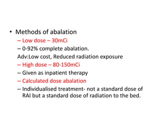
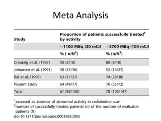
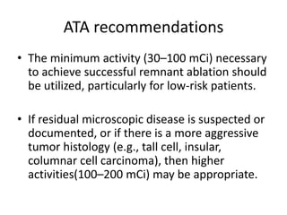
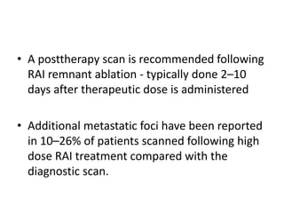
• Introduced by Seidlin et al. in 1946
• Physical half-life of 131I is 8.02 d.
• Mainly emit B rays– 90% of radioactivity of
131I, (and Gama rays)
• 131I is available for oral ingestion as sodium
iodine
• Most of the radiation dose is delivered by Bparticles
• B-particles do not penetrate deep into tissue(2
mm in depth, at most)
• Gama-radiation contributes only 10% of the total
radiation dose, fraction of gama-rays –absorbed
by functioning tyhroid tissue and vast majority
leaving via skin surface-Detected by a radiation
detector
• As liquid solution or in capsules.
• Each capsule accounts for 50 micro curie
• Capsules safer than liquid- less radioactivity
released into air during handling.
• Also result in less oral mucosal irritation
• Simultaneous ingestion large amounts of water
attenuates radiation dose emitted to gastric wall
• Rapidly and completely absorbed in the upper
intestine
• Picture of the capsule to be added
Iodine Metabolism
• Dietary iodine is absorbed in the GI tract, then taken
up by the thyroid gland (or removed from the body
by the kidneys).
• The transport of iodide into follicular cells is
dependent upon a Na+/I- cotransport system.
• Iodide taken up by the thyroid gland is oxidized by
peroxide in the lumen of the follicle:
peroxidase
I-

I+

• Oxidized iodine can then be used in production of
thyroid hormones.
Cancer vs normal cells
• Metabolism of radioiodine in papillary and
follicular carcinoma is profoundly altered when
compared with normal thyroid tissue.
• Several defects are present in cancer tissue:
– iodine uptake, via the sodium-iodide symporter (NIS),
is always decreased and is undetectable in about a
third of patients;
– Iodine organification is markedly reduced;
– Effective half-life of iodine in tumor tissue is always
shorter
• Tissues that often take up iodine and can be
misconstrued as metastases include the
– salivary glands in the mouth,
– esophagus (as a result of swallowing radioactive
saliva),
– thymus gland,
– breasts in some women,
– liver, stomach, colon, bladder
Prerequisites
• TSH of >30mU/L is associated with increased
RAI uptake in tumors
– withdrawal of LT-3for 2 weeks, or
– discontinuation of LT-4for 3 weeks
– rhTSH stimulation
Enhancing radioiodine uptake
• Low iodine diet
• Lithium - 10 mg/kg/day for 7 days. To keep S. Lithium levels at
0.8-1.2 mmol/L)

– Lithium had been shown to reduce exit of iodine from
normal thyroid cells
• Retinoic acid (1.2 mg/kg/day) – retinoids -apparently
redifferentiate thyroid cancer cells - enhanced
radioiodine uptake
Other agents:
• Based on increasing NIS expression in thyroid cancer cells
Histone deacetylate inhibitors
Demethylating agents
To be avoided
• FOOD SUPPPLEMENTS
• Iodised salt/Salty food-potato chips,nuts
• Sea food

•
•
•
•

DRUGS
1)iodex
2)amiodarone
3)Contrast medias(avoid CT scans)
• Post operative diagnostic I 131 scan –
– Doses less than 5 mCi – unlikely to miss out
treatable foci of disease
– Large dose causes stunning
We use 50 micro curie 131I(one capsule orally)
Do the neck uptake scan using a rectilinear scan.
If uptake <4% - proceed with WBS
WBS- 2 mci used as a solution. Drink with straw.
Stunning effect
• Stunning is defined as a reduction in uptake of
the 131I therapy dose induced by a
pretreatment diagnostic activity.
• Occurs most prominently with higher activities
(5–10 mCi) of 131I ,with increasing time
between the diagnostic dose and therapy
• Does not occur if the treatment dose is given
within 72 hours of the scanning dose.
ATA guidelines
• Based on Risk stratification of individual
patient,
• The primary goal of the first dose of RAI after
total thyroidectomy may be
– Remnant ablation (to facilitate detection of
recurrent disease and initial staging),
– Adjuvant therapy (to decrease risk of recurrence
and disease specific mortality by destroying
suspected, but unproven metastatic disease)
– RAI therapy (to treat known persistent disease).
Radio iodine abalation
Ablation of small amount of residual normal thyroid
remaining after surgery, left behind inadverently
or deliberately
May facilitate the
– early detection of recurrence based on serum Tg
– RAI WBS
ATA recommendations
• RAI ablation is recommended for all patients
with
– known distant metastases,
– gross extrathyroidal extension of the tumor
regardless of tumor size, or
– Primary tumor size >4 cm even in the absence of
other higher risk features
ATA recommendations
• RAI ablation is recommended for selected
patients with
– 1–4cm thyroid cancers confined to the thyroid,
– have documented lymph node metastases,
– other higher risk features( combination of age, tumor size, lymph
node status, and individual histology predicts an intermediate to high risk
of recurrence or death)

• histologic subtypes (such as tall cell, columnar, insular,
and solid variants, as well as poorly differentiated
thyroid cancer),
• the presence of intrathyroidal vascular invasion,
• the finding of gross or microscopic multifocal disease
ATA recommendations
• RAI ablation is not recommended for patients
with unifocal cancer <1 cm without other
higher risk features.

• RAI ablation is not recommended for patients
with multifocal cancer when all foci are <1 cm
in the absence other higher risk features.
ATA guidelines
MD anderson experience
• 1599 patient outcome analysis for various
treatment for differentiated thyroid Ca
• 46% had radioiodine therapy
• Treatment with radioiodine was the single most
powerful prognostic indicator for increased DFS
(p< 0.001)
• Its use significantly increased the survival both
low and high risk group
• Adult patients , females with intrathyroidal
papillary carcinoma treated with TT + RAI
between 20-59yrs – best prognosis
J Clin Endocrinol Metab. 1992 Sep;75(3):714-20.
• Decision analytic model -examine whether
apparently localized thyroid carcinoma pts
should receive RAI
– RAI modestly improves life expectancy by 2 to 15
months.
– benefit of reduction in likelihood of recurrence
outweighs risk of leukemia.
Wong et al ,Endocrinol Metab Clin North Am. 1990 Sep;19(3):741-60
Ablative radioactive iodine therapy for apparently localized thyroid
carcinoma,Massachusetts..
• 1004 dtc- Followed up with
– RAI – 151
– thyroid hormone alone 755
– no postoperative medical therapy -98
Tumor recurrence ~ threefold lower p < 0.001in RAI vs
other treatment
Fewer patients developed distant metastases (p < 0.002)
Significantly more pronounced in t>/=1.5cm
Mazzaferri EL et al: Thyroid remnant 131I ablation for papillary and
follicular thyroid carcinoma; Thyroid. 1997 Apr;7(2):265-71
• Methods of abalation
– Low dose – 30mCi
– 0-92% complete abalation.
Adv:Low cost, Reduced radiation exposure
– High dose – 80-150mCi
– Given as inpatient therapy
– Calculated dose abalation
– Individualised treatment- not a standard dose of
RAI but a standard dose of radiation to the bed.
National Thyroid Cancer Treatment Cooperative
Study Group (NTCTCSG)
• 2936 patients , median follow-up of 3years
• Near-total thyroidectomy followed by RAI therapy
and aggressive thyroid hormone suppression
therapy predicted improved overall survival of
patients with NTCTCSG stage III and IV disease
• Also beneficial for patients with NTCTCSG stage II
disease
• No impact of therapy was observed in patients
with stage I disease
Mayo clinic experience
• Review of more than 2,500 patients at the Mayo
Clinic between 1940 and 2000 having under gone
surgery and RRA
• RRA did not significantly improve the outcome
(either Cause specific mortality or Tumour
Reccurence ) in low-risk (MACIS < 6) patients
previously treated with initial near-total or total
thyroidectomy
• They discouraged the routine use of RRA in such
patients.
HiLo trial
• Multicentric study in UK
– Comparing low dose vs high dose radio iodine
– Thyrotropin alpha vs thyroid hormone withdrawal

• Inclusion Criteria
– stage T1 to T3N0/N+ but no distant metastasis
– total thyroidectomy, with or without central
lymph-node dissection
• 4 arms
– low-dose(220) or high-dose radioiodine(218),
– each combined with thyrotropin alfa (219)or
thyroid hormone withdrawal(219).
Thyroid hormone withdrawal, thyroxine was
discontinued 4 weeks before ablation
Thyrotropin alfa-intramuscular injection (0.9
mg)x2days prior to scan
• Preablation radionuclide scan technetium99m pertechnetate IV-to assess remnant size
• Whole-body iodine-131 scan- performed 3 to
7 days after ablation -gamma camera
• Diagnostic whole-body scan- performed 6 to 9
months after ablation – with iodine-131
• Primary end point
– success rate for ablation -defined as both a
negative scan (<0.1% uptake over the thyroid bed)
and a thyroglobulin level of less than 2.0 ng per
milliliter at 6 to 9 months.
– One of these criteria used if other not available

• Secondary end points
– were the number of days of hospitalization;
adverse events during ablation and 3 months after
ablation
Results
• Ablation was successful in
– 182 / 214 patients (85.0%) in low-dose radioiodine vs
– 184 / 207 patients (88.9%) in the high dose groups

• The difference in the success rate for this
comparison was
– −2.7 percentage points on the basis of scanning
results alone and
– −3.8 percentage points on the basis of both scanning
results and thyroglobulin level
Results
• Success rates were also similar in thyrotropin
alfa vs thyroid hormone withdrawal successful ablation
– 183 of 210 patients (87.1%) in the thyrotropin
alfa group versus
– 183 of 211 patients (86.7%) in the group
undergoing thyroid hormone withdrawal
• The treatment effects for stage T3 tumors or
lymph-node involvement were consistent with
those for all patients.
• −0.7 percentage points for patients with stage
T3 tumors and 4.9 percentage points for those
with lymph-node involvement
• Adverse events were 21% in the low-dose
group versus 33% in the high-dose group
(P=0.007)
• More patients in the high-dose group than in
the low-dose group were hospitalized for at
least 3 days (36.3% vs. 13.0%, P<0.001)
Draw backs
• Exclusion criteria
– the presence of aggressive malignant variants,
including tall-cell, insular, poorly differentiated,
and diffuse sclerosing thyroid cancer

• Results relate to ablation success at 6 to 9
months and do not address future recurrences
Meta Analysis
ATA recommendations
• The minimum activity (30–100 mCi) necessary
to achieve successful remnant ablation should
be utilized, particularly for low-risk patients.

• If residual microscopic disease is suspected or
documented, or if there is a more aggressive
tumor histology (e.g., tall cell, insular,
columnar cell carcinoma), then higher
activities(100–200 mCi) may be appropriate.
• A posttherapy scan is recommended following
RAI remnant ablation - typically done 2–10
days after therapeutic dose is administered

• Additional metastatic foci have been reported
in 10–26% of patients scanned following high
dose RAI treatment compared with the
diagnostic scan.
• Disease not visualized on DxWBS, regardless of
the activity of 131I employed, may occasionally
be visualized on RxWBS images.
• Following RAI ablation, when the posttherapy
scan does not reveal uptake outside the thyroid
bed, subsequent DxWBS have low sensitivity and
are usually not necessary in low-risk patients who
are clinically free of residual tumor and have an
undetectable serum Tg
• Post 1st RxWBS(post RAI), low-risk patients
with an undetectable Tg on thyroid hormone
with no Anti Tg antibodies , negative US do
not require routine DxWBS during follow-up
Radio iodine therapy
• For regional nodal metastases discovered on
DxWBS
• Surgery is typically used in the presence of bulky
disease and amenable to surgery on anatomic
imaging
• RAI may be employed –adjunctively following
surgery for regional nodal disease or aero
digestive invasion if residual RAI avid disease is
present or suspected.
• Three approaches to 131I therapy:
– empiric fixed amounts,
– therapy determined by the upper bound limit of
blood and body dosimetry and
– quantitative tumor dosimetry
Indications of RAI therapy
•
•
•
•
•

Inoperable tumour
Postoperative gross residual disease,
Extrathyroidal spread,
Locoregional spread to the nodes
Distant metastasis
• Max safe dose: <200 to blood
• Retained not more than 120mCi-whole body 48hrs
Cervical nodes
• Fixed dose format – 150 mci – upto 200
• Quantitative dosimetry
Pulmonary metastasis
Skeletal metastasis
• The most common distant metastatic sites are
lungs, spine, and appendicular bone.
Side effects
• Immediate
• Delayed
Immediate:
1)Neck swelling/edema 24-48 hrs
• More if there is substantial mass of thyroid
left behind
• Responds well to steroids
• Rarely may need tracheostomy, may develop
thyroid storm.
2) Sialadenitis –plenty of water, lemon.
Side effects
• Nausea, Loss of taste or dysgeusia- often last
few days
• Sialadenitis-pain and enlargement of salivary
glands, rarely progress to chronic xerostomia
– Prophylaxis -ingestion of large quantities of fluids
sialogogues- lemon juice or chewing gum

• Teratogenicity - recommended that
conception be delayed for 1 y after
therapeutic administrations of I131
Thyroid abalation

Thyroid abalation

  • 2.
    • Introduced bySeidlin et al. in 1946
  • 3.
    • Physical half-lifeof 131I is 8.02 d. • Mainly emit B rays– 90% of radioactivity of 131I, (and Gama rays) • 131I is available for oral ingestion as sodium iodine
  • 4.
    • Most ofthe radiation dose is delivered by Bparticles • B-particles do not penetrate deep into tissue(2 mm in depth, at most) • Gama-radiation contributes only 10% of the total radiation dose, fraction of gama-rays –absorbed by functioning tyhroid tissue and vast majority leaving via skin surface-Detected by a radiation detector
  • 5.
    • As liquidsolution or in capsules. • Each capsule accounts for 50 micro curie • Capsules safer than liquid- less radioactivity released into air during handling. • Also result in less oral mucosal irritation • Simultaneous ingestion large amounts of water attenuates radiation dose emitted to gastric wall • Rapidly and completely absorbed in the upper intestine
  • 6.
    • Picture ofthe capsule to be added
  • 7.
    Iodine Metabolism • Dietaryiodine is absorbed in the GI tract, then taken up by the thyroid gland (or removed from the body by the kidneys). • The transport of iodide into follicular cells is dependent upon a Na+/I- cotransport system. • Iodide taken up by the thyroid gland is oxidized by peroxide in the lumen of the follicle: peroxidase I- I+ • Oxidized iodine can then be used in production of thyroid hormones.
  • 9.
    Cancer vs normalcells • Metabolism of radioiodine in papillary and follicular carcinoma is profoundly altered when compared with normal thyroid tissue. • Several defects are present in cancer tissue: – iodine uptake, via the sodium-iodide symporter (NIS), is always decreased and is undetectable in about a third of patients; – Iodine organification is markedly reduced; – Effective half-life of iodine in tumor tissue is always shorter
  • 10.
    • Tissues thatoften take up iodine and can be misconstrued as metastases include the – salivary glands in the mouth, – esophagus (as a result of swallowing radioactive saliva), – thymus gland, – breasts in some women, – liver, stomach, colon, bladder
  • 11.
    Prerequisites • TSH of>30mU/L is associated with increased RAI uptake in tumors – withdrawal of LT-3for 2 weeks, or – discontinuation of LT-4for 3 weeks – rhTSH stimulation
  • 12.
    Enhancing radioiodine uptake •Low iodine diet • Lithium - 10 mg/kg/day for 7 days. To keep S. Lithium levels at 0.8-1.2 mmol/L) – Lithium had been shown to reduce exit of iodine from normal thyroid cells • Retinoic acid (1.2 mg/kg/day) – retinoids -apparently redifferentiate thyroid cancer cells - enhanced radioiodine uptake Other agents: • Based on increasing NIS expression in thyroid cancer cells Histone deacetylate inhibitors Demethylating agents
  • 13.
    To be avoided •FOOD SUPPPLEMENTS • Iodised salt/Salty food-potato chips,nuts • Sea food • • • • DRUGS 1)iodex 2)amiodarone 3)Contrast medias(avoid CT scans)
  • 14.
    • Post operativediagnostic I 131 scan – – Doses less than 5 mCi – unlikely to miss out treatable foci of disease – Large dose causes stunning We use 50 micro curie 131I(one capsule orally) Do the neck uptake scan using a rectilinear scan. If uptake <4% - proceed with WBS WBS- 2 mci used as a solution. Drink with straw.
  • 15.
    Stunning effect • Stunningis defined as a reduction in uptake of the 131I therapy dose induced by a pretreatment diagnostic activity. • Occurs most prominently with higher activities (5–10 mCi) of 131I ,with increasing time between the diagnostic dose and therapy • Does not occur if the treatment dose is given within 72 hours of the scanning dose.
  • 16.
    ATA guidelines • Basedon Risk stratification of individual patient, • The primary goal of the first dose of RAI after total thyroidectomy may be – Remnant ablation (to facilitate detection of recurrent disease and initial staging), – Adjuvant therapy (to decrease risk of recurrence and disease specific mortality by destroying suspected, but unproven metastatic disease) – RAI therapy (to treat known persistent disease).
  • 17.
    Radio iodine abalation Ablationof small amount of residual normal thyroid remaining after surgery, left behind inadverently or deliberately May facilitate the – early detection of recurrence based on serum Tg – RAI WBS
  • 18.
    ATA recommendations • RAIablation is recommended for all patients with – known distant metastases, – gross extrathyroidal extension of the tumor regardless of tumor size, or – Primary tumor size >4 cm even in the absence of other higher risk features
  • 19.
    ATA recommendations • RAIablation is recommended for selected patients with – 1–4cm thyroid cancers confined to the thyroid, – have documented lymph node metastases, – other higher risk features( combination of age, tumor size, lymph node status, and individual histology predicts an intermediate to high risk of recurrence or death) • histologic subtypes (such as tall cell, columnar, insular, and solid variants, as well as poorly differentiated thyroid cancer), • the presence of intrathyroidal vascular invasion, • the finding of gross or microscopic multifocal disease
  • 20.
    ATA recommendations • RAIablation is not recommended for patients with unifocal cancer <1 cm without other higher risk features. • RAI ablation is not recommended for patients with multifocal cancer when all foci are <1 cm in the absence other higher risk features.
  • 21.
  • 22.
    MD anderson experience •1599 patient outcome analysis for various treatment for differentiated thyroid Ca • 46% had radioiodine therapy • Treatment with radioiodine was the single most powerful prognostic indicator for increased DFS (p< 0.001) • Its use significantly increased the survival both low and high risk group • Adult patients , females with intrathyroidal papillary carcinoma treated with TT + RAI between 20-59yrs – best prognosis J Clin Endocrinol Metab. 1992 Sep;75(3):714-20.
  • 23.
    • Decision analyticmodel -examine whether apparently localized thyroid carcinoma pts should receive RAI – RAI modestly improves life expectancy by 2 to 15 months. – benefit of reduction in likelihood of recurrence outweighs risk of leukemia. Wong et al ,Endocrinol Metab Clin North Am. 1990 Sep;19(3):741-60 Ablative radioactive iodine therapy for apparently localized thyroid carcinoma,Massachusetts..
  • 24.
    • 1004 dtc-Followed up with – RAI – 151 – thyroid hormone alone 755 – no postoperative medical therapy -98 Tumor recurrence ~ threefold lower p < 0.001in RAI vs other treatment Fewer patients developed distant metastases (p < 0.002) Significantly more pronounced in t>/=1.5cm Mazzaferri EL et al: Thyroid remnant 131I ablation for papillary and follicular thyroid carcinoma; Thyroid. 1997 Apr;7(2):265-71
  • 25.
    • Methods ofabalation – Low dose – 30mCi – 0-92% complete abalation. Adv:Low cost, Reduced radiation exposure – High dose – 80-150mCi – Given as inpatient therapy – Calculated dose abalation – Individualised treatment- not a standard dose of RAI but a standard dose of radiation to the bed.
  • 26.
    National Thyroid CancerTreatment Cooperative Study Group (NTCTCSG) • 2936 patients , median follow-up of 3years • Near-total thyroidectomy followed by RAI therapy and aggressive thyroid hormone suppression therapy predicted improved overall survival of patients with NTCTCSG stage III and IV disease • Also beneficial for patients with NTCTCSG stage II disease • No impact of therapy was observed in patients with stage I disease
  • 28.
    Mayo clinic experience •Review of more than 2,500 patients at the Mayo Clinic between 1940 and 2000 having under gone surgery and RRA • RRA did not significantly improve the outcome (either Cause specific mortality or Tumour Reccurence ) in low-risk (MACIS < 6) patients previously treated with initial near-total or total thyroidectomy • They discouraged the routine use of RRA in such patients.
  • 29.
    HiLo trial • Multicentricstudy in UK – Comparing low dose vs high dose radio iodine – Thyrotropin alpha vs thyroid hormone withdrawal • Inclusion Criteria – stage T1 to T3N0/N+ but no distant metastasis – total thyroidectomy, with or without central lymph-node dissection
  • 30.
    • 4 arms –low-dose(220) or high-dose radioiodine(218), – each combined with thyrotropin alfa (219)or thyroid hormone withdrawal(219). Thyroid hormone withdrawal, thyroxine was discontinued 4 weeks before ablation Thyrotropin alfa-intramuscular injection (0.9 mg)x2days prior to scan
  • 32.
    • Preablation radionuclidescan technetium99m pertechnetate IV-to assess remnant size • Whole-body iodine-131 scan- performed 3 to 7 days after ablation -gamma camera • Diagnostic whole-body scan- performed 6 to 9 months after ablation – with iodine-131
  • 33.
    • Primary endpoint – success rate for ablation -defined as both a negative scan (<0.1% uptake over the thyroid bed) and a thyroglobulin level of less than 2.0 ng per milliliter at 6 to 9 months. – One of these criteria used if other not available • Secondary end points – were the number of days of hospitalization; adverse events during ablation and 3 months after ablation
  • 34.
    Results • Ablation wassuccessful in – 182 / 214 patients (85.0%) in low-dose radioiodine vs – 184 / 207 patients (88.9%) in the high dose groups • The difference in the success rate for this comparison was – −2.7 percentage points on the basis of scanning results alone and – −3.8 percentage points on the basis of both scanning results and thyroglobulin level
  • 35.
    Results • Success rateswere also similar in thyrotropin alfa vs thyroid hormone withdrawal successful ablation – 183 of 210 patients (87.1%) in the thyrotropin alfa group versus – 183 of 211 patients (86.7%) in the group undergoing thyroid hormone withdrawal
  • 36.
    • The treatmenteffects for stage T3 tumors or lymph-node involvement were consistent with those for all patients. • −0.7 percentage points for patients with stage T3 tumors and 4.9 percentage points for those with lymph-node involvement
  • 37.
    • Adverse eventswere 21% in the low-dose group versus 33% in the high-dose group (P=0.007) • More patients in the high-dose group than in the low-dose group were hospitalized for at least 3 days (36.3% vs. 13.0%, P<0.001)
  • 38.
    Draw backs • Exclusioncriteria – the presence of aggressive malignant variants, including tall-cell, insular, poorly differentiated, and diffuse sclerosing thyroid cancer • Results relate to ablation success at 6 to 9 months and do not address future recurrences
  • 39.
  • 40.
    ATA recommendations • Theminimum activity (30–100 mCi) necessary to achieve successful remnant ablation should be utilized, particularly for low-risk patients. • If residual microscopic disease is suspected or documented, or if there is a more aggressive tumor histology (e.g., tall cell, insular, columnar cell carcinoma), then higher activities(100–200 mCi) may be appropriate.
  • 41.
    • A posttherapyscan is recommended following RAI remnant ablation - typically done 2–10 days after therapeutic dose is administered • Additional metastatic foci have been reported in 10–26% of patients scanned following high dose RAI treatment compared with the diagnostic scan.
  • 42.
    • Disease notvisualized on DxWBS, regardless of the activity of 131I employed, may occasionally be visualized on RxWBS images. • Following RAI ablation, when the posttherapy scan does not reveal uptake outside the thyroid bed, subsequent DxWBS have low sensitivity and are usually not necessary in low-risk patients who are clinically free of residual tumor and have an undetectable serum Tg
  • 43.
    • Post 1stRxWBS(post RAI), low-risk patients with an undetectable Tg on thyroid hormone with no Anti Tg antibodies , negative US do not require routine DxWBS during follow-up
  • 44.
    Radio iodine therapy •For regional nodal metastases discovered on DxWBS • Surgery is typically used in the presence of bulky disease and amenable to surgery on anatomic imaging • RAI may be employed –adjunctively following surgery for regional nodal disease or aero digestive invasion if residual RAI avid disease is present or suspected.
  • 45.
    • Three approachesto 131I therapy: – empiric fixed amounts, – therapy determined by the upper bound limit of blood and body dosimetry and – quantitative tumor dosimetry
  • 46.
    Indications of RAItherapy • • • • • Inoperable tumour Postoperative gross residual disease, Extrathyroidal spread, Locoregional spread to the nodes Distant metastasis
  • 47.
    • Max safedose: <200 to blood • Retained not more than 120mCi-whole body 48hrs
  • 48.
    Cervical nodes • Fixeddose format – 150 mci – upto 200 • Quantitative dosimetry Pulmonary metastasis Skeletal metastasis
  • 49.
    • The mostcommon distant metastatic sites are lungs, spine, and appendicular bone.
  • 50.
  • 51.
    Immediate: 1)Neck swelling/edema 24-48hrs • More if there is substantial mass of thyroid left behind • Responds well to steroids • Rarely may need tracheostomy, may develop thyroid storm. 2) Sialadenitis –plenty of water, lemon.
  • 52.
    Side effects • Nausea,Loss of taste or dysgeusia- often last few days • Sialadenitis-pain and enlargement of salivary glands, rarely progress to chronic xerostomia – Prophylaxis -ingestion of large quantities of fluids sialogogues- lemon juice or chewing gum • Teratogenicity - recommended that conception be delayed for 1 y after therapeutic administrations of I131