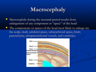
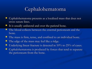
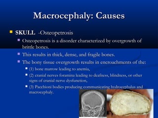
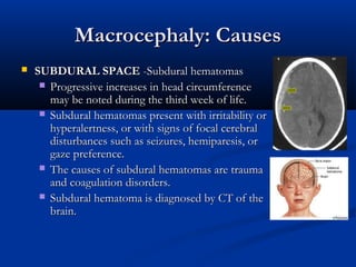
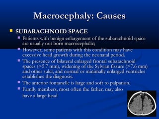
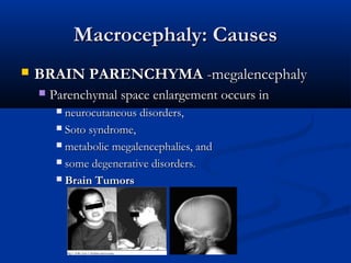
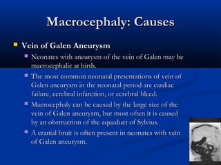
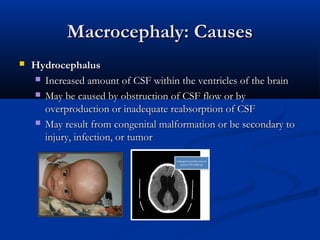
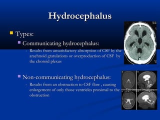
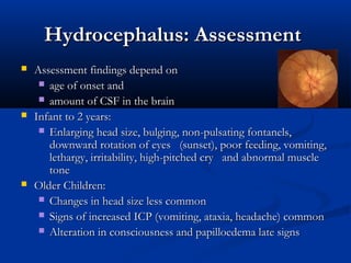
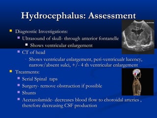
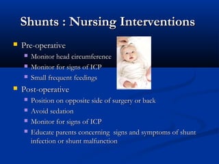
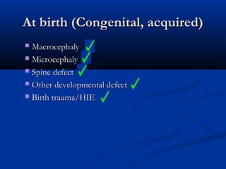
Neurological conditions
    and diseases


        Post Basic Paediatrics

       18 April 2012
Neurological conditions and diseases
   Part I
        At birth (Congenital, acquired)
             Macrocephaly
             Microcephaly
             Spine defect
             Other developmental defect
             Birth trauma/HIE
   Part II
        During development (Congenital, acquired)
             Meningitis
             Seizure
             Headache
             Stroke/Vascular
             Neoplasm/Tumour
             Trauma
             Coma
Neurological conditions
    and diseases

               Part I
     At birth (Congenital, acquired)
At birth (Congenital, acquired)
 Macrocephaly
 Microcephaly

 Spine defect

 Other developmental defect

 Birth trauma/HIE
Macrocephaly
   Macrocephaly during the neonatal period results from
    enlargement of any component or “space” of the head
   The components or spaces of the head most likely to enlarge are
    the scalp, skull, subdural space, subarachnoid space, brain
    parenchyma, intraparenchymal vessels, and ventricles.
Macrocephaly: Causes
   SCALP
     caput succedaneum,
     subgaleal hemorrhage, and

     cephalohematoma
Caput Succedaneum
   Caput succedaneum is due to edema between the skin and the
    epicranial aponeurosis.
   It presents as a mass, usually located in the vertex, that crosses
    the sutures and extends over several bones.
   The mass is soft, superficial, and pitting.
   The edema results from compression of the scalp by the uterus
    or suction on the scalp if a vacuum extractor was used during
    delivery
Subgaleal Hemorrhage
   Subgaleal hemorrhage is due to blood between the epicranial
    aponeurosis and the external periosteum.
   Subgaleal hemorrhage presents as an evenly spread mass
    throughout a large portion of the scalp.
   The mass is firm, fluctuant, crosses suture lines, and increases in
    size after birth (sometimes at an alarming speed)
Cephalohematoma
   Cephalohematoma presents as a localized mass that does not
    cross suture lines.
   It is usually unilateral and over the parietal bone.
   The blood collects between the external periosteum and the
    bone.
   The mass is firm, tense, and confined to an individual bone.
   The edge of the mass may feel like a ridge.
   Underlying linear fracture is detected in 10% to 25% of cases.
   Cephalohematoma is produced by forces that tend to separate
    the periosteum from the bone.
Macrocephaly: Causes
   SKULL -Osteopetrosis
       Osteopetrosis is a disorder characterized by overgrowth of
        brittle bones.
       This results in thick, dense, and fragile bones.
       The bony tissue overgrowth results in encroachments of the:
            (1) bone marrow leading to anemia,
            (2) cranial nerves foramina leading to deafness, blindness, or other
             signs of cranial nerve dysfunction,
            (3) Pacchioni bodies producing communicating hydrocephalus and
             macrocephaly.
Macrocephaly: Causes
   SUBDURAL SPACE -Subdural hematomas
      Progressive increases in head circumference
       may be noted during the third week of life.
      Subdural hematomas present with irritability or
       hyperalertness, or with signs of focal cerebral
       disturbances such as seizures, hemiparesis, or
       gaze preference.
      The causes of subdural hematomas are trauma
       and coagulation disorders.
      Subdural hematoma is diagnosed by CT of the
       brain.
Macrocephaly: Causes
   SUBARACHNOID SPACE
       Patients with benign enlargement of the subarachnoid space
        are usually not born macrocephalic;
       However, some patients with this condition may have
        excessive head growth during the neonatal period.
       The presence of bilateral enlarged frontal subarachnoid
        spaces (>5.7 mm), widening of the Sylvian fissure (>7.6 mm)
        and other sulci, and normal or minimally enlarged ventricles
        establishes the diagnosis.
       The anterior fontanelle is large and soft to palpation.
       Family members, most often the father, may also
        have a large head
Macrocephaly: Causes
   BRAIN PARENCHYMA -megalencephaly
       Parenchymal space enlargement occurs in
          neurocutaneous disorders,
          Soto syndrome,

          metabolic megalencephalies, and

          some degenerative disorders.

          Brain Tumors
Macrocephaly: Causes
   Vein of Galen Aneurysm
       Neonates with aneurysm of the vein of Galen may be
        macrocephalic at birth.
       The most common neonatal presentations of vein of
        Galen aneurysm in the neonatal period are cardiac
        failure, cerebral infarction, or cerebral bleed.
       Macrocephaly can be caused by the large size of the
        vein of Galen aneurysm, but most often it is caused
        by an obstruction of the aqueduct of Sylvius.
       A cranial bruit is often present in neonates with vein
        of Galen aneurysm.
Macrocephaly: Causes
   Hydrocephalus 
     Increased amount of CSF within the ventricles of the brain

     May be caused by obstruction of CSF flow or by
      overproduction or inadequate reabsorption of CSF
     May result from congenital malformation or be secondary to
      injury, infection, or tumor
Hydrocephalus
   Types:
       Communicating hydrocephalus:
         - Results from unsatisfactory absorption of CSF by the
           arachnoid gratulations or overproduction of CSF by
           the choroid plexus


       Non-communicating hydrocephalus:
         - Results from an obstruction to CSF flow , causing
           enlargement of only those ventricles proximal to the
           obstruction
Hydrocephalus: Causes
   Congenital
     Aqueductal anomalies

    - Primary aqueductal stenosis, or secondary to
      intrauterine infections i.e. varicella, mumps, TORCH
    - Dandy-Walker malformation
    - Chiari malformation
    - Myelomeningocele
Hydrocephalus: Causes
   Acquired
    -   Post meningitis
    -   Post hemorrhage- (SAH, IVH)
    -   Masses - vascular malformations, neoplastic
Clinical Presentation
   Increased head circumference
-   Irritability, lethargy, poor feeding, vomiting -infant
-   Headache, lethargy, vomiting- older child
-   Bulging anterior fontanelle
-   Widened cranial sutures
-   Cracked pot sound on cranial percussion
-   Scalp vein dilatation
Clinical Presentation
   Sunset sign - eyes deviate downward
-   Episodic bradycardia, apnea
-   Loss of color and peripheral vision(older child)
-   Cranial nerve palsies - e.g abnormal pupil size/reactivity, EOM’s,
    nystagmus
-   Spasticity limbs
-   Hyperreflexia, clonus
Hydrocephalus: Assessment
   Assessment findings depend on  
      age of onset and
      amount of CSF in the brain
   Infant to 2 years:
      Enlarging head size, bulging, non-pulsating fontanels,
       downward rotation of eyes   (sunset), poor feeding, vomiting,
       lethargy, irritability, high-pitched cry   and abnormal muscle
       tone
   Older Children:
      Changes in head size less common
      Signs of increased ICP (vomiting, ataxia, headache) common
      Alteration in consciousness and papilloedema late signs
Hydrocephalus: Assessment
   Diagnostic Investigations:
      Ultrasound of skull- through anterior fontanelle
          Shows ventricular enlargement
      CT of head
         - Shows ventricular enlargement, peri-ventricualr lucency,
           narrow/absent sulci, +/- 4 th ventricular enlargement
   Treatments:
      Serial Spinal taps
      Surgery- remove obstruction if possible
      Shunts
      Acetazolamide- decreases blood flow to choroidal arteries ,
       therefore decreasing CSF production
Hydrocephalus: Assessment
   Complications:
     - Shunt blockages
     - Infection of shunt
     - Over shunting
     - Seizures
     - Blindness
     - Cranial nerve dysfunction
     - ICP
     - Cognitive impairment
Shunts
   Insertion of a flexible tube into the lateral ventricle of the brain
   Catheter is the threaded under the skin and the distal end
    positioned in the peritoneum (common) or the right atrium
   Shunt drains excess CSF from the lateral ventricles; fluid is the
    absorbed by the peritoneum or absorbed in the general
    circulation via the right atrium
Shunts : Nursing Interventions
   Pre-operative
       Monitor head circumference
       Monitor for signs of ICP
       Small frequent feedings
   Post-operative
       Position on opposite side of surgery or back
       Avoid sedation
       Monitor for signs of ICP
       Educate parents concerning signs and symptoms of shunt
        infection or shunt malfunction
Macrocephaly: Causes
   Posthemorrhagic Hydrocephalus
       Posthemorrhagic hydrocephalus is the most common type of
        hydrocephalus in the neonatal period.
       Posthemorrhagic hydrocephalus may be communicating or
        noncommunicating.
       It is usually the consequence of intraventricular hemorrhage.
       Intraventricular hemorrhage usually occurs as a consequence
        of germinal matrix hemorrhage.
       Germinal matrix hemorrhages are unusual after 34 weeks
        gestational age.
Macrocephaly: Causes
   Posthemorrhagic Hydrocephalus
       Germinal matrix hemorrhages are classified based on brain
        ultrasound in four grades.
           Grade I intraventricular hemorrhage refers to the
             presence of subependymal bleed;
           Grade II intraventricular hemorrhage refers to
             extension of the subependymal bleed into the
             ventricles but without ventricular dilatation;
           Grade III intraventricular hemorrhage refers to
             subependymal bleed with extension of the bleed into
             the ventricles and hydrocephalus; and
           Grade IV intraventricular hemorrhage refers to
             subependymal bleed with extension of the bleed into
             the parenchyma as a result of venous infarcts
At birth (Congenital, acquired)
 Macrocephaly
 Microcephaly

 Spine defect

 Other developmental defect

 Birth trauma/HIE
Microcephaly
   Causes include:
     - Premature closure of skull sutures (craniosynostosis)
     - Microencephaly
        - small brain due to insult ( infectious, toxic, metabolic,
          vascular) sustained in the perinatal or early infancy period
          e.g rubella,CMV, Fetal alcohol syndrome, Genetic
          disorder - microencephaly vera, many syndromes and
          metabolic disorders
Anencephaly
   Defective closure of the rostral neural tube results in
    anencephaly or encephalocele
   Neonates with anencephaly have a rudimentary brainstem, or
    midrain , no cortex or cranium
   Rapidly fatal condition if born alive
At birth (Congenital, acquired)
 Macrocephaly
 Microcephaly

 Spine defect

 Other developmental defect

 Birth trauma/HIE
Neural Tube Defects
 Spina bifida
 Diastematomyelia
Spina Bifida (myelodysplasia)
   Neural tube defects that develop during the first trimester of
    fetal development
   Defect can occur at any place along the spinal canal
   Unknown etiology; thought to be associated with folic acid
    deficiency in mother’s diet prenatally
   Degree of disability dependent on location of the defect & if
    spinal nerves involved
Spina Bifida (myelodysplasia)
   Defective closure of the caudal end of NT at the end of 4th
    week of gestation
   Results in anomalies of the lumbar and sacral vertebrae or spinal
    cord
   Range of severity of CNS defect
   Preventable with pre-conceptual Folic acid supplements 0.4
    mg /day
Spina Bifida
   Occulta
   Meningocele
   Myelomeningocele
Spina bifida “Occulta"
   Spina bifida "occulta" (meaning "hidden" in latin)
   Posterior vertebral arches fail to fuse
   No herniation of meninges or spinal cord
   May have a tuft of hair or dimpling over the lumbarsacral area
   No loss of function
Meningocele
   Posterior vertebral arches fail to fuse
   Sac-like protrusion containing meninges and cerebral spinal fluid
   No spinal nerve involvement
Myelomeningocele
   Sac-like herniation containing meninges, CSF, and spinal nerves
    imbedded in the wall of the sac
   There may be no signs or symptoms
   The spinal arch has not closed, but the spinal cord underneath
    has retained its normal position and is not damaged
   Skin of back intact, small dimple or tuft of hair may be present
    over affected vertebrae
   A child could grow up and never know that he or she has the
    defect
Nursing Care – Spina Bifida
   Neurological status
   Assess degree of sensation at or below lesion
   Leg movement
   Neurogenic bladder
   Measure head circumference
   High risk of hydrocephalus
   High risk for infection
   High risk for impaired skin integrity
   Altered urinary elimination
   Bowel incontinence/constipation
   Impaired physical mobility
Nursing Care – Spina Bifida
   Sac
      Monitor for leakage of spinal fluid

      Monitor skin integrity of sac

      Assess for infection- Sac or systemic

      Position infant on side or abdomen

      Apply wet, sterile, saline dressing

      Do not allow sac to dry out
Nursing Care – Post-operative
   Defect/sac is surgically closed within 48 hours
   Observe for latex allergies
   Neurogenic bladder: straight catheterization
   Neurogenic bowel: bowel management program
   Monitor for signs/ symptoms of hydrocephalus
Diastematomyelia
- A bone or fibrous band divides spinal cord in two longitudinal
  sections
- Associated lipoma may be present, which tethers cord to
  vertebra
- Signs and symptoms include weakness, numbness in feet, urinary
  incontinence, decreased or absent reflexes in feet
- Treatment - surgery to free cord
At birth (Congenital, acquired)
 Macrocephaly
 Microcephaly

 Spine defect

 Other developmental defect

 Birth trauma/HIE
Encephalocele

   Skull defect with exposure of meninges alone or
    meninges and brain
   Sometimes defect can cause protrusion of
    frontal lobe through the nose
At birth (Congenital, acquired)
 Macrocephaly
 Microcephaly

 Spine defect

 Other developmental defect

 Birth trauma/HIE
Hypoxic-ischemic
                Encephalopathy 
   Hypoxic ischemic encephalopathy (HIE) refers to the CNS
    dysfunction associated with perinatal asphyxia.
   HIE is of foremost concern in an asphyxiated neonate because
    of its potential to cause serious long-term neuromotor sequelae
    among survivors.
   A simple and practical classification of HIE by severity of
    manifestations provided by Levene
Hypoxic-ischemic
                 Encephalopathy 
   Hypoxic-ischemic encephalopathy often involves the brain and
    the brainstem.
   Very severe hypoxic-ischemic encephalopathy may involve the
    brain, brainstem, spinal cord, and muscle.
   Magnetic resonance imaging of the brain in neonates with
    hypotonia due to hypoxic-ischemic encephalopathy shows loss
    of gray-white matter interface, cortical necrosis, or neuronal loss
    of the basal ganglia and thalamus. 
Mental Retardation
   Significant below average intellectual functioning
    which is associated with impaired learning
    difficulties
   Causes
     Pre-natal
     Perinatal

     Post-natal
Mental Retardation: Causes
•   Pre-natal -Genetic Disorders
     • Chromosomal aberrations- e.g. Down syndrome (trisomy
       21 )
     • Disorders with autosomal-dominant inheritance- e.g.
       Tuberous sclerosis
     • Disorders with autosomal-recessive inheritance-
       metabolic disorder; e.g. Phenylketonuria
     • X-linked mental retardation- Fragile X syndrome
     • Maternal infections- e.g. Rubella infection during the first
       month of pregnancy
     • Toxic substances- fetal alcohol syndrome
     • Toxemia of pregnancy and placental insufficiency
Mental Retardation: Causes
•   Perinatal (This period refers to 1 week before birth to 4 weeks
    after birth )
     • Infections -e.g. herpes simplex type 2
     • Delivery problems – e.g. birth asphyxia
     • Other perinatal problems
         • Retinopathy of prematurity
         • Hyperbilirubinemia
Mental Retardation: Causes
•   Postnatal
    •   Infections
         •   Bacterial and viral infections of the brain during childhood may cause
             meningitis and encephalitis and result in permanent damage
    •   Toxic substances –e.g. Lead poisoning
    •   Other postnatal causes
         •   Childhood malignancies, brain tumors
         •   Trauma
    •   Psychosocial problems –e.g. Severe maternal mental illness
    •   Unknown causes
         •   no cause can be identified in approximately 30% of cases of severe
             mental retardation and in 50% of cases of mild mental retardation
MR – Classifications
   Mild
      Slow learner, can work, marry, have children, may need
       assistance with crisis
   Moderate
      Needs life supervision

   Severe
      Needs a caretaker for basic needs

   Profound
Interventions
   Goal is to promote
     Optimal development
     Family support

     Community referrals
Cerebral Palsy
   A non-progressive motor disorder of the CNS resulting in
    alteration in movement and posture
   Cause is trauma, hemorrhage, anoxia or infection before, during
    or after birth
   1/3 of children have some degree of mental retardation
   Classified as:
       Spastic
       Spasticity (hypertonicity of muscle groups)
       Athetoid
       Worm-like movements of   extremities
       Ataxic
       Disturbed coordination
       Mixed
Cerebral Palsy – Assessment
   May have hypertonicity or hypotonia of varying degrees on
    different extremities
   May have scissoring of the legs
   Absence of expected reflexes or presence of reflexes that extend
    beyond expected age
   Failure to meet developmental milestones
   Difficulty swallowing
   Altered speech
Nursing Care
   Impaired physical mobility
   Self-care deficit
   Altered nutrition: less than body requirements
   High risk for injury related to neuromuscular,
    perceptual or cognitive impairments
Treatment
   Self-care is a goal for all children
   Team approach
   Nutrition
   Increased caloric intake
   Special feeding devices
   Community referrals
   Emotional support
At birth (Congenital, acquired)
 Macrocephaly
 Microcephaly

 Spine defect

 Other developmental defect

 Birth trauma/HIE
Thank You

Neurological Conditions and Diseases (At birth)

  • 1.
    Neurological conditions and diseases Post Basic Paediatrics 18 April 2012
  • 2.
    Neurological conditions anddiseases  Part I  At birth (Congenital, acquired)  Macrocephaly  Microcephaly  Spine defect  Other developmental defect  Birth trauma/HIE  Part II  During development (Congenital, acquired)  Meningitis  Seizure  Headache  Stroke/Vascular  Neoplasm/Tumour  Trauma  Coma
  • 3.
    Neurological conditions and diseases Part I At birth (Congenital, acquired)
  • 4.
    At birth (Congenital,acquired)  Macrocephaly  Microcephaly  Spine defect  Other developmental defect  Birth trauma/HIE
  • 5.
    Macrocephaly  Macrocephaly during the neonatal period results from enlargement of any component or “space” of the head  The components or spaces of the head most likely to enlarge are the scalp, skull, subdural space, subarachnoid space, brain parenchyma, intraparenchymal vessels, and ventricles.
  • 6.
    Macrocephaly: Causes  SCALP  caput succedaneum,  subgaleal hemorrhage, and  cephalohematoma
  • 7.
    Caput Succedaneum  Caput succedaneum is due to edema between the skin and the epicranial aponeurosis.  It presents as a mass, usually located in the vertex, that crosses the sutures and extends over several bones.  The mass is soft, superficial, and pitting.  The edema results from compression of the scalp by the uterus or suction on the scalp if a vacuum extractor was used during delivery
  • 8.
    Subgaleal Hemorrhage  Subgaleal hemorrhage is due to blood between the epicranial aponeurosis and the external periosteum.  Subgaleal hemorrhage presents as an evenly spread mass throughout a large portion of the scalp.  The mass is firm, fluctuant, crosses suture lines, and increases in size after birth (sometimes at an alarming speed)
  • 9.
    Cephalohematoma  Cephalohematoma presents as a localized mass that does not cross suture lines.  It is usually unilateral and over the parietal bone.  The blood collects between the external periosteum and the bone.  The mass is firm, tense, and confined to an individual bone.  The edge of the mass may feel like a ridge.  Underlying linear fracture is detected in 10% to 25% of cases.  Cephalohematoma is produced by forces that tend to separate the periosteum from the bone.
  • 10.
    Macrocephaly: Causes  SKULL -Osteopetrosis  Osteopetrosis is a disorder characterized by overgrowth of brittle bones.  This results in thick, dense, and fragile bones.  The bony tissue overgrowth results in encroachments of the:  (1) bone marrow leading to anemia,  (2) cranial nerves foramina leading to deafness, blindness, or other signs of cranial nerve dysfunction,  (3) Pacchioni bodies producing communicating hydrocephalus and macrocephaly.
  • 11.
    Macrocephaly: Causes  SUBDURAL SPACE -Subdural hematomas  Progressive increases in head circumference may be noted during the third week of life.  Subdural hematomas present with irritability or hyperalertness, or with signs of focal cerebral disturbances such as seizures, hemiparesis, or gaze preference.  The causes of subdural hematomas are trauma and coagulation disorders.  Subdural hematoma is diagnosed by CT of the brain.
  • 12.
    Macrocephaly: Causes  SUBARACHNOID SPACE  Patients with benign enlargement of the subarachnoid space are usually not born macrocephalic;  However, some patients with this condition may have excessive head growth during the neonatal period.  The presence of bilateral enlarged frontal subarachnoid spaces (>5.7 mm), widening of the Sylvian fissure (>7.6 mm) and other sulci, and normal or minimally enlarged ventricles establishes the diagnosis.  The anterior fontanelle is large and soft to palpation.  Family members, most often the father, may also have a large head
  • 13.
    Macrocephaly: Causes  BRAIN PARENCHYMA -megalencephaly  Parenchymal space enlargement occurs in  neurocutaneous disorders,  Soto syndrome,  metabolic megalencephalies, and  some degenerative disorders.  Brain Tumors
  • 14.
    Macrocephaly: Causes  Vein of Galen Aneurysm  Neonates with aneurysm of the vein of Galen may be macrocephalic at birth.  The most common neonatal presentations of vein of Galen aneurysm in the neonatal period are cardiac failure, cerebral infarction, or cerebral bleed.  Macrocephaly can be caused by the large size of the vein of Galen aneurysm, but most often it is caused by an obstruction of the aqueduct of Sylvius.  A cranial bruit is often present in neonates with vein of Galen aneurysm.
  • 15.
    Macrocephaly: Causes  Hydrocephalus   Increased amount of CSF within the ventricles of the brain  May be caused by obstruction of CSF flow or by overproduction or inadequate reabsorption of CSF  May result from congenital malformation or be secondary to injury, infection, or tumor
  • 16.
    Hydrocephalus  Types:  Communicating hydrocephalus: - Results from unsatisfactory absorption of CSF by the arachnoid gratulations or overproduction of CSF by the choroid plexus  Non-communicating hydrocephalus: - Results from an obstruction to CSF flow , causing enlargement of only those ventricles proximal to the obstruction
  • 17.
    Hydrocephalus: Causes  Congenital  Aqueductal anomalies - Primary aqueductal stenosis, or secondary to intrauterine infections i.e. varicella, mumps, TORCH - Dandy-Walker malformation - Chiari malformation - Myelomeningocele
  • 18.
    Hydrocephalus: Causes  Acquired - Post meningitis - Post hemorrhage- (SAH, IVH) - Masses - vascular malformations, neoplastic
  • 19.
    Clinical Presentation  Increased head circumference - Irritability, lethargy, poor feeding, vomiting -infant - Headache, lethargy, vomiting- older child - Bulging anterior fontanelle - Widened cranial sutures - Cracked pot sound on cranial percussion - Scalp vein dilatation
  • 20.
    Clinical Presentation  Sunset sign - eyes deviate downward - Episodic bradycardia, apnea - Loss of color and peripheral vision(older child) - Cranial nerve palsies - e.g abnormal pupil size/reactivity, EOM’s, nystagmus - Spasticity limbs - Hyperreflexia, clonus
  • 21.
    Hydrocephalus: Assessment  Assessment findings depend on    age of onset and  amount of CSF in the brain  Infant to 2 years:  Enlarging head size, bulging, non-pulsating fontanels, downward rotation of eyes   (sunset), poor feeding, vomiting, lethargy, irritability, high-pitched cry   and abnormal muscle tone  Older Children:  Changes in head size less common  Signs of increased ICP (vomiting, ataxia, headache) common  Alteration in consciousness and papilloedema late signs
  • 22.
    Hydrocephalus: Assessment  Diagnostic Investigations:  Ultrasound of skull- through anterior fontanelle  Shows ventricular enlargement  CT of head - Shows ventricular enlargement, peri-ventricualr lucency, narrow/absent sulci, +/- 4 th ventricular enlargement  Treatments:  Serial Spinal taps  Surgery- remove obstruction if possible  Shunts  Acetazolamide- decreases blood flow to choroidal arteries , therefore decreasing CSF production
  • 23.
    Hydrocephalus: Assessment  Complications: - Shunt blockages - Infection of shunt - Over shunting - Seizures - Blindness - Cranial nerve dysfunction - ICP - Cognitive impairment
  • 24.
    Shunts  Insertion of a flexible tube into the lateral ventricle of the brain  Catheter is the threaded under the skin and the distal end positioned in the peritoneum (common) or the right atrium  Shunt drains excess CSF from the lateral ventricles; fluid is the absorbed by the peritoneum or absorbed in the general circulation via the right atrium
  • 25.
    Shunts : NursingInterventions  Pre-operative  Monitor head circumference  Monitor for signs of ICP  Small frequent feedings  Post-operative  Position on opposite side of surgery or back  Avoid sedation  Monitor for signs of ICP  Educate parents concerning signs and symptoms of shunt infection or shunt malfunction
  • 26.
    Macrocephaly: Causes  Posthemorrhagic Hydrocephalus  Posthemorrhagic hydrocephalus is the most common type of hydrocephalus in the neonatal period.  Posthemorrhagic hydrocephalus may be communicating or noncommunicating.  It is usually the consequence of intraventricular hemorrhage.  Intraventricular hemorrhage usually occurs as a consequence of germinal matrix hemorrhage.  Germinal matrix hemorrhages are unusual after 34 weeks gestational age.
  • 27.
    Macrocephaly: Causes  Posthemorrhagic Hydrocephalus  Germinal matrix hemorrhages are classified based on brain ultrasound in four grades.  Grade I intraventricular hemorrhage refers to the presence of subependymal bleed;  Grade II intraventricular hemorrhage refers to extension of the subependymal bleed into the ventricles but without ventricular dilatation;  Grade III intraventricular hemorrhage refers to subependymal bleed with extension of the bleed into the ventricles and hydrocephalus; and  Grade IV intraventricular hemorrhage refers to subependymal bleed with extension of the bleed into the parenchyma as a result of venous infarcts
  • 28.
    At birth (Congenital,acquired)  Macrocephaly  Microcephaly  Spine defect  Other developmental defect  Birth trauma/HIE
  • 29.
    Microcephaly  Causes include: - Premature closure of skull sutures (craniosynostosis) - Microencephaly - small brain due to insult ( infectious, toxic, metabolic, vascular) sustained in the perinatal or early infancy period e.g rubella,CMV, Fetal alcohol syndrome, Genetic disorder - microencephaly vera, many syndromes and metabolic disorders
  • 30.
    Anencephaly  Defective closure of the rostral neural tube results in anencephaly or encephalocele  Neonates with anencephaly have a rudimentary brainstem, or midrain , no cortex or cranium  Rapidly fatal condition if born alive
  • 31.
    At birth (Congenital,acquired)  Macrocephaly  Microcephaly  Spine defect  Other developmental defect  Birth trauma/HIE
  • 32.
    Neural Tube Defects Spina bifida  Diastematomyelia
  • 33.
    Spina Bifida (myelodysplasia)  Neural tube defects that develop during the first trimester of fetal development  Defect can occur at any place along the spinal canal  Unknown etiology; thought to be associated with folic acid deficiency in mother’s diet prenatally  Degree of disability dependent on location of the defect & if spinal nerves involved
  • 34.
    Spina Bifida (myelodysplasia)  Defective closure of the caudal end of NT at the end of 4th week of gestation  Results in anomalies of the lumbar and sacral vertebrae or spinal cord  Range of severity of CNS defect  Preventable with pre-conceptual Folic acid supplements 0.4 mg /day
  • 35.
    Spina Bifida  Occulta  Meningocele  Myelomeningocele
  • 36.
    Spina bifida “Occulta"  Spina bifida "occulta" (meaning "hidden" in latin)  Posterior vertebral arches fail to fuse  No herniation of meninges or spinal cord  May have a tuft of hair or dimpling over the lumbarsacral area  No loss of function
  • 37.
    Meningocele  Posterior vertebral arches fail to fuse  Sac-like protrusion containing meninges and cerebral spinal fluid  No spinal nerve involvement
  • 38.
    Myelomeningocele  Sac-like herniation containing meninges, CSF, and spinal nerves imbedded in the wall of the sac  There may be no signs or symptoms  The spinal arch has not closed, but the spinal cord underneath has retained its normal position and is not damaged  Skin of back intact, small dimple or tuft of hair may be present over affected vertebrae  A child could grow up and never know that he or she has the defect
  • 39.
    Nursing Care –Spina Bifida  Neurological status  Assess degree of sensation at or below lesion  Leg movement  Neurogenic bladder  Measure head circumference  High risk of hydrocephalus  High risk for infection  High risk for impaired skin integrity  Altered urinary elimination  Bowel incontinence/constipation  Impaired physical mobility
  • 40.
    Nursing Care –Spina Bifida  Sac  Monitor for leakage of spinal fluid  Monitor skin integrity of sac  Assess for infection- Sac or systemic  Position infant on side or abdomen  Apply wet, sterile, saline dressing  Do not allow sac to dry out
  • 41.
    Nursing Care –Post-operative  Defect/sac is surgically closed within 48 hours  Observe for latex allergies  Neurogenic bladder: straight catheterization  Neurogenic bowel: bowel management program  Monitor for signs/ symptoms of hydrocephalus
  • 42.
    Diastematomyelia - A boneor fibrous band divides spinal cord in two longitudinal sections - Associated lipoma may be present, which tethers cord to vertebra - Signs and symptoms include weakness, numbness in feet, urinary incontinence, decreased or absent reflexes in feet - Treatment - surgery to free cord
  • 43.
    At birth (Congenital,acquired)  Macrocephaly  Microcephaly  Spine defect  Other developmental defect  Birth trauma/HIE
  • 44.
    Encephalocele  Skull defect with exposure of meninges alone or meninges and brain  Sometimes defect can cause protrusion of frontal lobe through the nose
  • 45.
    At birth (Congenital,acquired)  Macrocephaly  Microcephaly  Spine defect  Other developmental defect  Birth trauma/HIE
  • 46.
    Hypoxic-ischemic Encephalopathy   Hypoxic ischemic encephalopathy (HIE) refers to the CNS dysfunction associated with perinatal asphyxia.  HIE is of foremost concern in an asphyxiated neonate because of its potential to cause serious long-term neuromotor sequelae among survivors.  A simple and practical classification of HIE by severity of manifestations provided by Levene
  • 47.
    Hypoxic-ischemic Encephalopathy   Hypoxic-ischemic encephalopathy often involves the brain and the brainstem.  Very severe hypoxic-ischemic encephalopathy may involve the brain, brainstem, spinal cord, and muscle.  Magnetic resonance imaging of the brain in neonates with hypotonia due to hypoxic-ischemic encephalopathy shows loss of gray-white matter interface, cortical necrosis, or neuronal loss of the basal ganglia and thalamus. 
  • 48.
    Mental Retardation  Significant below average intellectual functioning which is associated with impaired learning difficulties  Causes  Pre-natal  Perinatal  Post-natal
  • 49.
    Mental Retardation: Causes • Pre-natal -Genetic Disorders • Chromosomal aberrations- e.g. Down syndrome (trisomy 21 ) • Disorders with autosomal-dominant inheritance- e.g. Tuberous sclerosis • Disorders with autosomal-recessive inheritance- metabolic disorder; e.g. Phenylketonuria • X-linked mental retardation- Fragile X syndrome • Maternal infections- e.g. Rubella infection during the first month of pregnancy • Toxic substances- fetal alcohol syndrome • Toxemia of pregnancy and placental insufficiency
  • 50.
    Mental Retardation: Causes • Perinatal (This period refers to 1 week before birth to 4 weeks after birth ) • Infections -e.g. herpes simplex type 2 • Delivery problems – e.g. birth asphyxia • Other perinatal problems • Retinopathy of prematurity • Hyperbilirubinemia
  • 51.
    Mental Retardation: Causes • Postnatal • Infections • Bacterial and viral infections of the brain during childhood may cause meningitis and encephalitis and result in permanent damage • Toxic substances –e.g. Lead poisoning • Other postnatal causes • Childhood malignancies, brain tumors • Trauma • Psychosocial problems –e.g. Severe maternal mental illness • Unknown causes • no cause can be identified in approximately 30% of cases of severe mental retardation and in 50% of cases of mild mental retardation
  • 52.
    MR – Classifications  Mild  Slow learner, can work, marry, have children, may need assistance with crisis  Moderate  Needs life supervision  Severe  Needs a caretaker for basic needs  Profound
  • 53.
    Interventions  Goal is to promote  Optimal development  Family support  Community referrals
  • 54.
    Cerebral Palsy  A non-progressive motor disorder of the CNS resulting in alteration in movement and posture  Cause is trauma, hemorrhage, anoxia or infection before, during or after birth  1/3 of children have some degree of mental retardation  Classified as:  Spastic  Spasticity (hypertonicity of muscle groups)  Athetoid  Worm-like movements of   extremities  Ataxic  Disturbed coordination  Mixed
  • 55.
    Cerebral Palsy –Assessment  May have hypertonicity or hypotonia of varying degrees on different extremities  May have scissoring of the legs  Absence of expected reflexes or presence of reflexes that extend beyond expected age  Failure to meet developmental milestones  Difficulty swallowing  Altered speech
  • 56.
    Nursing Care  Impaired physical mobility  Self-care deficit  Altered nutrition: less than body requirements  High risk for injury related to neuromuscular, perceptual or cognitive impairments
  • 57.
    Treatment  Self-care is a goal for all children  Team approach  Nutrition  Increased caloric intake  Special feeding devices  Community referrals  Emotional support
  • 58.
    At birth (Congenital,acquired)  Macrocephaly  Microcephaly  Spine defect  Other developmental defect  Birth trauma/HIE
  • 59.