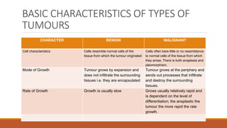
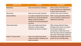
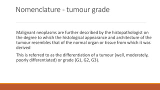
neoplasms
MRS MUNANG’ANDU MUFUNGWE
introduction
The term neoplasm refers to the tumour mass itself
The process that occurs to produce a neoplasm is called neoplasia
Willis defined a neoplasm as “an abnormal mass of tissue, the
growth of which exceeds and is uncoordinated with that of the
surrounding normal tissues, and persists in the same excessive
manner after cessation of the stimuli which evoked the change”.
(For example, once a lung cancer has developed, it doesn’t go away
if the individual stops smoking.)
Introduction…..
Neoplasms have an increase in cell numbers that must be
distinguished from the reversible increase in cell number
(hyperplasia such as in enlarged prostates of older men) or cell size
(hypertrophy such as in the enlarged uterus of pregnancy) that may
also be seen in larger organs
Definition of terms
Hyperplasia: This is an increase in the number of cells of a tissue. It
is a common proliferation process during periods of rapid growth
(e.g. foetal and adolescent growth) and during epithelial and bone
marrow regeneration
This is a normal cellular response when a physiological demand
exists and an abnormal response when growth exceeds the
physiological demand
This usually leads to an increase in the size of a tissue or organ
Definition of terms……
Metaplasia: This occurs when one type of mature cells is converted
to another type by means of an outside stimulus that affects the
parent stem cell. Metaplastic changes may be reversible or may
progress to dysplasia
Definition of terms……
Dysplasia: This is a bizarre cell growth resulting in cells that differ in
size, shape or arrangement from other cells of the same type of
tissue
It can occur from chemicals, radiation, or chronic inflammation or
irritation. It can be reversible or can proceed to an irreversible
neoplastic change
Definition of terms……..
cells (differentiation is the extent to which cells differ from their cells
of origin and their maturity)
Anaplastic cells lack normal cellular characteristics and are nearly
always malignant
Definition of terms…….
Neoplasia: This is uncontrolled cell growth that follows no
physiological demand and can be benign or malignant (cancerous)
Anaplasia: This is a lower degree of differentiation of dysplastic
HOW DO TUMOURS DEVELOP?
There has to be a change to DNA
The change must cause an alteration in cell growth and behaviour
The change must be non-lethal and be passed onto daughter cells
Alteration is to more than one gene
Genes concerned are oncogenes/tumour suppressor genes
Sequence of gene alterations from normal to benign to malignant
Intrinsic and extrinsic / inheritance and environment key factors
Essential features of neoplasia or tumour
growth
Tumours are composed of living cells, which differ from the cells of
the normal organ from which the tumour is derived in that the cell
division, cell death and cell differentiation controls operating in that
tissue or organ have been deregulated or lost
The control of cell division, death and differentiation may have
been lost to the extent that the tumour:
Essential features of neoplasia or tumour
growth ……
(a) loses partially or totally specialised functions (cell appearance
changes)
(b) acquires new functions such as invasion – the capacity to
infiltrate the surrounding tissues and organs and metastasis – the
ability to spread to and proliferate in distant parts of the body after
tumour cells have been transported by lymph or blood or along
body spaces
PATHOPHYSIOLOGY OF CANCER
Cancer is disease that begins when abnormal cells arise from
normal body cells as a result of some poorly understood
mechanisms of change
As the disease progresses, these abnormal cells proliferate still
within a local area. However, a stage is reached in which the cells
acquire invasive characteristics and changes occur in the
surrounding tissues or areas as well.
PATHOPHYSIOLOGY OF CANCER……
Although the disease process can be described in the general terms,
cancer is not a single disease with a single cause; rather it is a group
of distinct diseases with different causes, manifestations and
prognoses
AETIOLOGICAL FACTORS OF TUMOURS
AND CANCER
Viruses
Viral causation in human cancers is very difficult to ascertain because
the isolation of viruses is difficult
Viruses are thought to incorporate themselves in the genetic structure
of cells. For example, the Herpes Stein Bar Virus (HBV) is highly suspect
in causing Burkitt’s lymphoma and nasopharyngeal cancers, Herpes
Simplex Type 2 virus, Cytomegalovirus and papilloma viruses have been
associated with dysplasia and malignancy of the cervix, Hepatitis B
Virus with hepatocellular carcinoma, HIV with Kaposi’s sarcoma etc.
AETIOLOGICAL FACTORS OF TUMOURS
AND CANCER
Physical Agents
Factors that are physical and associated with carcinogenesis include
persistent exposure to sunlight or radiation of the sun and chronic
irritation or inflammation
These agents are thought to interfere with the cellular mitotic stages
over time
AETIOLOGICAL FACTORS OF TUMOURS
AND CANCER
Chemical Agents
Many chemical agents found in work places have proven to be
carcinogenic or co – carcinogens in the cancer development process.
The liver and kidneys are the organs most often affected chemical
related cancers presumably because of their roles in the
detoxification of chemicals
AETIOLOGICAL FACTORS OF TUMOURS
AND CANCER
Genetic and Familial Factors
Some adult and childhood cancers display familial predisposition.
These tend to occur at an early age and at multiple sites in one
organ or one pair of organs.
Dietary Factors
Dietary factors are thought to be related to 40 – 60% of all
environmental cancers
AETIOLOGICAL FACTORS OF TUMOURS
AND CANCER
Dietary substances can either be protective or carcinogenic
The risk of cancer increases over long term ingestion of carcinogenic
or co – carcinogenic substances or the chronic absence of protective
substances in the diet one ingests
AETIOLOGICAL FACTORS OF TUMOURS
AND CANCER
Hormonal Factors
Tumour growth may be promoted or encouraged by the
disturbance in the hormonal balance by either the body’s own
hormone production (endogenous) or by administration of
exogenous substances
Thus cancers of the breast, prostate and uterus are considered to
be dependent on endogenous hormonal levels of growth
Administration of oral contraceptive diethylstilbestrol (DES) has
been associated with hepatocellular carcinomas and vaginal
carcinomas respectively
Benign neoplasms
Benign neoplasms do not invade or metastasise (spread)
Benign tumours are those which proliferate and divide but do not
invade the surrounding tissues, nor metastasize
Such benign neoplasms or tumours and the use of the term benign
implies that their growth is not life-threatening, and their clinical
course is predictable
They may cause problems due to pressure, obstruction or excessive
hormone production
Malignant neoplasms
Malignant neoplasms show evidence of invasion (or infiltration) into
adjacent tissues, often in a destructive way and many will undergo
metastasis (spread to other sites in the body to form secondary
tumours – the original is often called a primary tumour)
Malignant neoplasms…..
Their clinical course is progressive; unless adequate therapy is
available the patient will eventually die as a result of the disease.
Cancers show destructive invasive growth behaviour and can produce
signs and symptoms such as:
Blood loss – ulceration and haemorrhage
Pressure and destruction of adjacent tissue
Obstruction or constriction of flow in vital organs
Malignant neoplasms…..
Metabolic effects: general – cachexia (severe weight loss including a
reduction in muscle mass); and specific – production/secretion of
tumour-specific products/hormones
BASIC CHARACTERISTICS OF TYPES OF
TUMOURS
CHARACTER BENIGN MALIGNANT
Cell characteristics Cells resemble normal cells of the
tissue from which the tumour originated
Cells often bare little or no resemblance
to normal cells of the tissue from which
they arose. There is both anaplasia and
pleomorphism.
Mode of Growth Tumour grows by expansion and
does not infiltrate the surrounding
tissues i.e. they are encapsulated
Tumour grows at the periphery and
sends out processes that infiltrate
and destroy the surrounding
tissues.
Rate of Growth Growth is usually slow Grows usually relatively rapid and
is dependent on the level of
differentiation; the anaplastic the
tumour the more rapid the rate
growth.
CHARACTER BENIGN MALIGNANT
Metastasis Does not spread by metastasis Gains access to the blood and
lymph channels and metastasizes
to other areas of the body.
Recurrence Does not recur when removed. Tends to recur when removed.
General Effects Is usually a localized phenomenon
that does not cause generalized
effects unless by location it
interferes with vital physiological
functions.
Often causes generalized effects
such as anaemia, weakness and
weight loss (cachexia).
Tissue Destruction Does not usually cause tissue
destruction unless location
interferes with blood circulation.
Often causes extensive tissue
damage as the tumour out grows
its own blood supply or it
encroaches on blood flow to the
area may also produce substances
that cause cell damage (cytotoxic).
Ability To Cause Death Does not usually cause death
unless its location interferes with
blood circulation.
Will always cause death unless its
growth can be controlled early.
Nomenclature - types of neoplasm
Benign neoplasm of glandular tissue is an adenoma; malignant is an
adenocarcinoma
Benign neoplasm of squamous epithelium is a papilloma; malignant
is a squamous cell carcinoma
Benign neoplasm of fat is a lipoma; malignant is a liposarcoma
Benign neoplasm of smooth muscle is a leiomyoma; malignant is a
leiomyosarcom
Benign tumours of connective tissue
Adipocytes – lipoma
Fibrocytes – fibroma
Chondrocytes – chondroma
Smooth muscle – leiomyoma
Striated muscle – rhabdomyoma
Bone – osteoma
Malignant tumours arising from connective
tissues
Malignant tumours arising from connective tissues are called
SARCOMAS and further classified by the cell of origin:
Fibrocytes – fibrosarcoma
Osteoblasts – osteosarcoma
Chondrocytes – chondrosarcoma
Smooth muscle – leiomyosarcoma
Striated muscle – rhabdomyosarcoma
Tumour Nomenclature Exceptions
Sometimes names persist for historical reasons, such as melanoma
of the skin, or seminoma of the testis, or lymphoma in the lymph
nodes, all of which are malignant and life-threatening (despite
ending in –oma)
Nomenclature - tumour grade
Malignant neoplasms are further described by the histopathologist on
the degree to which the histological appearance and architecture of the
tumour resembles that of the normal organ or tissue from which it was
derived
This is referred to as the differentiation of a tumour (well, moderately,
poorly differentiated) or grade (G1, G2, G3).
Nomenclature - tumour grade……
This information is of use in determining treatment and prognosis
Poorly differentiated (high (G3) grade) tumours usually demonstrate
a more aggressive behaviour
CANCER STAGING AND GRADING
Two main types of staging systems are used by doctors
The tumor, nodes and metastases (TNM) system and number
systems
Some cancers have their own staging systems, such as Dukes' staging
for bowel cancer
CANCER STAGING AND GRADING…….
Staging is a way of describing the size of a cancer and how far it has
grown
When doctors first diagnose a cancer, they carry out tests to
investigate the extent of the cancer locally and to see whether it
has spread to another part of the body
This is different to the grade of cancer, which describes how similar
a cancer cell is to a normal cell
Importance of staging
Staging is important because it usually tells the specialist which
treatments a patient needs. If a cancer is just in one place, then a
local treatment such as surgery or radiotherapy could be enough to
get rid of it completely
A local treatment treats only one area of the body
Importance of staging…..
If a cancer has spread, then local treatment alone will not be enough
A systemic treatment will be needed as well. Systemic means
treating the whole body
Chemotherapy, hormone therapy and other drug treatments are
systemic treatments because they circulate throughout the body in
the bloodstream
Importance of staging…..
Sometimes doctors aren't sure if a cancer has spread to another
part of the body or not
They look at the lymph nodes near to the cancer. If there are cancer
cells in these nodes, it is a sign that the cancer has begun to spread
Cancer doctors call this having positive lymph nodes
The cells have broken away from the original cancer and got
trapped in the lymph nodes
Importance of staging…….
But we can't always tell if they have gone anywhere else
In this situation, doctors usually suggest adjuvant treatment
This means treatment alongside the treatment for the main primary
tumour (chemotherapy after surgery, for example)
The aim is to kill any cancer cells that have broken away from the
primary tumour
The 'TNM' staging system
This system can describe the size of a primary tumour, whether
the cancer has spread to the lymph nodes and whether the cancer
has spread to a different part of the body (metastasised)
The system uses numbers to describe the cancer
'T' refers to the size of the cancer and how far it has spread into
nearby tissue - it can be 1, 2, 3 or 4, with 1 being small and 4 large
The 'TNM' staging system……
'N' refers to whether the cancer has spread to the lymph nodes -
it can be between 0 (no lymph nodes containing cancer cells)
and 3 (lots of lymph nodes containing cancer cells)
'M' refers to whether the cancer has spread to another part of the
body - it can either be 0 (the cancer hasn't spread) or 1 (the cancer
has spread)
The 'TNM' staging system…..
Sometimes the letters a, b or c are used to further divide the
categories. For example, stage
M1a lung cancer (the cancer has spread to the other lung) and
stage M1b lung cancer (the cancer has spread to other parts of the
body)
As well as T1 - T4, you can get Tis
This means carcinoma in situ, which is a very small and very early
stage cancer. It is such an early stage that it is sometimes called pre-
cancer
The 'TNM' staging system……
P is sometimes used before the letters TNM
This stands for pathological stage. It means that the stage is based on
examining cancer cells in the lab after surgery to remove a cancer
C is sometimes used before the letters TNM
This stands for clinical stage. It means the stage is based on what the
doctor knows about the cancer before surgery. The stage is based on
clinical information from examining the patient and looking at his test
results
The 'TNM' staging system……
So for example, a small cancer that has spread to the lymph nodes
but not to anywhere else in the body may be T2N1M0
Or a more advanced cancer that has spread may be T4N3M1
Number systems
These usually have a scale of 1 to 4
Less often, the scale is A to D. '1' typically means a small tumour that
has not spread and there is no cancer in the lymph nodes
'4' would mean that the cancer has spread to other major organs in
the body
Sometimes clinicians use the letters A,B or C to further divide the
number categories - for example, stage 3B cervical cancer
Overall Stage Grouping
This is also referred to as Roman Numeral Staging. This
system uses numerals I, II, III, and IV (plus the 0) to
describe the progression of cancer.
Stage 0: carcinoma in situ
Stage I: cancers are localized to one part of the body.
Stage I cancer can be surgically removed if small enough
Stage II: cancers are locally advanced. Stage II cancer can
be treated by chemo, radiation, or surgery
•
Overall Stage Grouping…..
Stage III: cancers are also locally advanced. Whether a cancer is
designated as Stage II or Stage III can depend on the specific type of
cancer; for example, in Hodgkin's Disease, Stage II indicates affected
lymph nodes on only one side of the diaphragm, whereas Stage III
indicates affected lymph nodes above and below the diaphragm
The specific criteria for Stages II and III therefore differ according to
diagnosis. Stage III can be treated by chemo, radiation, or surgery
•
Overall Stage Grouping…….
Stage IV: cancers have often metastasized, or spread to other organs
or throughout the body
Stage IV cancer can be treated by chemotherapy, radiation, and
surgery
MANIFESTATIONS AND EFFECTS OF CANCER
These depend on the location and whether they are in the
secondary stage e.g. haemorrhage, ulceration etc
Most neoplasms are space occupying, they may compress blood
vessels, neighbouring tissues or exert pressure on the regional
nerves causing pain or even paralysis or produce malfunctioning by
invasion and replacement of normal tissue
IMPORTANT EARLY WARNING SIGNS AND
SYMPTOMS OF CANCER
Unusual bleeding or discharge from anybody orifice
A lamp or thickening in the breast or elsewhere
A sore that does not heal
A persistent change in bowel or urinary bladder habits
A persistent cough or hoarse voice
Persistent indigestion or difficult in swallowing
A change in a wart or mole
MANAGEMENT OF A PATIENT WITH
CANCER/TUMOUR
MEDICAL
INVESTIGATIONS
Patient’s Health History
Get the patient’s signs and symptoms as mentioned under early
warning signs and symptoms
Examine the patient’s medical history for clues e.g. allergies,
chemotherapy, ionizing radiation etc.
Ask the patient about family history
Physical Assessment
Take vital signs noting the range of temperature as fever is
common in cancer cases
Inspect the face for signs of paralysis, anaemia, jaundice and
ears for discharge
Observe the breast symmetry; dimpling, flattening, oedema,
ulceration etc palpate the breasts for lumps
Inspect the patient’s chest for respiratory sounds; weak or
absence of sounds suggests fluid, a mass or obstruction of
the pleural space
Physical Assessment
Inspect the abdomen considering the shape, tone and symmetry by
palpating to reveal abnormal masses and lymph nodes for any
lymphadenopathy that may indicate spreading cancer (metastasis)
Inspect the extremities to note any signs of immobility or fractures
which indicate bone metastasis
Inspect the genitalia for discharge and ulcers or lesions and
inflammation
Perform a neurologic assessment with the sitting upright as cancer
patients commonly experience various neurological problems such as
spinal cord compression and peripheral neuropathies
Diagnostic tests
Useful tests for detecting cancerous lesions include;
Tissue biopsy is the single most important tool for diagnosing
cancer
X ray
Lymphangiography
Mammography
Endoscopy
GENERAL TREATMENT OF CANCER –
TREATMENT MODALITIES
There are three forms of cancer treatment available
In each patient treatment depends on a number of situations that
include; the cancer type, its stage, location and localization,
patient’s responsiveness and his limitations
The three types of treatment of cancer are;
use of surgery, chemotherapy (drugs) and radiation
These may be used independently or in combination depending on
the extent and objective of treatment
SUREGRY
The surgical treatment of cancer involves removal of
the tumour involved as much as possible the
surrounding normal tissues
This form of treatment is mainly effective in localized
and small tumours
The ability of tumour cells to move from the original
(mother) tumour presents restrictions
However, surgery is employed to meet a variety of
goals as follows;
SUREGRY……
Prevention – It can be used to eliminate or eradicate cancer
development especially in situations where the patient is in pre
cancer stage (cancer in situ) at which time the cells involved are just
beginning to undergo the change
The treatment is effective and can guarantee total cure for the
patient
SUREGRY……
Cure and Control – Several principles are applicable when surgery is
used to cure and control cancer. Thus they include;
Only as much tissue as necessary is removed
Preventive measures are used to reduce the surgical seeding of
cancer cells
The usual sites of regional spread may be surgically removed to
evaluate the presence of microscopic disease or to minimize the risk
of recurrence
CHEMOTHERAPY
Chemotherapy involves the use of drugs systemically to treat
cancer
These drugs are targeted at cancer cells as they are cytotoxic
and they destroy them
The goal is to destroy as many tumour cells as possible with
minimal effects on healthy cells
This form of cancer treatment can be used to cure and control
cancer and for palliative purposes
Classification of Chemotherapeutic Agents
Against Cancer
There are two major categories of chemotherapeutic drugs that are
commonly used.
Cell Cycle Non Specifiers: – These drugs have their effect on the
cells that are in the process of cellular replication and proliferation
as well as on the cells that are in the resting phase.
Cell Cycle Specifiers: – These drugs are effective only on one specific
phase of cell cycle
Principles of Chemotherapy
Combination chemotherapy is far more superior to single
chemotherapy
Complete remission is the minimum requisite for cure and even
survival
The first round chemotherapy offers the best chance for significant
benefit
Maximum dose of drugs is used to attain maximum tumour cell kill;
“dose reduction to minimize toxicity has been called ‘Killing patients
with kindness.’ ”
Principles of Chemotherapy…..
Induction chemotherapy is always recommended for some specific
cancers
Chemoprevention shows promise in the prevention of some second
primary cancer
Drugs Commonly Used
Alkalating Agents: e.g. Bulsufan (Myleran)
Dose: - 4 – 8mg orally daily up to 12mg maximum dose
Action: - it crosses links of cellular DNA and interferes with
RNA transcription leading to cell death.
Side Effects: Unusual tiredness, weakness, anorexia, dry
mouth etc.
Nursing Implications:
Monitor the patient’s appetite and advise him to take a lot of
fluids to avoid dehydration.
Drugs Commonly Used…….
Antimetabolites: e.g. Pentostatin (2 Deoxicoformycin)
Dose: - 4mg/m
Route: - Intravenously
Action: - It leads to cell damage and is useful in the treatment of
leukaenmias.
Side Effects: - Anxiety, confusion, headache, depression,
arrhythmias
Drugs Commonly Used…..
Nursing Implications
Use cautiously and only under supervision of a qualified medical
doctor
Anti tumour Antibiotics: Mitomycin
(Mutamycin)
Dose: - 20mg/m
Route: - Intravenously
Action: - Binds directly to the DNA hence interfering with
ribonucleic transcription through inhibition of the synthesis of
DNA
Side Effects: - nausea, vomiting, fatigue, thrombocytopaenia
Nursing Implications
•Do not give intramuscularly or subcutaneously
•Warn the patient to watch for signs of infection
•Stop infusing immediately if extravasation occurs
Hormonal Agents: Corticosteroids –
Dexamethasone
Dose: - 10mg
Route: - intravenously
Action: - It inhibits mitosis, disrupts cell membranes and increases a sense of
wellbeing
Side Effects: - menstrual irregularities, peptic ulceration, delayed wound
healing etc
Nursing Implications
•Use cautiously in patients with gastrointestinal tract ulcers
•Know that the drug makes the elderly susceptible to osteoporosis
Vinca Alkaloids: Vincristine
Dose: - 1.4mg/m squared
Route: - Intravenously
Action: - It arrests mitosis in metaphase, blocks cell division
Side Effects: - peripheral neuropathy, headache, ataxia, visual
disturbance, diarrhoea etc.
Nursing Implications
•Use cautiously in patients with hepatic dysfunction
•Monitor intake and output of fluid to avoid overload
•Drug should not be given more than once a week
•Take care to avoid infusion
RADIOTHERAPY
Radiotherapy is the treatment by the use of high ionizing energy rays
to treat a variety of cancers
These destroy the cell’s ability to reproduce by damaging the cell’s
DNA as this is the major target
Rapidly dividing cells are more vulnerable than slow dividing cells
TYPES OF RADIOTHERAPY
External radiation
In this type of radiotherapy radiation is given externally from a source
placed at a distance from the target site. It is usually given by a high X –
ray machine
Advantage: It has a skin sparing effect i.e. radiation occurs in the tumour
and not the skin surface
TYPES OF RADIOTHERAPY……
Internal Radiation
This type of radiotherapy involves placement of specifically prepared
isotopes directly into the tumour itself
This is known as Brachy Therapy
There are two types of internal radiation and these include;
•Sealed Source: This is a type of internal radiation where the radioactive
material is enclosed in a sealed container
This is used in intracavity and intestinal therapy.
TYPES OF RADIOTHERAPY……..
•Unsealed Source: This is a type of internal radiation where systemic
therapy is used
•Radioisotopes are administered intravenously
Goals of Radiation
•Cure: Is a goal in which radiotherapy is used alone a curative modality for
treating patients with basal cell carcinoma of the skin
•Control: Radiation is used when controlling the disease process of cancer and
is thus considered a reasonable goal in some situations
The treatment is usually given at the time of diagnosis and as additional
treatment each time the symptoms occur
•Palliation: Radiation is used to control distressing symptoms that are
occurring as a result of the disease process. It may be used to relieve pain
associated with bone metastasis, spinal cord compression, bronchial or
tracheal obstruction and bleeding
Radiation Safety Precautions
•Distance: The greater the distance from the radiation source the less
exposure dose of ionizing rays
•Time: There should be promotion of minimal exposure to radiation
even though the patient’s cure needs must be met
•Shielding: The dose of X – rays and gamma rays is reduced as the
thickness of the lead shield is increased
ADDITIONAL GUIDELINES FOR CARING FOR
CLIENTS UNDERGOING RADIATION THERAPY
•Place the client in a private room to prevent undue exposure to other clients
•Do not spend more than 30 minutes with the client
•Stand either at the patient’s shoulder side or at the foot end to avoid close
contact with unshielded areas
•Use a lead shield in case of exposure to the patient’s unshielded parts
•Do not care for more than one client with radiation implant at a time
•When caring for the patient, all health personnel should wear protective
devices
.
ADDITIONAL GUIDELINES FOR CARING FOR
CLIENTS UNDERGOING RADIATION THERAPY
•Check all linen for the presence of implants which may pose danger to
people found in the laundry
•Keep lead shield and lead handled forceps in close range when an
implant is taking place
•The rooms should be clearly marked with signs to notify others of the
presence of radiation
•Do not allow children less than 18 years or pregnant mothers to visit
clients under radiotherapy
GENERAL NURSING CARE OF A PATIENT WITH
MALIGNANT DISEASE
Objectives
To treat and care for a patient with cancer efforts will be directed
towards;
•Suppression of production of abnormal cells
•Prevention of common infections and haemorrhage
•Supporting the patient psychologically and physiologically
ENVIRONMENT
The patient should be nursed in a side ward isolated for fear of the
patient acquiring infections – Reverse Isolation
The room should be well ventilated to support breathing since there
can be anaemia in this condition. It should be dust free since dust is
a good medium of respiratory tract infections
If possible air entering the room can be filtered to filter
microorganisms
ENVIRONMENT…..
Nurses and other staff attending to the patient are expected to put
on face masks and gowns in order to prevent transfer of organisms
to the patient
People with infectious diseases are not allowed to come into contact
with the patient
When the patient is under radiation therapy the room should be
clearly labeled and identified for people to see
PSYCHOLOGICAL CARE
All patients with malignant disease are often depressed, frightened
and lonely and as such as caring nurses we should contribute to the
comfort of the patient by explaining the importance of reverse
isolation and procedures being done on him to alley anxiety
Explain the disease process and prognosis by being honest with the
patient about the aspects of his condition to promote self sufficiency
and self esteem
PSYCHOLOGICAL CARE……
Also explain to the patient about the long term chemotherapy and
the side effects like alopecia
Encourage the patient to select a wig prior to the time of hair loss
and begin to wear it before the hair loss
In males encourage cutting of hair. Encourage him to express his
feelings and thoughts about the illness e.g when he complains the
best response may be, “Will you say how you feel perhaps
something can be done about it?”
PSYCHOLOGICAL CARE…….
If the patient is indicated for surgery, create a good nurse – patient
relationship, be open to them and verbalise their worries
Explain the hospital routines, procedures and theatre environment
Explain the type of operation to be done, the type of anaesthesia to
be used, the expected outcomes and that the operation is life saving
or improve their state rather than damage
Explain the post operative expectations such as where he will be
taken to after surgery such e.g. the recovery room or surgical ward
and he should be told about the family being allowed to visit him
PSYCHOLOGICAL CARE…….
Spiritual care is given to help the patient to have hope for improvement
and also do provide the diversional therapy like magazines, song books etc
to keep their mind busy and away from fear and worries
NUTRITION
Nutritional problems that most frequently occur in cancer patients
are malnutrition, anorexia and altered taste sensation
These problems can be caused by a combination of many factors
such as drug toxicity, effects of radiation therapy, tumour
involvement, recent surgery, and emotional stress or difficult with
ingestion or digestion of food or the client is inadequately nourished
Normal cells are depressed due to depletion of protein stores
NUTRITION……
Provide a balanced diet including the basic foods with increased
calorie intake
Provide small amounts of appetizing food every few hours
The patient is greatly encouraged to eat but nagging must be
avoided at meal times
Teach the patient what to eat rather than stressing the fact that
more food should be eaten
Home prepared meals are often more appealing
NUTRITION……
If the patient is from surgery, for him to recover quickly after surgery, it is
important that his nutritional status be improved i.e. foods rich in protein for
tissue repair
underweight patients have to be put on a high calorie diet while obese
patients have to be put on low calorie diet to reduce weight for it delays
wound healing
It also gives more load on the heart
Give a lot of fluids especially to dehydrated patients to replace fluid and
electrolytes and combat volume deficit
Patients on chemotherapy may easily be nauseated and vomit
It is therefore important if this becomes serious that the patient be given
antiemetics
HYGIENE
Daily baths with antiseptic soaps to maintain the skin clean is
needed if the patient is weak
Oral toilet should be encouraged to keep the mouth clean, fresh and
prevent oral lesions as well as to promote appetite
Oral toilet is also necessary to prevent oral infections like stomatitis
Encourage the use of a soft tooth brush since hard tooth brushes
may cause bleeding from the gums
Encourage nail care so as to avoid injuries due to scratching causing
bleeding
HYGIENE…..
Pressure area care should be done gently and frequently
If the patient is confined in bed, prevent decubitus ulcers
Change bed linen frequently to prevent infection and to promote the
patient’s comfort
Hair care should be done to promote a positive body image of the
patient despite him having alopecia due to cytotoxic drugs
Prepare the patient psychologically to accept the status
HYGIENE…..
If the patient is indicated for surgery immediately skin care be done
i.e. shave the part to be operated on extensively or do it according to
the surgeon’s order and preference
Cleanse the site with soap and water and a disinfectant like savlon
1:100 to reduce microorganisms
OBSERVATIONS
Do vital signs for baseline data
These should include checking of the blood pressure, pulse rate, respiratory
rate and temperateure
These are done to rule out the patient developing heart and respiratory
problems as well as to detect early sings of infections as patients with
advanced cancer have a higher chance of frequent infections due to their
lowered immunities
Weight should also be checked to assess the patient’s nutritional status and
progress
Thus daily weighing should be done or they should have a schedule such as
weekly checking in place
OBSERVATIONS….
This is so because the effect of the disease process has a disturbance
on the patient’s appetite as well as the chemotherapy
Weight loss is frequently associated with nausea caused by cytotoxic
drugs
Observe the patient’s perception and response to the disease and
treatment
COMPLICATIONS OF CANCER
Some of the common complications that cancer patients experience
are secondary conditions and these include the following;
•Anaemia
•Frequent infection due to lowered immunity
•Loss of weight over time due chronic nausea and vomiting
•Metastasis
•Chronic nausea and vomiting
COMPLICATIONS OF CANCER….
•Mental and emotional disturbances e.g. mood disorders like anxiety
and depression
•Pain
•Lymphoedema
•Malignant pleural effusion
•Sexual dysfunction and vaginal dryness
•Hypercalcaemia
PREVENTION OF CANCER AND HEALTH
PROMOTION
At least one-third of all cancer cases are preventable
Prevention offers the most cost-effective long-term strategy for the control of
cancer
Avoidance of Tobacco Smoking
Tobacco use is the single greatest avoidable risk factor for cancer mortality
worldwide, causing an estimated 22% of cancer deaths per year
In 2004, 1.6 million of the 7.4 million cancer deaths were due to tobacco use
Tobacco smoking causes many types of cancer, including cancers of the lung,
esophagus, larynx (voice box), mouth, throat, kidney, bladder, pancreas,
stomach and cervix
PREVENTION OF CANCER AND HEALTH
PROMOTION….
About 70% of the lung cancer burden can be attributed to smoking
alone
Second-hand smoke (SHS), also known as environmental tobacco
smoke, has been proven to cause lung cancer in nonsmoking adults
Smokeless tobacco (also called oral tobacco, chewing tobacco or
snuff) causes oral, esophageal and pancreatic cancer
PREVENTION OF CANCER AND HEALTH
PROMOTION….
Physical inactivity, dietary factors, obesity and being overweight
Dietary modification is another important approach to cancer
control
There is a link between overweight and obesity to many types of
cancer such as oesophagus, colorectum, breast, endometrium and
kidney
Diets high in fruits and vegetables may have a protective effect
against many cancers
Conversely, excess consumption of red and preserved meat may be
associated with an increased risk of colorectal cancer
PREVENTION OF CANCER AND HEALTH
PROMOTION….
In addition, healthy eating habits that prevent the development of diet-
associated cancers will also lower the risk of cardiovascular disease
Regular physical activity and the maintenance of a healthy body weight,
along with a healthy diet, will considerably reduce cancer risk
National policies and programmes should be implemented to raise
awareness and reduce exposure to cancer risk factors, and to ensure that
people are provided with the information and support they need to
adopt healthy lifestyles
PREVENTION OF CANCER AND HEALTH
PROMOTION….
Reducing Alcohol Use and Abuse
Alcohol use is a risk factor for many cancer types including cancer of the
oral cavity, pharynx, larynx, oesophagus, liver, colorectum and breast
Risk of cancer increases with the amount of alcohol consumed
The risk from heavy drinking for several cancer types (e.g. oral cavity,
pharynx, larynx and oesophagus) substantially increases if the person is
also a heavy smoker
PREVENTION OF CANCER AND HEALTH
PROMOTION….
Attributable fractions vary between men and women for certain
types of alcohol-related cancer, mainly because of differences in
average levels of consumption
For example, 22% of mouth and oropharynx cancers in men are
attributable to alcohol whereas in women the attributable burden
drops to 9%
A similar sex difference exists for oesophageal and liver cancers
(Rehm et al., 2004)
PREVENTION OF CANCER AND HEALTH
PROMOTION….
Avoidance of Infections
Infectious agents are responsible for almost 22% of cancer deaths in the
developing world and 6% in industrialized countries
Viral hepatitis B and C cause cancer of the liver; human papilloma virus
infection causes cervical cancer; the bacterium Helicobacter pylori increases
the risk of stomach cancer
In some countries the parasitic infection schistosomiasis increases the risk of
bladder cancer and in other countries the liver fluke increases the risk of
cholangiocarcinoma of the bile ducts
Preventive measures include vaccination and prevention of infection and
infestation
PREVENTION OF CANCER AND HEALTH
PROMOTION….
Avoidance of Environmental Pollution
Environmental pollution of air, water and soil with carcinogenic
chemicals accounts for 1–4% of all cancers (IARC/WHO, 2003)
Exposure to carcinogenic chemicals in the environment can occur
through drinking water or pollution of indoor and ambient air
Exposure to carcinogens also occurs via the contamination of food by
chemicals, such as afl atoxins or dioxins
PREVENTION OF CANCER AND HEALTH
PROMOTION….
Indoor air pollution from coal fires doubles the risk of lung cancer,
particularly among non-smoking women (Smith, Mehta & Feuz,
2004)
Worldwide, indoor air pollution from domestic coal fires is
responsible for approximately 1.5% of all lung cancer deaths
PREVENTION OF CANCER AND HEALTH
PROMOTION….
Avoidance of Contact with Occupational Carcinogens
More than 40 agents, mixtures and exposure circumstances in the
working environment are carcinogenic to humans and are classified
as occupational carcinogens (Siemiatycki et al., 2004)
That occupational carcinogens are causally related to cancer of the
lung, bladder, larynx and skin, leukaemia and nasopharyngeal cancer
is well documented
Mesothelioma (cancer of the outer lining of the lung or chest cavity)
is to a large extent caused by work-related exposure to asbestos
PREVENTION OF CANCER AND HEALTH
PROMOTION….
Occupational cancers are concentrated among specific groups of the
working population, for whom the risk of developing a particular
form of cancer may be much higher than for the general population
About 20–30% of the male and 5–20% of the female working-age
population (people aged 15–64 years) may have been exposed to
lung carcinogens during their working lives, accounting for about
10% of lung cancers worldwide
About 2% of leukaemia cases worldwide are attributable to
occupational exposures
PREVENTION OF CANCER AND HEALTH
PROMOTION….
Avoidance of Ionising Radiation
Ionizing radiation is carcinogenic to humans. Knowledge on radiation risk has
been mainly acquired from epidemiological studies of the Japanese A-bomb
survivors as well as from studies of medical and occupational radiation
exposure cohorts
Ionizing radiation can induce leukaemia and a number of solid tumours, with
higher risks at young age at exposure
Residential exposure to radon gas from soil and building materials is
estimated to cause between 3% and 14% of all lung cancers, making it the
second cause of lung cancer after tobacco smoke
PREVENTION OF CANCER AND HEALTH
PROMOTION….
Ionizing radiation is an essential diagnostic and therapeutic tool
To guarantee that benefits exceed potential radiation risks
radiological medical procedures should be appropriately prescribed
and properly performed, to reduce unnecessary radiation doses,
particularly in children
Ultraviolet (UV) radiation, and in particular solar radiation, is
carcinogenic to humans, causing all major types of skin cancer, such
as basal cell carcinoma (BCC), squamous cell carcinoma (SCC) and
melanoma
Globally in 2000, over 200 000 cases of melanoma were diagnosed
and there were 65 000 melanoma-associated deaths
PREVENTION OF CANCER AND HEALTH
PROMOTION….
Avoiding excessive exposure, use of sunscreen and protective
clothing are effective preventive measuresU
V-emitting tanning devices are now also classified as carcinogenic to
humans based on their association with skin and ocular melanoma
cancers
neoplasms.pptx

neoplasms.pptx

  • 1.
  • 2.
    introduction The term neoplasmrefers to the tumour mass itself The process that occurs to produce a neoplasm is called neoplasia Willis defined a neoplasm as “an abnormal mass of tissue, the growth of which exceeds and is uncoordinated with that of the surrounding normal tissues, and persists in the same excessive manner after cessation of the stimuli which evoked the change”. (For example, once a lung cancer has developed, it doesn’t go away if the individual stops smoking.)
  • 3.
    Introduction….. Neoplasms have anincrease in cell numbers that must be distinguished from the reversible increase in cell number (hyperplasia such as in enlarged prostates of older men) or cell size (hypertrophy such as in the enlarged uterus of pregnancy) that may also be seen in larger organs
  • 4.
    Definition of terms Hyperplasia:This is an increase in the number of cells of a tissue. It is a common proliferation process during periods of rapid growth (e.g. foetal and adolescent growth) and during epithelial and bone marrow regeneration This is a normal cellular response when a physiological demand exists and an abnormal response when growth exceeds the physiological demand This usually leads to an increase in the size of a tissue or organ
  • 5.
    Definition of terms…… Metaplasia:This occurs when one type of mature cells is converted to another type by means of an outside stimulus that affects the parent stem cell. Metaplastic changes may be reversible or may progress to dysplasia
  • 6.
    Definition of terms…… Dysplasia:This is a bizarre cell growth resulting in cells that differ in size, shape or arrangement from other cells of the same type of tissue It can occur from chemicals, radiation, or chronic inflammation or irritation. It can be reversible or can proceed to an irreversible neoplastic change
  • 7.
    Definition of terms…….. cells(differentiation is the extent to which cells differ from their cells of origin and their maturity) Anaplastic cells lack normal cellular characteristics and are nearly always malignant
  • 8.
    Definition of terms……. Neoplasia:This is uncontrolled cell growth that follows no physiological demand and can be benign or malignant (cancerous) Anaplasia: This is a lower degree of differentiation of dysplastic
  • 9.
    HOW DO TUMOURSDEVELOP? There has to be a change to DNA The change must cause an alteration in cell growth and behaviour The change must be non-lethal and be passed onto daughter cells Alteration is to more than one gene Genes concerned are oncogenes/tumour suppressor genes Sequence of gene alterations from normal to benign to malignant Intrinsic and extrinsic / inheritance and environment key factors
  • 10.
    Essential features ofneoplasia or tumour growth Tumours are composed of living cells, which differ from the cells of the normal organ from which the tumour is derived in that the cell division, cell death and cell differentiation controls operating in that tissue or organ have been deregulated or lost The control of cell division, death and differentiation may have been lost to the extent that the tumour:
  • 11.
    Essential features ofneoplasia or tumour growth …… (a) loses partially or totally specialised functions (cell appearance changes) (b) acquires new functions such as invasion – the capacity to infiltrate the surrounding tissues and organs and metastasis – the ability to spread to and proliferate in distant parts of the body after tumour cells have been transported by lymph or blood or along body spaces
  • 12.
    PATHOPHYSIOLOGY OF CANCER Canceris disease that begins when abnormal cells arise from normal body cells as a result of some poorly understood mechanisms of change As the disease progresses, these abnormal cells proliferate still within a local area. However, a stage is reached in which the cells acquire invasive characteristics and changes occur in the surrounding tissues or areas as well.
  • 13.
    PATHOPHYSIOLOGY OF CANCER…… Althoughthe disease process can be described in the general terms, cancer is not a single disease with a single cause; rather it is a group of distinct diseases with different causes, manifestations and prognoses
  • 14.
    AETIOLOGICAL FACTORS OFTUMOURS AND CANCER Viruses Viral causation in human cancers is very difficult to ascertain because the isolation of viruses is difficult Viruses are thought to incorporate themselves in the genetic structure of cells. For example, the Herpes Stein Bar Virus (HBV) is highly suspect in causing Burkitt’s lymphoma and nasopharyngeal cancers, Herpes Simplex Type 2 virus, Cytomegalovirus and papilloma viruses have been associated with dysplasia and malignancy of the cervix, Hepatitis B Virus with hepatocellular carcinoma, HIV with Kaposi’s sarcoma etc.
  • 15.
    AETIOLOGICAL FACTORS OFTUMOURS AND CANCER Physical Agents Factors that are physical and associated with carcinogenesis include persistent exposure to sunlight or radiation of the sun and chronic irritation or inflammation These agents are thought to interfere with the cellular mitotic stages over time
  • 16.
    AETIOLOGICAL FACTORS OFTUMOURS AND CANCER Chemical Agents Many chemical agents found in work places have proven to be carcinogenic or co – carcinogens in the cancer development process. The liver and kidneys are the organs most often affected chemical related cancers presumably because of their roles in the detoxification of chemicals
  • 17.
    AETIOLOGICAL FACTORS OFTUMOURS AND CANCER Genetic and Familial Factors Some adult and childhood cancers display familial predisposition. These tend to occur at an early age and at multiple sites in one organ or one pair of organs. Dietary Factors Dietary factors are thought to be related to 40 – 60% of all environmental cancers
  • 18.
    AETIOLOGICAL FACTORS OFTUMOURS AND CANCER Dietary substances can either be protective or carcinogenic The risk of cancer increases over long term ingestion of carcinogenic or co – carcinogenic substances or the chronic absence of protective substances in the diet one ingests
  • 19.
    AETIOLOGICAL FACTORS OFTUMOURS AND CANCER Hormonal Factors Tumour growth may be promoted or encouraged by the disturbance in the hormonal balance by either the body’s own hormone production (endogenous) or by administration of exogenous substances Thus cancers of the breast, prostate and uterus are considered to be dependent on endogenous hormonal levels of growth Administration of oral contraceptive diethylstilbestrol (DES) has been associated with hepatocellular carcinomas and vaginal carcinomas respectively
  • 20.
    Benign neoplasms Benign neoplasmsdo not invade or metastasise (spread) Benign tumours are those which proliferate and divide but do not invade the surrounding tissues, nor metastasize Such benign neoplasms or tumours and the use of the term benign implies that their growth is not life-threatening, and their clinical course is predictable They may cause problems due to pressure, obstruction or excessive hormone production
  • 21.
    Malignant neoplasms Malignant neoplasmsshow evidence of invasion (or infiltration) into adjacent tissues, often in a destructive way and many will undergo metastasis (spread to other sites in the body to form secondary tumours – the original is often called a primary tumour)
  • 22.
    Malignant neoplasms….. Their clinicalcourse is progressive; unless adequate therapy is available the patient will eventually die as a result of the disease. Cancers show destructive invasive growth behaviour and can produce signs and symptoms such as: Blood loss – ulceration and haemorrhage Pressure and destruction of adjacent tissue Obstruction or constriction of flow in vital organs
  • 23.
    Malignant neoplasms….. Metabolic effects:general – cachexia (severe weight loss including a reduction in muscle mass); and specific – production/secretion of tumour-specific products/hormones
  • 24.
    BASIC CHARACTERISTICS OFTYPES OF TUMOURS CHARACTER BENIGN MALIGNANT Cell characteristics Cells resemble normal cells of the tissue from which the tumour originated Cells often bare little or no resemblance to normal cells of the tissue from which they arose. There is both anaplasia and pleomorphism. Mode of Growth Tumour grows by expansion and does not infiltrate the surrounding tissues i.e. they are encapsulated Tumour grows at the periphery and sends out processes that infiltrate and destroy the surrounding tissues. Rate of Growth Growth is usually slow Grows usually relatively rapid and is dependent on the level of differentiation; the anaplastic the tumour the more rapid the rate growth.
  • 25.
    CHARACTER BENIGN MALIGNANT MetastasisDoes not spread by metastasis Gains access to the blood and lymph channels and metastasizes to other areas of the body. Recurrence Does not recur when removed. Tends to recur when removed. General Effects Is usually a localized phenomenon that does not cause generalized effects unless by location it interferes with vital physiological functions. Often causes generalized effects such as anaemia, weakness and weight loss (cachexia). Tissue Destruction Does not usually cause tissue destruction unless location interferes with blood circulation. Often causes extensive tissue damage as the tumour out grows its own blood supply or it encroaches on blood flow to the area may also produce substances that cause cell damage (cytotoxic). Ability To Cause Death Does not usually cause death unless its location interferes with blood circulation. Will always cause death unless its growth can be controlled early.
  • 26.
    Nomenclature - typesof neoplasm Benign neoplasm of glandular tissue is an adenoma; malignant is an adenocarcinoma Benign neoplasm of squamous epithelium is a papilloma; malignant is a squamous cell carcinoma Benign neoplasm of fat is a lipoma; malignant is a liposarcoma Benign neoplasm of smooth muscle is a leiomyoma; malignant is a leiomyosarcom
  • 27.
    Benign tumours ofconnective tissue Adipocytes – lipoma Fibrocytes – fibroma Chondrocytes – chondroma Smooth muscle – leiomyoma Striated muscle – rhabdomyoma Bone – osteoma
  • 28.
    Malignant tumours arisingfrom connective tissues Malignant tumours arising from connective tissues are called SARCOMAS and further classified by the cell of origin: Fibrocytes – fibrosarcoma Osteoblasts – osteosarcoma Chondrocytes – chondrosarcoma Smooth muscle – leiomyosarcoma Striated muscle – rhabdomyosarcoma
  • 29.
    Tumour Nomenclature Exceptions Sometimesnames persist for historical reasons, such as melanoma of the skin, or seminoma of the testis, or lymphoma in the lymph nodes, all of which are malignant and life-threatening (despite ending in –oma)
  • 30.
    Nomenclature - tumourgrade Malignant neoplasms are further described by the histopathologist on the degree to which the histological appearance and architecture of the tumour resembles that of the normal organ or tissue from which it was derived This is referred to as the differentiation of a tumour (well, moderately, poorly differentiated) or grade (G1, G2, G3).
  • 31.
    Nomenclature - tumourgrade…… This information is of use in determining treatment and prognosis Poorly differentiated (high (G3) grade) tumours usually demonstrate a more aggressive behaviour
  • 32.
    CANCER STAGING ANDGRADING Two main types of staging systems are used by doctors The tumor, nodes and metastases (TNM) system and number systems Some cancers have their own staging systems, such as Dukes' staging for bowel cancer
  • 33.
    CANCER STAGING ANDGRADING……. Staging is a way of describing the size of a cancer and how far it has grown When doctors first diagnose a cancer, they carry out tests to investigate the extent of the cancer locally and to see whether it has spread to another part of the body This is different to the grade of cancer, which describes how similar a cancer cell is to a normal cell
  • 34.
    Importance of staging Stagingis important because it usually tells the specialist which treatments a patient needs. If a cancer is just in one place, then a local treatment such as surgery or radiotherapy could be enough to get rid of it completely A local treatment treats only one area of the body
  • 35.
    Importance of staging….. Ifa cancer has spread, then local treatment alone will not be enough A systemic treatment will be needed as well. Systemic means treating the whole body Chemotherapy, hormone therapy and other drug treatments are systemic treatments because they circulate throughout the body in the bloodstream
  • 36.
    Importance of staging….. Sometimesdoctors aren't sure if a cancer has spread to another part of the body or not They look at the lymph nodes near to the cancer. If there are cancer cells in these nodes, it is a sign that the cancer has begun to spread Cancer doctors call this having positive lymph nodes The cells have broken away from the original cancer and got trapped in the lymph nodes
  • 37.
    Importance of staging……. Butwe can't always tell if they have gone anywhere else In this situation, doctors usually suggest adjuvant treatment This means treatment alongside the treatment for the main primary tumour (chemotherapy after surgery, for example) The aim is to kill any cancer cells that have broken away from the primary tumour
  • 38.
    The 'TNM' stagingsystem This system can describe the size of a primary tumour, whether the cancer has spread to the lymph nodes and whether the cancer has spread to a different part of the body (metastasised) The system uses numbers to describe the cancer 'T' refers to the size of the cancer and how far it has spread into nearby tissue - it can be 1, 2, 3 or 4, with 1 being small and 4 large
  • 39.
    The 'TNM' stagingsystem…… 'N' refers to whether the cancer has spread to the lymph nodes - it can be between 0 (no lymph nodes containing cancer cells) and 3 (lots of lymph nodes containing cancer cells) 'M' refers to whether the cancer has spread to another part of the body - it can either be 0 (the cancer hasn't spread) or 1 (the cancer has spread)
  • 40.
    The 'TNM' stagingsystem….. Sometimes the letters a, b or c are used to further divide the categories. For example, stage M1a lung cancer (the cancer has spread to the other lung) and stage M1b lung cancer (the cancer has spread to other parts of the body) As well as T1 - T4, you can get Tis This means carcinoma in situ, which is a very small and very early stage cancer. It is such an early stage that it is sometimes called pre- cancer
  • 41.
    The 'TNM' stagingsystem…… P is sometimes used before the letters TNM This stands for pathological stage. It means that the stage is based on examining cancer cells in the lab after surgery to remove a cancer C is sometimes used before the letters TNM This stands for clinical stage. It means the stage is based on what the doctor knows about the cancer before surgery. The stage is based on clinical information from examining the patient and looking at his test results
  • 42.
    The 'TNM' stagingsystem…… So for example, a small cancer that has spread to the lymph nodes but not to anywhere else in the body may be T2N1M0 Or a more advanced cancer that has spread may be T4N3M1
  • 43.
    Number systems These usuallyhave a scale of 1 to 4 Less often, the scale is A to D. '1' typically means a small tumour that has not spread and there is no cancer in the lymph nodes '4' would mean that the cancer has spread to other major organs in the body Sometimes clinicians use the letters A,B or C to further divide the number categories - for example, stage 3B cervical cancer
  • 44.
    Overall Stage Grouping Thisis also referred to as Roman Numeral Staging. This system uses numerals I, II, III, and IV (plus the 0) to describe the progression of cancer. Stage 0: carcinoma in situ Stage I: cancers are localized to one part of the body. Stage I cancer can be surgically removed if small enough Stage II: cancers are locally advanced. Stage II cancer can be treated by chemo, radiation, or surgery •
  • 45.
    Overall Stage Grouping….. StageIII: cancers are also locally advanced. Whether a cancer is designated as Stage II or Stage III can depend on the specific type of cancer; for example, in Hodgkin's Disease, Stage II indicates affected lymph nodes on only one side of the diaphragm, whereas Stage III indicates affected lymph nodes above and below the diaphragm The specific criteria for Stages II and III therefore differ according to diagnosis. Stage III can be treated by chemo, radiation, or surgery •
  • 46.
    Overall Stage Grouping……. StageIV: cancers have often metastasized, or spread to other organs or throughout the body Stage IV cancer can be treated by chemotherapy, radiation, and surgery
  • 47.
    MANIFESTATIONS AND EFFECTSOF CANCER These depend on the location and whether they are in the secondary stage e.g. haemorrhage, ulceration etc Most neoplasms are space occupying, they may compress blood vessels, neighbouring tissues or exert pressure on the regional nerves causing pain or even paralysis or produce malfunctioning by invasion and replacement of normal tissue
  • 48.
    IMPORTANT EARLY WARNINGSIGNS AND SYMPTOMS OF CANCER Unusual bleeding or discharge from anybody orifice A lamp or thickening in the breast or elsewhere A sore that does not heal A persistent change in bowel or urinary bladder habits A persistent cough or hoarse voice Persistent indigestion or difficult in swallowing A change in a wart or mole
  • 49.
    MANAGEMENT OF APATIENT WITH CANCER/TUMOUR MEDICAL INVESTIGATIONS Patient’s Health History Get the patient’s signs and symptoms as mentioned under early warning signs and symptoms Examine the patient’s medical history for clues e.g. allergies, chemotherapy, ionizing radiation etc. Ask the patient about family history
  • 50.
    Physical Assessment Take vitalsigns noting the range of temperature as fever is common in cancer cases Inspect the face for signs of paralysis, anaemia, jaundice and ears for discharge Observe the breast symmetry; dimpling, flattening, oedema, ulceration etc palpate the breasts for lumps Inspect the patient’s chest for respiratory sounds; weak or absence of sounds suggests fluid, a mass or obstruction of the pleural space
  • 51.
    Physical Assessment Inspect theabdomen considering the shape, tone and symmetry by palpating to reveal abnormal masses and lymph nodes for any lymphadenopathy that may indicate spreading cancer (metastasis) Inspect the extremities to note any signs of immobility or fractures which indicate bone metastasis Inspect the genitalia for discharge and ulcers or lesions and inflammation Perform a neurologic assessment with the sitting upright as cancer patients commonly experience various neurological problems such as spinal cord compression and peripheral neuropathies
  • 52.
    Diagnostic tests Useful testsfor detecting cancerous lesions include; Tissue biopsy is the single most important tool for diagnosing cancer X ray Lymphangiography Mammography Endoscopy
  • 53.
    GENERAL TREATMENT OFCANCER – TREATMENT MODALITIES There are three forms of cancer treatment available In each patient treatment depends on a number of situations that include; the cancer type, its stage, location and localization, patient’s responsiveness and his limitations The three types of treatment of cancer are; use of surgery, chemotherapy (drugs) and radiation These may be used independently or in combination depending on the extent and objective of treatment
  • 54.
    SUREGRY The surgical treatmentof cancer involves removal of the tumour involved as much as possible the surrounding normal tissues This form of treatment is mainly effective in localized and small tumours The ability of tumour cells to move from the original (mother) tumour presents restrictions However, surgery is employed to meet a variety of goals as follows;
  • 55.
    SUREGRY…… Prevention – Itcan be used to eliminate or eradicate cancer development especially in situations where the patient is in pre cancer stage (cancer in situ) at which time the cells involved are just beginning to undergo the change The treatment is effective and can guarantee total cure for the patient
  • 56.
    SUREGRY…… Cure and Control– Several principles are applicable when surgery is used to cure and control cancer. Thus they include; Only as much tissue as necessary is removed Preventive measures are used to reduce the surgical seeding of cancer cells The usual sites of regional spread may be surgically removed to evaluate the presence of microscopic disease or to minimize the risk of recurrence
  • 57.
    CHEMOTHERAPY Chemotherapy involves theuse of drugs systemically to treat cancer These drugs are targeted at cancer cells as they are cytotoxic and they destroy them The goal is to destroy as many tumour cells as possible with minimal effects on healthy cells This form of cancer treatment can be used to cure and control cancer and for palliative purposes
  • 58.
    Classification of ChemotherapeuticAgents Against Cancer There are two major categories of chemotherapeutic drugs that are commonly used. Cell Cycle Non Specifiers: – These drugs have their effect on the cells that are in the process of cellular replication and proliferation as well as on the cells that are in the resting phase. Cell Cycle Specifiers: – These drugs are effective only on one specific phase of cell cycle
  • 59.
    Principles of Chemotherapy Combinationchemotherapy is far more superior to single chemotherapy Complete remission is the minimum requisite for cure and even survival The first round chemotherapy offers the best chance for significant benefit Maximum dose of drugs is used to attain maximum tumour cell kill; “dose reduction to minimize toxicity has been called ‘Killing patients with kindness.’ ”
  • 60.
    Principles of Chemotherapy….. Inductionchemotherapy is always recommended for some specific cancers Chemoprevention shows promise in the prevention of some second primary cancer
  • 61.
    Drugs Commonly Used AlkalatingAgents: e.g. Bulsufan (Myleran) Dose: - 4 – 8mg orally daily up to 12mg maximum dose Action: - it crosses links of cellular DNA and interferes with RNA transcription leading to cell death. Side Effects: Unusual tiredness, weakness, anorexia, dry mouth etc. Nursing Implications: Monitor the patient’s appetite and advise him to take a lot of fluids to avoid dehydration.
  • 62.
    Drugs Commonly Used……. Antimetabolites:e.g. Pentostatin (2 Deoxicoformycin) Dose: - 4mg/m Route: - Intravenously Action: - It leads to cell damage and is useful in the treatment of leukaenmias. Side Effects: - Anxiety, confusion, headache, depression, arrhythmias
  • 63.
    Drugs Commonly Used….. NursingImplications Use cautiously and only under supervision of a qualified medical doctor
  • 64.
    Anti tumour Antibiotics:Mitomycin (Mutamycin) Dose: - 20mg/m Route: - Intravenously Action: - Binds directly to the DNA hence interfering with ribonucleic transcription through inhibition of the synthesis of DNA Side Effects: - nausea, vomiting, fatigue, thrombocytopaenia Nursing Implications •Do not give intramuscularly or subcutaneously •Warn the patient to watch for signs of infection •Stop infusing immediately if extravasation occurs
  • 65.
    Hormonal Agents: Corticosteroids– Dexamethasone Dose: - 10mg Route: - intravenously Action: - It inhibits mitosis, disrupts cell membranes and increases a sense of wellbeing Side Effects: - menstrual irregularities, peptic ulceration, delayed wound healing etc Nursing Implications •Use cautiously in patients with gastrointestinal tract ulcers •Know that the drug makes the elderly susceptible to osteoporosis
  • 66.
    Vinca Alkaloids: Vincristine Dose:- 1.4mg/m squared Route: - Intravenously Action: - It arrests mitosis in metaphase, blocks cell division Side Effects: - peripheral neuropathy, headache, ataxia, visual disturbance, diarrhoea etc. Nursing Implications •Use cautiously in patients with hepatic dysfunction •Monitor intake and output of fluid to avoid overload •Drug should not be given more than once a week •Take care to avoid infusion
  • 67.
    RADIOTHERAPY Radiotherapy is thetreatment by the use of high ionizing energy rays to treat a variety of cancers These destroy the cell’s ability to reproduce by damaging the cell’s DNA as this is the major target Rapidly dividing cells are more vulnerable than slow dividing cells
  • 68.
    TYPES OF RADIOTHERAPY Externalradiation In this type of radiotherapy radiation is given externally from a source placed at a distance from the target site. It is usually given by a high X – ray machine Advantage: It has a skin sparing effect i.e. radiation occurs in the tumour and not the skin surface
  • 69.
    TYPES OF RADIOTHERAPY…… InternalRadiation This type of radiotherapy involves placement of specifically prepared isotopes directly into the tumour itself This is known as Brachy Therapy There are two types of internal radiation and these include; •Sealed Source: This is a type of internal radiation where the radioactive material is enclosed in a sealed container This is used in intracavity and intestinal therapy.
  • 70.
    TYPES OF RADIOTHERAPY…….. •UnsealedSource: This is a type of internal radiation where systemic therapy is used •Radioisotopes are administered intravenously
  • 71.
    Goals of Radiation •Cure:Is a goal in which radiotherapy is used alone a curative modality for treating patients with basal cell carcinoma of the skin •Control: Radiation is used when controlling the disease process of cancer and is thus considered a reasonable goal in some situations The treatment is usually given at the time of diagnosis and as additional treatment each time the symptoms occur •Palliation: Radiation is used to control distressing symptoms that are occurring as a result of the disease process. It may be used to relieve pain associated with bone metastasis, spinal cord compression, bronchial or tracheal obstruction and bleeding
  • 72.
    Radiation Safety Precautions •Distance:The greater the distance from the radiation source the less exposure dose of ionizing rays •Time: There should be promotion of minimal exposure to radiation even though the patient’s cure needs must be met •Shielding: The dose of X – rays and gamma rays is reduced as the thickness of the lead shield is increased
  • 73.
    ADDITIONAL GUIDELINES FORCARING FOR CLIENTS UNDERGOING RADIATION THERAPY •Place the client in a private room to prevent undue exposure to other clients •Do not spend more than 30 minutes with the client •Stand either at the patient’s shoulder side or at the foot end to avoid close contact with unshielded areas •Use a lead shield in case of exposure to the patient’s unshielded parts •Do not care for more than one client with radiation implant at a time •When caring for the patient, all health personnel should wear protective devices .
  • 74.
    ADDITIONAL GUIDELINES FORCARING FOR CLIENTS UNDERGOING RADIATION THERAPY •Check all linen for the presence of implants which may pose danger to people found in the laundry •Keep lead shield and lead handled forceps in close range when an implant is taking place •The rooms should be clearly marked with signs to notify others of the presence of radiation •Do not allow children less than 18 years or pregnant mothers to visit clients under radiotherapy
  • 75.
    GENERAL NURSING CAREOF A PATIENT WITH MALIGNANT DISEASE Objectives To treat and care for a patient with cancer efforts will be directed towards; •Suppression of production of abnormal cells •Prevention of common infections and haemorrhage •Supporting the patient psychologically and physiologically
  • 76.
    ENVIRONMENT The patient shouldbe nursed in a side ward isolated for fear of the patient acquiring infections – Reverse Isolation The room should be well ventilated to support breathing since there can be anaemia in this condition. It should be dust free since dust is a good medium of respiratory tract infections If possible air entering the room can be filtered to filter microorganisms
  • 77.
    ENVIRONMENT….. Nurses and otherstaff attending to the patient are expected to put on face masks and gowns in order to prevent transfer of organisms to the patient People with infectious diseases are not allowed to come into contact with the patient When the patient is under radiation therapy the room should be clearly labeled and identified for people to see
  • 78.
    PSYCHOLOGICAL CARE All patientswith malignant disease are often depressed, frightened and lonely and as such as caring nurses we should contribute to the comfort of the patient by explaining the importance of reverse isolation and procedures being done on him to alley anxiety Explain the disease process and prognosis by being honest with the patient about the aspects of his condition to promote self sufficiency and self esteem
  • 79.
    PSYCHOLOGICAL CARE…… Also explainto the patient about the long term chemotherapy and the side effects like alopecia Encourage the patient to select a wig prior to the time of hair loss and begin to wear it before the hair loss In males encourage cutting of hair. Encourage him to express his feelings and thoughts about the illness e.g when he complains the best response may be, “Will you say how you feel perhaps something can be done about it?”
  • 80.
    PSYCHOLOGICAL CARE……. If thepatient is indicated for surgery, create a good nurse – patient relationship, be open to them and verbalise their worries Explain the hospital routines, procedures and theatre environment Explain the type of operation to be done, the type of anaesthesia to be used, the expected outcomes and that the operation is life saving or improve their state rather than damage Explain the post operative expectations such as where he will be taken to after surgery such e.g. the recovery room or surgical ward and he should be told about the family being allowed to visit him
  • 81.
    PSYCHOLOGICAL CARE……. Spiritual careis given to help the patient to have hope for improvement and also do provide the diversional therapy like magazines, song books etc to keep their mind busy and away from fear and worries
  • 82.
    NUTRITION Nutritional problems thatmost frequently occur in cancer patients are malnutrition, anorexia and altered taste sensation These problems can be caused by a combination of many factors such as drug toxicity, effects of radiation therapy, tumour involvement, recent surgery, and emotional stress or difficult with ingestion or digestion of food or the client is inadequately nourished Normal cells are depressed due to depletion of protein stores
  • 83.
    NUTRITION…… Provide a balanceddiet including the basic foods with increased calorie intake Provide small amounts of appetizing food every few hours The patient is greatly encouraged to eat but nagging must be avoided at meal times Teach the patient what to eat rather than stressing the fact that more food should be eaten Home prepared meals are often more appealing
  • 84.
    NUTRITION…… If the patientis from surgery, for him to recover quickly after surgery, it is important that his nutritional status be improved i.e. foods rich in protein for tissue repair underweight patients have to be put on a high calorie diet while obese patients have to be put on low calorie diet to reduce weight for it delays wound healing It also gives more load on the heart Give a lot of fluids especially to dehydrated patients to replace fluid and electrolytes and combat volume deficit Patients on chemotherapy may easily be nauseated and vomit It is therefore important if this becomes serious that the patient be given antiemetics
  • 85.
    HYGIENE Daily baths withantiseptic soaps to maintain the skin clean is needed if the patient is weak Oral toilet should be encouraged to keep the mouth clean, fresh and prevent oral lesions as well as to promote appetite Oral toilet is also necessary to prevent oral infections like stomatitis Encourage the use of a soft tooth brush since hard tooth brushes may cause bleeding from the gums Encourage nail care so as to avoid injuries due to scratching causing bleeding
  • 86.
    HYGIENE….. Pressure area careshould be done gently and frequently If the patient is confined in bed, prevent decubitus ulcers Change bed linen frequently to prevent infection and to promote the patient’s comfort Hair care should be done to promote a positive body image of the patient despite him having alopecia due to cytotoxic drugs Prepare the patient psychologically to accept the status
  • 87.
    HYGIENE….. If the patientis indicated for surgery immediately skin care be done i.e. shave the part to be operated on extensively or do it according to the surgeon’s order and preference Cleanse the site with soap and water and a disinfectant like savlon 1:100 to reduce microorganisms
  • 88.
    OBSERVATIONS Do vital signsfor baseline data These should include checking of the blood pressure, pulse rate, respiratory rate and temperateure These are done to rule out the patient developing heart and respiratory problems as well as to detect early sings of infections as patients with advanced cancer have a higher chance of frequent infections due to their lowered immunities Weight should also be checked to assess the patient’s nutritional status and progress Thus daily weighing should be done or they should have a schedule such as weekly checking in place
  • 89.
    OBSERVATIONS…. This is sobecause the effect of the disease process has a disturbance on the patient’s appetite as well as the chemotherapy Weight loss is frequently associated with nausea caused by cytotoxic drugs Observe the patient’s perception and response to the disease and treatment
  • 90.
    COMPLICATIONS OF CANCER Someof the common complications that cancer patients experience are secondary conditions and these include the following; •Anaemia •Frequent infection due to lowered immunity •Loss of weight over time due chronic nausea and vomiting •Metastasis •Chronic nausea and vomiting
  • 91.
    COMPLICATIONS OF CANCER…. •Mentaland emotional disturbances e.g. mood disorders like anxiety and depression •Pain •Lymphoedema •Malignant pleural effusion •Sexual dysfunction and vaginal dryness •Hypercalcaemia
  • 92.
    PREVENTION OF CANCERAND HEALTH PROMOTION At least one-third of all cancer cases are preventable Prevention offers the most cost-effective long-term strategy for the control of cancer Avoidance of Tobacco Smoking Tobacco use is the single greatest avoidable risk factor for cancer mortality worldwide, causing an estimated 22% of cancer deaths per year In 2004, 1.6 million of the 7.4 million cancer deaths were due to tobacco use Tobacco smoking causes many types of cancer, including cancers of the lung, esophagus, larynx (voice box), mouth, throat, kidney, bladder, pancreas, stomach and cervix
  • 93.
    PREVENTION OF CANCERAND HEALTH PROMOTION…. About 70% of the lung cancer burden can be attributed to smoking alone Second-hand smoke (SHS), also known as environmental tobacco smoke, has been proven to cause lung cancer in nonsmoking adults Smokeless tobacco (also called oral tobacco, chewing tobacco or snuff) causes oral, esophageal and pancreatic cancer
  • 94.
    PREVENTION OF CANCERAND HEALTH PROMOTION…. Physical inactivity, dietary factors, obesity and being overweight Dietary modification is another important approach to cancer control There is a link between overweight and obesity to many types of cancer such as oesophagus, colorectum, breast, endometrium and kidney Diets high in fruits and vegetables may have a protective effect against many cancers Conversely, excess consumption of red and preserved meat may be associated with an increased risk of colorectal cancer
  • 95.
    PREVENTION OF CANCERAND HEALTH PROMOTION…. In addition, healthy eating habits that prevent the development of diet- associated cancers will also lower the risk of cardiovascular disease Regular physical activity and the maintenance of a healthy body weight, along with a healthy diet, will considerably reduce cancer risk National policies and programmes should be implemented to raise awareness and reduce exposure to cancer risk factors, and to ensure that people are provided with the information and support they need to adopt healthy lifestyles
  • 96.
    PREVENTION OF CANCERAND HEALTH PROMOTION…. Reducing Alcohol Use and Abuse Alcohol use is a risk factor for many cancer types including cancer of the oral cavity, pharynx, larynx, oesophagus, liver, colorectum and breast Risk of cancer increases with the amount of alcohol consumed The risk from heavy drinking for several cancer types (e.g. oral cavity, pharynx, larynx and oesophagus) substantially increases if the person is also a heavy smoker
  • 97.
    PREVENTION OF CANCERAND HEALTH PROMOTION…. Attributable fractions vary between men and women for certain types of alcohol-related cancer, mainly because of differences in average levels of consumption For example, 22% of mouth and oropharynx cancers in men are attributable to alcohol whereas in women the attributable burden drops to 9% A similar sex difference exists for oesophageal and liver cancers (Rehm et al., 2004)
  • 98.
    PREVENTION OF CANCERAND HEALTH PROMOTION…. Avoidance of Infections Infectious agents are responsible for almost 22% of cancer deaths in the developing world and 6% in industrialized countries Viral hepatitis B and C cause cancer of the liver; human papilloma virus infection causes cervical cancer; the bacterium Helicobacter pylori increases the risk of stomach cancer In some countries the parasitic infection schistosomiasis increases the risk of bladder cancer and in other countries the liver fluke increases the risk of cholangiocarcinoma of the bile ducts Preventive measures include vaccination and prevention of infection and infestation
  • 99.
    PREVENTION OF CANCERAND HEALTH PROMOTION…. Avoidance of Environmental Pollution Environmental pollution of air, water and soil with carcinogenic chemicals accounts for 1–4% of all cancers (IARC/WHO, 2003) Exposure to carcinogenic chemicals in the environment can occur through drinking water or pollution of indoor and ambient air Exposure to carcinogens also occurs via the contamination of food by chemicals, such as afl atoxins or dioxins
  • 100.
    PREVENTION OF CANCERAND HEALTH PROMOTION…. Indoor air pollution from coal fires doubles the risk of lung cancer, particularly among non-smoking women (Smith, Mehta & Feuz, 2004) Worldwide, indoor air pollution from domestic coal fires is responsible for approximately 1.5% of all lung cancer deaths
  • 101.
    PREVENTION OF CANCERAND HEALTH PROMOTION…. Avoidance of Contact with Occupational Carcinogens More than 40 agents, mixtures and exposure circumstances in the working environment are carcinogenic to humans and are classified as occupational carcinogens (Siemiatycki et al., 2004) That occupational carcinogens are causally related to cancer of the lung, bladder, larynx and skin, leukaemia and nasopharyngeal cancer is well documented Mesothelioma (cancer of the outer lining of the lung or chest cavity) is to a large extent caused by work-related exposure to asbestos
  • 102.
    PREVENTION OF CANCERAND HEALTH PROMOTION…. Occupational cancers are concentrated among specific groups of the working population, for whom the risk of developing a particular form of cancer may be much higher than for the general population About 20–30% of the male and 5–20% of the female working-age population (people aged 15–64 years) may have been exposed to lung carcinogens during their working lives, accounting for about 10% of lung cancers worldwide About 2% of leukaemia cases worldwide are attributable to occupational exposures
  • 103.
    PREVENTION OF CANCERAND HEALTH PROMOTION…. Avoidance of Ionising Radiation Ionizing radiation is carcinogenic to humans. Knowledge on radiation risk has been mainly acquired from epidemiological studies of the Japanese A-bomb survivors as well as from studies of medical and occupational radiation exposure cohorts Ionizing radiation can induce leukaemia and a number of solid tumours, with higher risks at young age at exposure Residential exposure to radon gas from soil and building materials is estimated to cause between 3% and 14% of all lung cancers, making it the second cause of lung cancer after tobacco smoke
  • 104.
    PREVENTION OF CANCERAND HEALTH PROMOTION…. Ionizing radiation is an essential diagnostic and therapeutic tool To guarantee that benefits exceed potential radiation risks radiological medical procedures should be appropriately prescribed and properly performed, to reduce unnecessary radiation doses, particularly in children Ultraviolet (UV) radiation, and in particular solar radiation, is carcinogenic to humans, causing all major types of skin cancer, such as basal cell carcinoma (BCC), squamous cell carcinoma (SCC) and melanoma Globally in 2000, over 200 000 cases of melanoma were diagnosed and there were 65 000 melanoma-associated deaths
  • 105.
    PREVENTION OF CANCERAND HEALTH PROMOTION…. Avoiding excessive exposure, use of sunscreen and protective clothing are effective preventive measuresU V-emitting tanning devices are now also classified as carcinogenic to humans based on their association with skin and ocular melanoma cancers