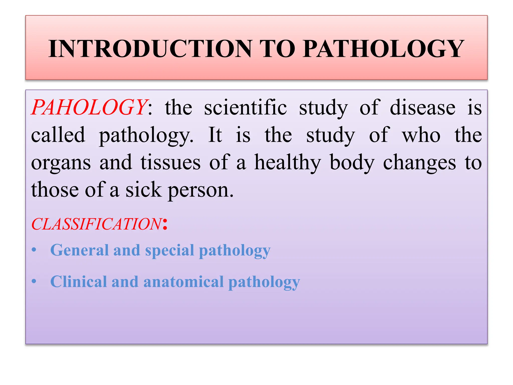
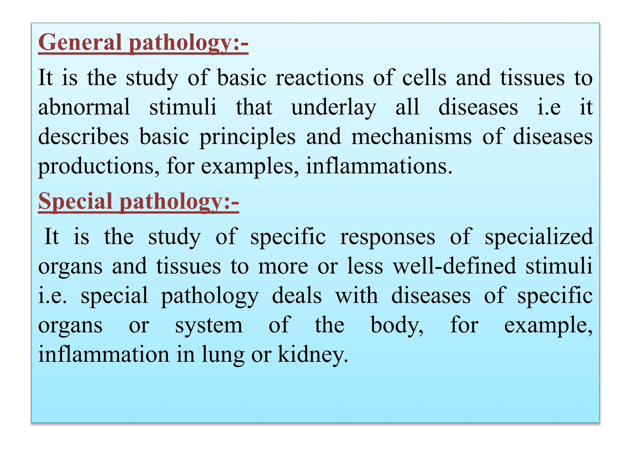
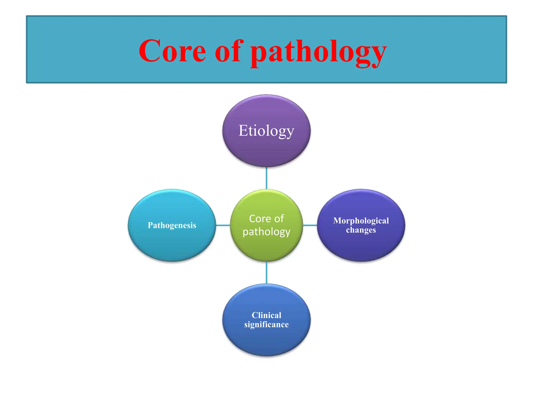
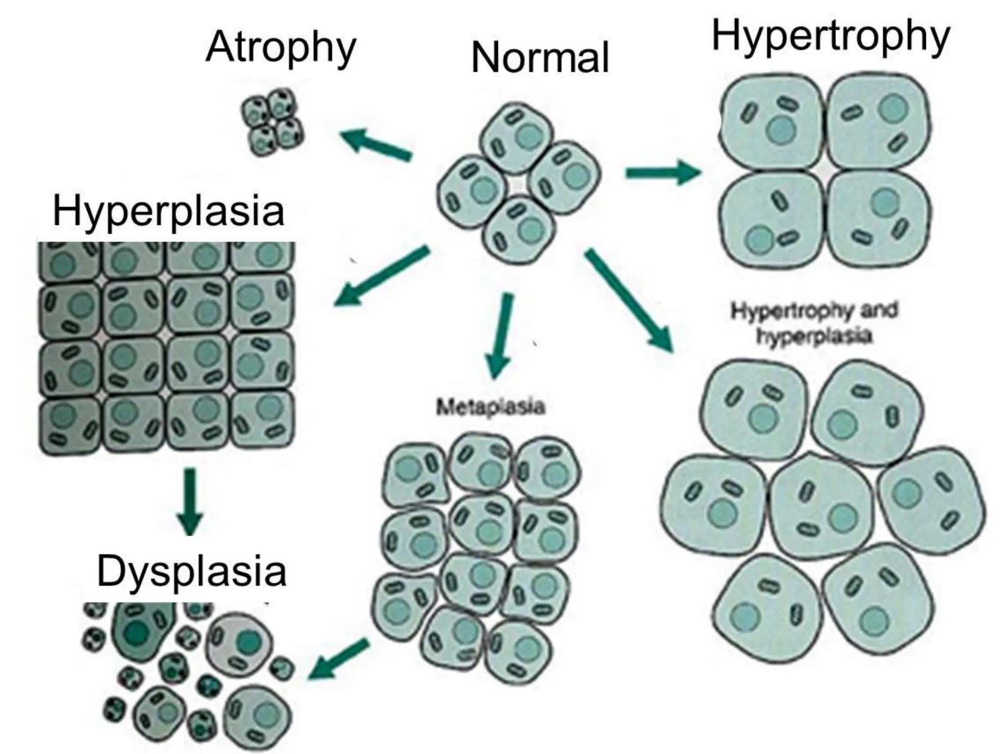
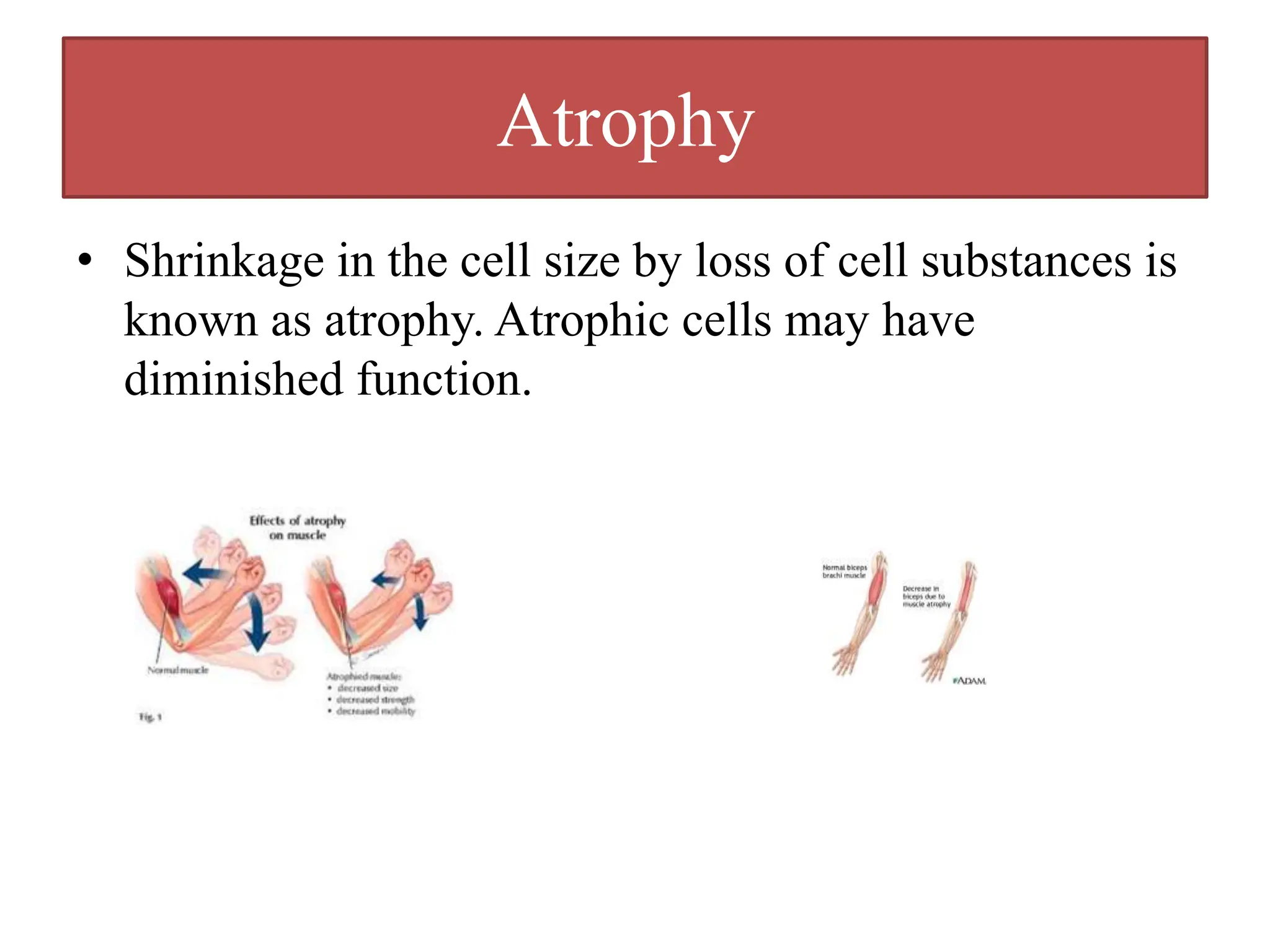
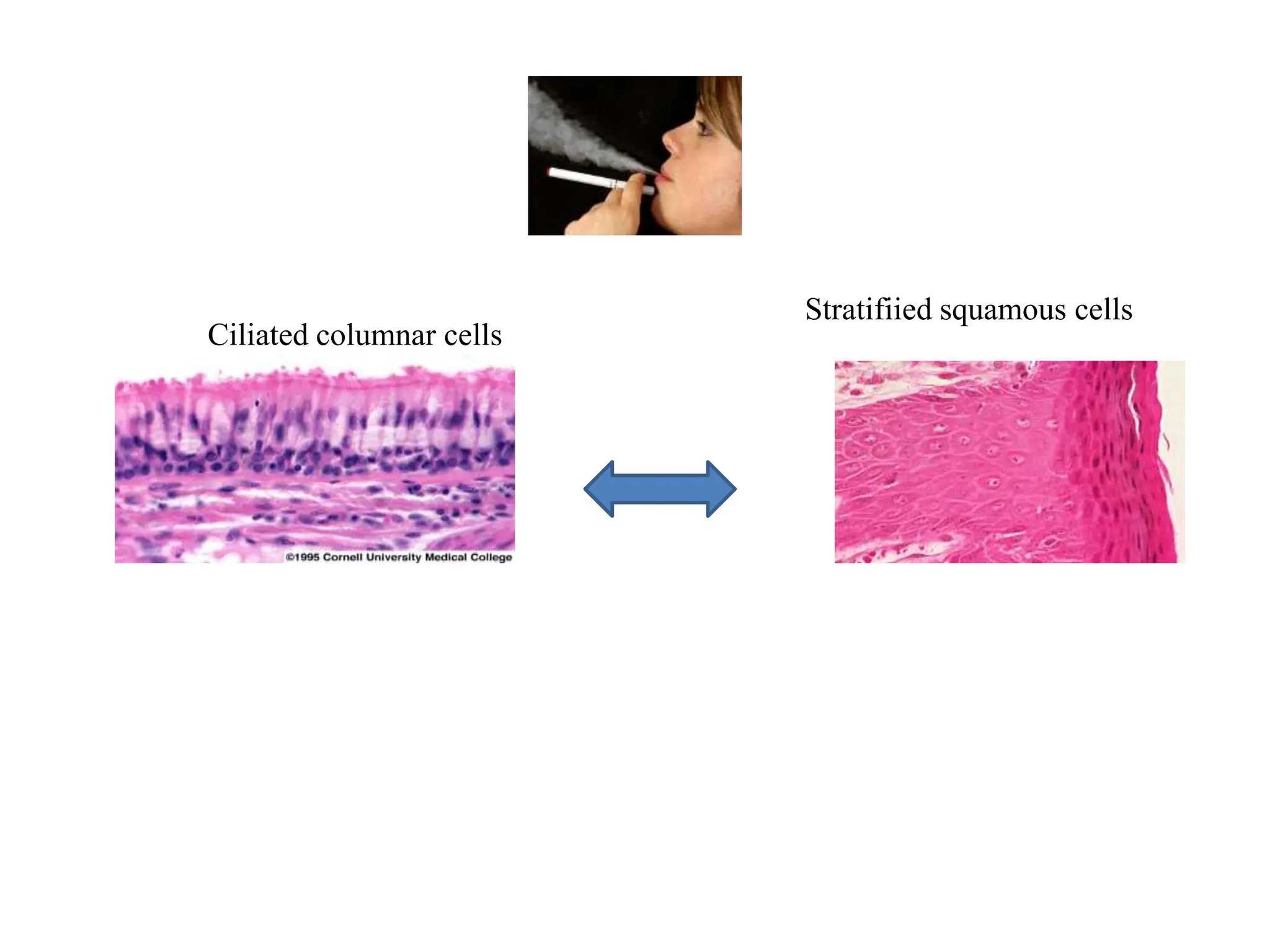
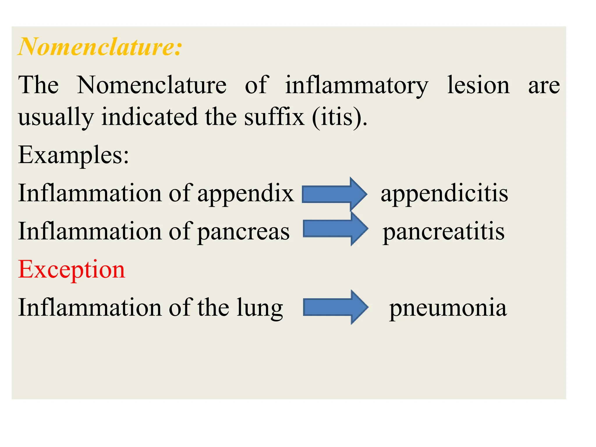
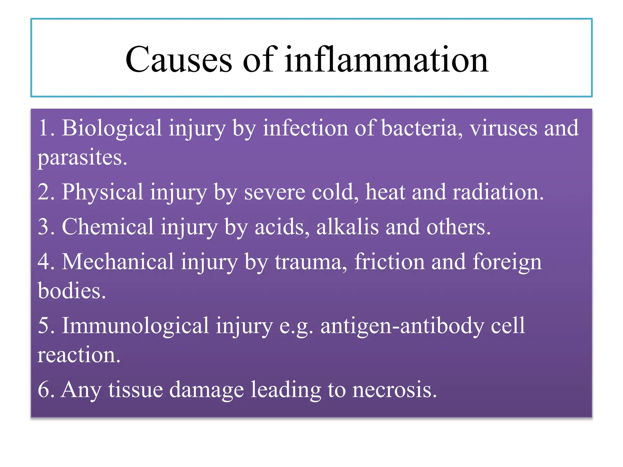
PATHOLOGY
Dr/ Ammar Omar
Assistant professor of Histopathology
Department of Pathology and Histology
Thamar university
INTRODUCTION TO PATHOLOGY
PAHOLOGY: the scientific study of disease is
called pathology. It is the study of who the
organs and tissues of a healthy body changes to
those of a sick person.
CLASSIFICATION:
• General and special pathology
• Clinical and anatomical pathology
General pathology:-
It is the study of basic reactions of cells and tissues to
abnormal stimuli that underlay all diseases i.e it
describes basic principles and mechanisms of diseases
productions, for examples, inflammations.
Special pathology:-
It is the study of specific responses of specialized
organs and tissues to more or less well-defined stimuli
i.e. special pathology deals with diseases of specific
organs or system of the body, for example,
inflammation in lung or kidney.
Core of pathology
Core of
pathology
Etiology
Morphological
changes
Clinical
significance
Pathogenesis
Etiology-course of disease
Etiology
Genetic
Acquired
Genetic:-
Abnormalities of chromosomes called mutations or defect
in genes.
Acquired:-
• Physical agents
• Chemical poisons
• Nutritional deficiencies
• Infections
• Abnormal immunological reactions
• Psychological factors
Pathogenesis
Pathogenesis means the sequences of events in the cells or tissues
or the whole organism in response to the causative agent- from
the initial stimulus to the ultimate expression of the
manifestations of the diseases.
Morphological changes
This refers to the structural and associated functional
changes in the cells or tissues that are either characteristics
of the diseases or diagnostic of the etiological process.
Clinical significance
It refers to sings, symptoms, course and prognosis of the
disease.
Cell injury
When the limits of adaptive capacity are
exceeded or when no adaptive response is
possible, a consequences of events follows,
termed as cell injury.
Reversible cell injury
It donates pathological changes
that can be reversed when the
stimulus (or stress) is removed
or if the cause of injury is mild.
Irreversible cell injury
It donates pathological changes
that are permanent and cause
cell death.
Causes of cell
injury
Hypoxia
Physical
agents
Chemical
agents
Immunological
reactions
Genetic
defects
Microbiologi
cal agents
Nutritional
imbalance
Mechanisms of cell injury
1. Impaired cell membrane function by
• Production of free radicals
• Loss of calcium homeostasis
• Activations of complements
• Lysis of enzymes
• Direct membrane Lysis by viruses, heats, cold
and chemicals
2. Decrease ATP production due to
• Hypoxia
• Hypoglycemia
3. Genetics alterations
4. Metabolic derangements
• Exposure to exogenous injurious agents
• Accumulation of some endogenous substances
Features of irreversible cell injury
Nuclear changes
Nucleus may show one of the three patterns of
changes.
• Pyknosis
• Karyolysis
• Karyorrhexis
Cytoplasmic changes
• Irreversible damage to mitochondria manifested by
sever vacuolization
• Extensive damage to the plasma membrane
• Massive calcium influx acting as a poisons for
mitochondria
• Loss of enzyme and proteins due to increased
membrane permeability
• Lysosomal swelling and leakage of enzymes
Necrosis
Definitions
1- Necrosis refers to a sequence of morphological
changes that follow cell death in livening tissue.
2- The morphological changes caused by progressive
degradative actions on dead cells is called necrosis.
3- Necrosis is the sum of intracellular degradative
reactions occurring after the death of individual cell
within a living organism.
Necrosis = cell death + morphological changes.
Mechanism of necrosis
Mechanism
of necrosis
Enzymatic
degradation of cell
Denaturation
of protein
Types of Necrosis
Types of
necrosis
Liquefactive
necrosis
Gangrenous
necrosis
Fat necrosis
Fibrinoid
necrosis
Coagulative
necrosis
Coagulative necrosis
• in this type of necrosis, the necrotic cell retains
in cellular outline for several days. Coagulative
necrosis typically occur in solid organ, such as
kidney, heart, and adrenal gland as a result of
deficient blood supply and anoxia.
Liquefactive necrosis
Liquefactive necrosis:- Liquefactive necrosis is
characterized by digestion of tissue. It shows
softening& liquefaction of tissue. It
characteristically results from ischemic injury to
the CNS. It also occurs in superlative infections
characterized by formation of pus.
Caseous necrosis
• Caseous necrosis has a cheese-like (caseous,
white) appearance to the naked eye. And it
appears as an amorphous eosinophilic
material on microscopic examination. Caseous
necrosis is typical of tuberculosis.
Fat necrosis
• Fat necrosis can be caused by trauma to tissue with high fat
content, such as the breast or it can also be caused by acute
hemorrhagic pancreatitis in which pancreatic enzymes diffuse
into the inflamed pancreatic tissue & digest it. The fatty acids
released from the digestion form calcium salts (soap formation
or dystrophic calcification). In addition, the elastase enzyme
digests the blood vessels & cause the hemorrhage inside the
pancreas, hence the name hemorrhagic pancreatitis.
Fibrinoid necrosis
• Fibrinoid necrosis is a special form of necrosis
usually caused by immunemediated vascular damage.
It is marked by complexes of antigen and antibodies,
sometimes referred to as ―immune complexes‖.
Gangrenous necrosis
Gangrenous
necrosis
Dry Gangrenous Gas Gangrenous
Wet Gangrenous
Dry Gangrenous
. Commonly occur in limbs due to ischemia leading to
Coagulative necrosis involving multiple tissue layers.
. The organ is dry, shrunken and limited putrefaction.
. Not complicated with bacterial infection.
Wet Gangrenous
• occurs due to ischemia ( see in moist tissue e.g
intestine).
• The organ is moist, ,soft, swollen, and dark
with marked putrefaction.
• Coplicated with bacterial infections.
Gas Gangrenous
• Caused by bacteria (clostridium perferingens.
• commonly occur in muscles tissues.
Apoptosis
• Definition: apoptosis is a distinctive morphologic pattern of cell death affecting a
single or small group of cell. Apoptosis is energy- dependent programmed cell
death for removal of unwanted individual cell. Apoptosis in Greak means
*dropping off*
• Death by apoptosis is a normal phenomenon that serve to eliminate cell that are no
longer needed and to maintain a steady number of cells in tissue.
• The main event in apoptosis is the activation of enzymes called caspases
Comparison between necrosis and apoptosis
Necrosis Apoptosis
Stimuli Hypoxia & toxins Physiological &
pathological
Histological detection Easy Difficult
Cell affected Large group Single cell or small group
Nucleus Pyknosis,
karyorrhexis&karyolysis
Condensation &
fragmentation of chromatin
Cytoplasm Cytomegaly Shrinkage
Tissue reaction Inflammation No inflammation
Cellular Adaptations
• Adaptations are reversible changes in the
number, size, phenotypes, metabolic activity,
or function of cells in response to changes in
their environment.
Following are the important adaptive changes:
 Atrophy: decrease in cell size.
 Hypertrophy: increase in cell size.
 Hyperplasia: increase in cell number.
 Metaplasia: change in cell type.
Atrophy
• Shrinkage in the cell size by loss of cell substances is
known as atrophy. Atrophic cells may have
diminished function.
Atrophy
Disuse
atrophy
Denervation
atrophy
Pressure
atrophy
Atrophy due
to endocrine
deficiency
Atrophy due
to lack of
nutrients
Senile
atrophy
Ischemic
atrophy
Hypertrophy
• is increase in the size of cells resulting in size of organ.
1. Physiologic hypertrophy e.g increased functional
demand (striated muscle cells in skeletal muscles and
heart) or hormonal stimulations (enlargement of
uterus during pregnancy).
2. Pathologic hypertrophy e.g cardiac enlargement due
to hypertension or aortic valve diseases.
Hyperplasia
• is an increase in the number of cells. It can
lead to an increase in the size of the organ. It is
usually caused by hormonal stimulation. It can
be physiological as in enlargement of the
breast during pregnancy or it can pathological
as in wound healing.
Metaplasia
• Transformation of differentiated mature cells
into another type of cells of the same group.
• Metaplasia occurs in cells to adapt them to the
change in their environment or their function.
Metaplasia may be a precancerous lesion.
Ciliated columnar cells
Stratifiied squamous cells
Dysplasia
• disordered epithelial cellular proliferation commonly
in association with chronic irritation or chronic
inflammation.
• Loss of normal arrangement. Cell show pleomorphism, hyperchromatism
and occasional mitosis.
• Pleomorphism: cells variable in size and shape in the tissue.
• Dysplasia may be:
• -Mild (basal third of the epithelium).
• Moderate (lower two third).
• Severe (whole epithelial thickness).
• Mild dysplasia is commonly reversible, severe dysplasia is a precancerous
lesion.
Anaplasia
• Cells differentiate to a more IMMATURE or
embryonic form.
• Malignant tumors are characterized by anaplastic cell
growth.
• Ovarian cancer cells dividing
INFLAMMATION
Definition:
Inflammation is a protective involving host cells,
B.V, proteins and other mediators that is intended
to:
Eliminate the initial cause of cell injury.
Removal of the necrotic cells and tissue.
Initiate the process of repair.
Nomenclature:
The Nomenclature of inflammatory lesion are
usually indicated the suffix (itis).
Examples:
Inflammation of appendix appendicitis
Inflammation of pancreas pancreatitis
Exception
Inflammation of the lung pneumonia
Causes of inflammation
1. Biological injury by infection of bacteria, viruses and
parasites.
2. Physical injury by severe cold, heat and radiation.
3. Chemical injury by acids, alkalis and others.
4. Mechanical injury by trauma, friction and foreign
bodies.
5. Immunological injury e.g. antigen-antibody cell
reaction.
6. Any tissue damage leading to necrosis.
Cardinal signs of inflammation:
1. Heat (calor)
2. Redness ( rubor)
3. Pain (dolor)
4. Loss of function ( functio lesa)
5. Swelling (tumor)
Types of inflammation
Acute inflammation
is an inflammation of short duration characterized by
formation of inflammation exudate.
Chronic inflammation
which is an inflammation of prolonged duration
characterized by tissue destruction and healing of the
tissue.
Acute inflammation
• In acute inflammation the tissue response is
rapid i.e. sudden onset. It lasts for days of few
weeks and is characterized by the presence of
fluid exudate, fibrin threads and
polymorphonuclear leucocytes (neutrophil).
ACUTE INFLAMMATION:
The classical signs are: REDNESS (rubor)
HEAT (calor)
SWELLING (tumour)
PAIN (dolor)
LOSS OF FUNCTION (functio laesa).
These gross signs are explained by changes occurring at
microscopic level. Three essential features are:
1. HYPERAEMIA
2. EXUDATION OF FLUID
3. EMIGRATION OF LEUCOCYTES.
• HYPERAEMIA: The hyperaemia in inflammation is
associated with the well known microvascular changes.
EXUDATION: Exudation is the increased passage of protein-rich
fluid through the vessel wall into the interstitial tissue.
• EMIGRATION OF LEUCOCYTES :
Neutrophils and mononuclears pass between the endothelial cell
junctions by amoeboid movement through the venule wall into
the tissue spaces. In this process both neutrophils and endothelial
cells are activated and both express cell adhesion molecules,
initially SELECTINS and then INTEGRINS.
CHEMOTAXIS:
The initial margination of neutrophils and mononuclears
is potentiated by slowing of blood flow and by
increased ‘stickiness’ of the endothelial surface. After
penetration of the vessel wall, the subsequent movement
of the leucocytes is controlled by CHEMOTAXIS. The
cell moves in response to an increasing concentration
gradient of the particular chemotactic agent, usually a
protein or polypeptide
• Important examples of chemotactic agents are:
Fractions of the COMPLEMENT SYSTEM (esp. C3a) Factors
derived from arachidonic acid by the neutrophils –
LEUKOTRIENES (e.g. LTB4) Factors derived from pathogenic
BACTERIA Factors derived from sensitised lymphocytes –
CYTOKINES (e.g. IL-8). The leucocytes move by extension of
an anterior pseudopod with attachment to extracellular matrix
molecules such as fibronectin using cell adhesion molecules. The
cell body is then pulled forward by actin and myosin filaments.
PHAGOCYTOSIS:
This is the process by which neutrophils and
macrophages clear the injurious agent. It is an
important defence mechanism in bacterial
infections particularly.
There are 3 families of OPSONIN.
1. Immunoglobulin, especially IgG – recognized by Fc receptors
on neutrophil surface.
2. Complement, especially C3b – recognized by C3b receptors on
neutrophil surface.
3. Carbohydrate binding proteins, or lectins – bind sugar residues
on bacterial cell walls. The opsonic activity is enhanced when it is
confined within a solid organ or rigid medium such as a fibrin
network; where conditions are looser and more fluid, activity is
diminished.
• CHEMICAL MEDIATORS:
Various chemical mediators have roles in the inflammatory
process. They may be circulating in plasma and require activation
or they may be secreted by inflammatory cells. Many of these
mediators have overlapping actions.
Mediators derived from:
1. Inflammatory cells
2. Plasma. These pathways are all interrelated.
SEQUELS OF ACUTE INFLAMMATION
Types of acute inflammation
1. Suppurative inflammation: associated with pus
formation.
• Localized: abscess.
• Diffuse: cellulitis.
cellulitis
2. Non suppurative inflammation: with no pus
formation. It includes:
Catarrhal inflammation Catarrha rhinitis
Membranous inflammation Diphtheria
Fibrinous inflammation Lobar pneumonia
Serous inflammation Mild burns
Sero- fibrinous inflammation Pleurisy
Haemorrhagic inflammation Streptococcal
Necrotizing inflammation Infective gangrene
Allergic inflammation Bronchial asthma
Catarrhal inflammation
Serous inflammation
Pleurisy
Sero- fibrinous inflammation
A-Right cranial lobe, lobar pneumonia (arrow), dorsal appearance, B-Right cranial
lobe with medial lobe, lobar pneumonia (arrow), dorsal appearance, C-Medial lobe
lobular pneumonia (arrow), adhesion on visceral pleura (thin arrow), dorsal
appearance, D-.The cut section of pneumonic areas
Membranous inflammation
Diphtheria
Haemorrhagic inflammation
Streptococcal
Necrotizing inflammation
Allergic inflammation Bronchial asthma
CHRONIC INFLAMMATION
Definition:
Chronic inflammation can be defined as a prolonged
inflammatory process (weeks or months) where an
active inflammation, tissue destruction and attempts at
repair are proceeding simultaneously.
Causes of chronic inflammation:
 Persistent infections Certain microorganisms associated with
intracellular infection such as tuberculosis, leprosy, certain
fungi etc characteristically cause chronic inflammation.
 These organisms are of low toxicity and evoke delayed
hypersensitivity reactions.
 Prolonged exposure to nondegradable but partially toxic
substances either endogenous lipid components which result
in atherosclerosis or exogenous substances such as silica,
asbestos.
 Progression from acute inflammation: Acute inflammation
almost always progresses to chronic inflammation following:
a. Persistent suppuration as a result of uncollapsed abscess
cavities, foreign body materials (dirt, cloth, wool, etc),
sequesterum in osteomylitis, or a sinus/fistula from chronic
abscesses.
 Autoimmuniy. Autoimmune diseases such as rheumatoid
arthritis and systemic lupus erythematosis are chronic
inflammations from the outset.
Cells of chronic inflammation:
Classification of chronic inflammation:
1. Non specific chronic inflammation:
This involves a diffuse accumulation of macrophages and lymphocytes at
site of injury that is usually productive with new fibrous tissue formations.
E.g. Chronic cholecystitis.
2. Specific inflammation (granulomatous inflammation):
Definition: Granulomatous inflammation is characterized by the presence
of granuloma. A granuloma is a microscopic aggregate of epithelioid cells.
Epithelioid cell is an activated macrophage, with a modified epithelial cell-
like appearance (hence the name epithelioid). The epitheloid cells can fuse
with each other & form multinucleated giant cells. So, even though, a
granuloma is basically a collection of epithelioid cells, it also usually
contains multinucleated giant cell & is usually surrounded by a cuff of
lymphocytes and occasional plasma cells. There are two types of giant
cells:
a. Foreign body-type giant cells which have irregularly
scattered nuclei in presence of indigestible materials.
b. Langhans giant cells in which the nuclei are arranged
peripherally in a horse -shoe pattern which is seen
typically in tuberculosis, sarcoidosis etc… Giant cells are
formed by fusion of macrophages perhaps by a concerted
attempt of two or more cells to engulf a single particle.
Pathogenesis:
There are two types of granulomas, which differ
in their pathogenesis.
A. Foreign body granuloma
These granulomas are initiated by inert foreign
bodies such as talc, sutures (nonabsorbable),
fibers, etc… that are large enough to preclude
phagocytosis by a single macrophage and do not
incite an immune response.
B. Immune granulomas
Antigen presenting cells (macrophages) engulf a poorly
soluble inciting agent. Then, the macrophage processes and
presents part of the antigen (in association with MHC type2
molecules) to CD4+T helper 1 cells which become activated.
The activated CD4+ T-cells produce cytokines (IL-2 and
interferon gamma).The IL-2 activates other CD4+T helper
cells and perpetuates the response while IFN-γ is important in
transforming macrophages into epitheloid cells and
multinucleated giant cells. The cytokines have been implicated
not only in the formation but also in the maintenance of
granuloma.
Macrophage inhibitory factor helps to localize activated
macrophages and epitheloid cells.
SYSTEMIC EFFECTS OF INFLAMMATIONS
• The systemic effects of inflammation include:
a. Fever
b. Endocrine & metabolic responses
c. Autonomic responses
d. Behavioral responses
e. Leukocytosis
f. Leukopenia
g. Weight loss
Repair
Definition: repair is the replacement of the
damaged tissue by new healthy one.
Types:
1. Regeneration: replacement of damaged cells by
cells of the same kind. Regeneration means the
ability of the cells to divide and reproduce
themselves.
2. Fibrosis: replacement of damaged tissue by
granulation tissue which matures to fibrous tissue.
HEALING
I. Definition of healing
The word healing, used in a pathological context, refers to
the body‘s replacement of destroyed tissue by living tissue.
II. Processes of healing
The healing process involves two distinct processes:
- Regeneration, the replacement of lost tissue by tissues
similar in type and
- Repair (healing by scaring), the replacement of lost tissue
by granulation tissue which matures to form scar tissue.
Healing by fibrosis is inevitable when the surrounding
specialized cells do not possess the capacity to proliferate.
• Tissue → Inflammation → Removal of dead
tissue & Damage
• Processes of healing: Removal of dead tissue &
injurious agent and replacement occur simultaneously.
Whether healing takes place by regeneration or by repair
(scarring) is determined partly by the type of cells in the
damaged organ & partly by the destruction or the
intactness of the stromal frame work of the organ. Hence,
it is important to know the types of cells in the body.
• Types of cells:
Based on their proliferative capacity there are three types
of cells.
1. Labile cells
These are cells which have a continuous turn over by
programmed division of stem cells. They are found in the
surface epithelium of the gastrointestinal tract, urinary
tract or the skin. The cells of lymphoid and haemopoietic
systems are further examples of labile cells. The chances
of regeneration are excellent.
2. Stable cells
Tissues which have such type of cells have normally a much
lower level of replication and there are few stem cells.
However, the cells of such tissues can undergo rapid
division in response to injury. For example, mesenchymal
cells such as smooth muscle cells, fibroblasts, osteoblasts
and endothelial cells are stable cells which can proliferate.
Liver, endocrine glands and renal tubular epithelium has
also such type of cells which can regenerate. Their chances
of regeneration are good.
3. Permanent cells
These are non-dividing cells. If lost, permanent cells
cannot be replaced, because they don not have the
capacity to proliferate. For example: adult neurons,
striated muscle cells, and cells of the lens.
• REGENERATION involves Two PROCESSES:
1. PROLIFERATION of SURVIVING CELLS to
replace lost tissue.
2. MIGRATION of SURVIVING CELLS into the
vacant space.
The FACTORS which CONTROL healing and repair are
complex: they include the production of a large variety of growth
factors.
WOUND HEALING
Healing of a wound shows both epithelial
regeneration (healing of the epidermis) and
repair by scarring (healing of the dermis). Two
patterns are described depending on the amount
of tissue damage. These are the same process
varying only in amount.
1. Healing by first intention (primary union)
This occurs in clean, incised wounds with good
apposition of the edges – particularly planned
surgical incisions.
2. Healing by second intention (secondary union) This
occurs in open wounds, particularly when there has
been significant loss of tissue, necrosis or infection.
FIBROSIS: is the end result of WOUND HEALING, CHRONIC
INFLAMMATION and ORGANISATION.
Formation of fibrous tissue:
REMODELLING follows: Action of
COLLAGENASE SCAR TISSUE + secretion of
COLLAGEN
REMODELLING follows: Action of COLLAGENASE SCAR TISSUE
+ secretion of COLLAGEN
NEOPLASIA
• NEOPLASIA definition:
NEOPLASIA = new growth.
Oncology = onco =tumor = neoplasm. Logy: science
Neoplasm: abnormal mass of tissue due to uncontrolled
proliferation of the cells.
Benign neoplasm : innocent behavior
Malignant: cancer ( behave like crab)
Nomenclature
• Neoplasms are named based upon two factors
1. The histologic types : mesenchymal and
epithelial.
2. Behavioral patterns : benign and malignant
neoplasms.
• Benign neoplasms
Thus, the suffix -oma denotes a benign neoplasm.
Benign mesenchymal neoplasms originating from muscle, bone,
fat, blood vessel nerve, fibrous tissue and cartilages are named as
Rhabdomyoma, osteoma, lipoma, hemangioma, neuroma, fibroma
and chondroma respectively. Benign epithelial neoplasms are
classified on the basis of cell of origin for example adenoma is the
term for benign epithelial neoplasm that form glandular pattern or
on basis of microscopic or macroscopic patterns for example
visible finger like or warty projection from epithelial surface are
referred to as papillomas.
• This nomenclature has, however, some
exceptions (I) Non neoplastic misnomers
hematoma, granuloma, hamartoma.
(II) Malignant misnomers melanoma, lymphoma,
seminoma, glioma, hepatoma.
• Malignant neoplasm nomenclature:
essentially follows the same scheme used for benign
neoplasm with certain additions. Malignant neoplasms
arising from mesenchymal tissues are called sarcomas
(Greed sar =fleshy). Thus, it is a fleshy tumour. These
neoplasms are named as fibrosarcoma, liposarcoma,
osteosarcoma, hemangiosarcoma etc.
• Malignant neoplasms of epithelial cell origin derived
from any of the three germ layers are called carcinomas.
Eg. Ectodermal origin: skin (epidermis squamous cell carcinoma, basal
cell carcinoma)Mesodermal origin: renal tubules (renal cell
carcinoma).Endodermal origin: linings of the gastrointestinal tract
(colonic carcinoma) Carcinomas can be furtherly classified those
producing glandular microscopic pictures are called Aden carcinomas
and those producing recognizable squamous cells are designated as
squamous cell carcinoma etc furthermore, when possible the carcinoma
can be specified by naming the origin of the tumour such as renal cell
adenocarcinoma etc.
• Tumors that arise from more than tissue components:-
Teratomas contain representative of parenchyma cells of more
than one germ layer, usually all three layers. They arise from
totipotential cells and so are principally encountered in ovary
and testis.
- Mixed tumors containing both epithelial and mesenchymal
components Examples include pleomorphic adenoma and
fibroadenoma
III. Characteristics of Benign and Malignant
Neoplasms
• The difference in characteristics of these
neoplasms can be conveniently discussed
under the following headings:
1. Differentiation & anaplasia
2. Rate of growth
3. Local invasion
4. Metastasis
Microscopical features of malignant cells
1- pleomorphism ( marked variation in shape and size of
cell).
2- hyperchromatic ( dark staining) and large nuclei.
3- increase nuclear cytoplasmic ratio 1:1 ( normal 1:4 or
1:6).
4- numerous atypical mitosis.
5- giant cell.
6- lose of orientation to each other.
• Sequential steps in mechanisms of tumor
invasion & metastasis:
a. Carcinoma in-situ b. Malignant cell surface receptors bind to
basement membrane components (ex laminin). c. Malignant
cell disrupt and invade basement membrane by releasing
collagenase type IV and other protease. d. Invasion of the
extracellular matrix e. Detachment f. Embolization g.
Survival in the circulation h. Arrest i. Extravasation j. Evasion
of host defense k. Progressive growth l. Metastasis
Epidemiology of cancer
The incidence of cancer varies with:
1. Age
2. Race
3. Geographic
4. Genetic factors.
Etiology of neoplasm ( carcinogenic agents)
• Four classes of carcinogenic agent have been
identified:
1. Genetic causes
2. Chemical causes
3. Radiation
4. Microbial agents ( viruses)
Clinical effects of neoplasm
• Clinical effects of benign neoplasms:
 pressure atrophy
Obstructions
Hemorrhage
Hormonal over secretion
 deformity
• Clinical effects of malignant neoplasms:
 Pressure atrophy
 Obstruction
 Ulceration
 Distraction of tissue
 Hemorrhage
 Hormonal over secretion
 Infection
 Starvation and anemia
 Pain
 Carcinomatous syndrome
 Cachaxia or wasting
 Effect of metastasis
Staging of malignancy
• Staging: measuring the size of neoplasm and extent of
spread.
• Staging is the functions of physician, surgeon,
pathologist and oncologist.
• Staging based on:
1) Size of primary lesion
2) Regional lymph node
3) Distant of metastasis
• Significance of staging: assessing prognosis and
treatment.
• System of staging:
 TNM system: T = tumor N= lymph node
statues
M = metastasis
So the stage take the following degree I, II, III,
IV)
2- AJC American joint committee
Example for staging of malignancy
Stage Definition
T0 In situ, non-invasive (confined to epithelium)
T1 Small, minimally invasive within primary organ site
T2 Large, more invasive within primary organ
T3 Large and invasive beyond primary organ.
T4 Very large and or very invasive , spread to adjacent
organ
N0 No lymph node involvement
N1 Regional lymph node involvement
N2 extensive regional LN involvement
N3 more distance LN involvement
M0 No distances metastasis
Grading of malignancy:
Grading: measuring the extent and degree of
differentiation. It is the function of pathologist.
Significance of grading: assessing aggressiveness
of neoplasm.
Grading based in:
1- degree of differentiation
2- pleomorphism
3- mitotic activities
• System of grading:
1. Grade I, II, III, IV.
2. Low grade, intermediate grade and high grade
3. Well- differentiated (WD), moderately
differentiated (MD), poorly differentiated (PD)
and undifferentiated – anaplastic (UD).
Staging: progression or spread in the body.
Grading: cell differentiation and rate of growth –
microscopy.
Diagnosis of neoplasm
• Clinical data, radiology, and ultrasound
• Cytology
• Histological study
• Biochemical study( seromarkers)
• ICC or IHC
• Flow cytometers
• Molecular diagnosis
Cancer prevention
1. Avoid tobacco 2- avoid alcohol
3- limit fat and calories 4- avoid khat
5- protect yourself from excessive sunlight
6- breast, prostate, ovarian, colorectal, and
cervical cancer screening ( early detection)
7- consumed fruited and vegetable
8- avoid cancer viruses
9- treat bacteria predispose to gastric cancer.

PATHOLOGY -Dr- Ammar Omar -Assistant professor of Histopathology -Department of Pathology and Histology -Thamar university-1 (3).pptx

  • 1.
    PATHOLOGY Dr/ Ammar Omar Assistantprofessor of Histopathology Department of Pathology and Histology Thamar university
  • 2.
    INTRODUCTION TO PATHOLOGY PAHOLOGY:the scientific study of disease is called pathology. It is the study of who the organs and tissues of a healthy body changes to those of a sick person. CLASSIFICATION: • General and special pathology • Clinical and anatomical pathology
  • 3.
    General pathology:- It isthe study of basic reactions of cells and tissues to abnormal stimuli that underlay all diseases i.e it describes basic principles and mechanisms of diseases productions, for examples, inflammations. Special pathology:- It is the study of specific responses of specialized organs and tissues to more or less well-defined stimuli i.e. special pathology deals with diseases of specific organs or system of the body, for example, inflammation in lung or kidney.
  • 4.
    Core of pathology Coreof pathology Etiology Morphological changes Clinical significance Pathogenesis
  • 5.
  • 6.
    Genetic:- Abnormalities of chromosomescalled mutations or defect in genes. Acquired:- • Physical agents • Chemical poisons • Nutritional deficiencies • Infections • Abnormal immunological reactions • Psychological factors
  • 7.
    Pathogenesis Pathogenesis means thesequences of events in the cells or tissues or the whole organism in response to the causative agent- from the initial stimulus to the ultimate expression of the manifestations of the diseases. Morphological changes This refers to the structural and associated functional changes in the cells or tissues that are either characteristics of the diseases or diagnostic of the etiological process.
  • 8.
    Clinical significance It refersto sings, symptoms, course and prognosis of the disease.
  • 9.
    Cell injury When thelimits of adaptive capacity are exceeded or when no adaptive response is possible, a consequences of events follows, termed as cell injury.
  • 11.
    Reversible cell injury Itdonates pathological changes that can be reversed when the stimulus (or stress) is removed or if the cause of injury is mild. Irreversible cell injury It donates pathological changes that are permanent and cause cell death.
  • 12.
  • 13.
    Mechanisms of cellinjury 1. Impaired cell membrane function by • Production of free radicals • Loss of calcium homeostasis • Activations of complements • Lysis of enzymes • Direct membrane Lysis by viruses, heats, cold and chemicals
  • 14.
    2. Decrease ATPproduction due to • Hypoxia • Hypoglycemia 3. Genetics alterations 4. Metabolic derangements • Exposure to exogenous injurious agents • Accumulation of some endogenous substances
  • 15.
    Features of irreversiblecell injury Nuclear changes Nucleus may show one of the three patterns of changes. • Pyknosis • Karyolysis • Karyorrhexis
  • 16.
    Cytoplasmic changes • Irreversibledamage to mitochondria manifested by sever vacuolization • Extensive damage to the plasma membrane • Massive calcium influx acting as a poisons for mitochondria • Loss of enzyme and proteins due to increased membrane permeability • Lysosomal swelling and leakage of enzymes
  • 17.
    Necrosis Definitions 1- Necrosis refersto a sequence of morphological changes that follow cell death in livening tissue. 2- The morphological changes caused by progressive degradative actions on dead cells is called necrosis. 3- Necrosis is the sum of intracellular degradative reactions occurring after the death of individual cell within a living organism. Necrosis = cell death + morphological changes.
  • 18.
    Mechanism of necrosis Mechanism ofnecrosis Enzymatic degradation of cell Denaturation of protein
  • 19.
    Types of Necrosis Typesof necrosis Liquefactive necrosis Gangrenous necrosis Fat necrosis Fibrinoid necrosis Coagulative necrosis
  • 20.
    Coagulative necrosis • inthis type of necrosis, the necrotic cell retains in cellular outline for several days. Coagulative necrosis typically occur in solid organ, such as kidney, heart, and adrenal gland as a result of deficient blood supply and anoxia.
  • 21.
    Liquefactive necrosis Liquefactive necrosis:-Liquefactive necrosis is characterized by digestion of tissue. It shows softening& liquefaction of tissue. It characteristically results from ischemic injury to the CNS. It also occurs in superlative infections characterized by formation of pus.
  • 23.
    Caseous necrosis • Caseousnecrosis has a cheese-like (caseous, white) appearance to the naked eye. And it appears as an amorphous eosinophilic material on microscopic examination. Caseous necrosis is typical of tuberculosis.
  • 24.
    Fat necrosis • Fatnecrosis can be caused by trauma to tissue with high fat content, such as the breast or it can also be caused by acute hemorrhagic pancreatitis in which pancreatic enzymes diffuse into the inflamed pancreatic tissue & digest it. The fatty acids released from the digestion form calcium salts (soap formation or dystrophic calcification). In addition, the elastase enzyme digests the blood vessels & cause the hemorrhage inside the pancreas, hence the name hemorrhagic pancreatitis.
  • 25.
    Fibrinoid necrosis • Fibrinoidnecrosis is a special form of necrosis usually caused by immunemediated vascular damage. It is marked by complexes of antigen and antibodies, sometimes referred to as ―immune complexes‖.
  • 26.
  • 27.
    Dry Gangrenous . Commonlyoccur in limbs due to ischemia leading to Coagulative necrosis involving multiple tissue layers. . The organ is dry, shrunken and limited putrefaction. . Not complicated with bacterial infection.
  • 28.
    Wet Gangrenous • occursdue to ischemia ( see in moist tissue e.g intestine). • The organ is moist, ,soft, swollen, and dark with marked putrefaction. • Coplicated with bacterial infections.
  • 29.
    Gas Gangrenous • Causedby bacteria (clostridium perferingens. • commonly occur in muscles tissues.
  • 30.
    Apoptosis • Definition: apoptosisis a distinctive morphologic pattern of cell death affecting a single or small group of cell. Apoptosis is energy- dependent programmed cell death for removal of unwanted individual cell. Apoptosis in Greak means *dropping off* • Death by apoptosis is a normal phenomenon that serve to eliminate cell that are no longer needed and to maintain a steady number of cells in tissue. • The main event in apoptosis is the activation of enzymes called caspases
  • 32.
    Comparison between necrosisand apoptosis Necrosis Apoptosis Stimuli Hypoxia & toxins Physiological & pathological Histological detection Easy Difficult Cell affected Large group Single cell or small group Nucleus Pyknosis, karyorrhexis&karyolysis Condensation & fragmentation of chromatin Cytoplasm Cytomegaly Shrinkage Tissue reaction Inflammation No inflammation
  • 33.
  • 34.
    • Adaptations arereversible changes in the number, size, phenotypes, metabolic activity, or function of cells in response to changes in their environment.
  • 35.
    Following are theimportant adaptive changes:  Atrophy: decrease in cell size.  Hypertrophy: increase in cell size.  Hyperplasia: increase in cell number.  Metaplasia: change in cell type.
  • 37.
    Atrophy • Shrinkage inthe cell size by loss of cell substances is known as atrophy. Atrophic cells may have diminished function.
  • 38.
  • 39.
    Hypertrophy • is increasein the size of cells resulting in size of organ. 1. Physiologic hypertrophy e.g increased functional demand (striated muscle cells in skeletal muscles and heart) or hormonal stimulations (enlargement of uterus during pregnancy). 2. Pathologic hypertrophy e.g cardiac enlargement due to hypertension or aortic valve diseases.
  • 40.
    Hyperplasia • is anincrease in the number of cells. It can lead to an increase in the size of the organ. It is usually caused by hormonal stimulation. It can be physiological as in enlargement of the breast during pregnancy or it can pathological as in wound healing.
  • 42.
    Metaplasia • Transformation ofdifferentiated mature cells into another type of cells of the same group. • Metaplasia occurs in cells to adapt them to the change in their environment or their function. Metaplasia may be a precancerous lesion.
  • 43.
  • 44.
    Dysplasia • disordered epithelialcellular proliferation commonly in association with chronic irritation or chronic inflammation.
  • 45.
    • Loss ofnormal arrangement. Cell show pleomorphism, hyperchromatism and occasional mitosis. • Pleomorphism: cells variable in size and shape in the tissue. • Dysplasia may be: • -Mild (basal third of the epithelium). • Moderate (lower two third). • Severe (whole epithelial thickness). • Mild dysplasia is commonly reversible, severe dysplasia is a precancerous lesion.
  • 46.
    Anaplasia • Cells differentiateto a more IMMATURE or embryonic form. • Malignant tumors are characterized by anaplastic cell growth. • Ovarian cancer cells dividing
  • 47.
    INFLAMMATION Definition: Inflammation is aprotective involving host cells, B.V, proteins and other mediators that is intended to: Eliminate the initial cause of cell injury. Removal of the necrotic cells and tissue. Initiate the process of repair.
  • 48.
    Nomenclature: The Nomenclature ofinflammatory lesion are usually indicated the suffix (itis). Examples: Inflammation of appendix appendicitis Inflammation of pancreas pancreatitis Exception Inflammation of the lung pneumonia
  • 49.
    Causes of inflammation 1.Biological injury by infection of bacteria, viruses and parasites. 2. Physical injury by severe cold, heat and radiation. 3. Chemical injury by acids, alkalis and others. 4. Mechanical injury by trauma, friction and foreign bodies. 5. Immunological injury e.g. antigen-antibody cell reaction. 6. Any tissue damage leading to necrosis.
  • 50.
    Cardinal signs ofinflammation: 1. Heat (calor) 2. Redness ( rubor) 3. Pain (dolor) 4. Loss of function ( functio lesa) 5. Swelling (tumor)
  • 51.
    Types of inflammation Acuteinflammation is an inflammation of short duration characterized by formation of inflammation exudate. Chronic inflammation which is an inflammation of prolonged duration characterized by tissue destruction and healing of the tissue.
  • 53.
    Acute inflammation • Inacute inflammation the tissue response is rapid i.e. sudden onset. It lasts for days of few weeks and is characterized by the presence of fluid exudate, fibrin threads and polymorphonuclear leucocytes (neutrophil).
  • 54.
    ACUTE INFLAMMATION: The classicalsigns are: REDNESS (rubor) HEAT (calor) SWELLING (tumour) PAIN (dolor) LOSS OF FUNCTION (functio laesa). These gross signs are explained by changes occurring at microscopic level. Three essential features are: 1. HYPERAEMIA 2. EXUDATION OF FLUID 3. EMIGRATION OF LEUCOCYTES.
  • 55.
    • HYPERAEMIA: Thehyperaemia in inflammation is associated with the well known microvascular changes.
  • 56.
    EXUDATION: Exudation isthe increased passage of protein-rich fluid through the vessel wall into the interstitial tissue.
  • 58.
    • EMIGRATION OFLEUCOCYTES : Neutrophils and mononuclears pass between the endothelial cell junctions by amoeboid movement through the venule wall into the tissue spaces. In this process both neutrophils and endothelial cells are activated and both express cell adhesion molecules, initially SELECTINS and then INTEGRINS.
  • 61.
    CHEMOTAXIS: The initial marginationof neutrophils and mononuclears is potentiated by slowing of blood flow and by increased ‘stickiness’ of the endothelial surface. After penetration of the vessel wall, the subsequent movement of the leucocytes is controlled by CHEMOTAXIS. The cell moves in response to an increasing concentration gradient of the particular chemotactic agent, usually a protein or polypeptide
  • 63.
    • Important examplesof chemotactic agents are: Fractions of the COMPLEMENT SYSTEM (esp. C3a) Factors derived from arachidonic acid by the neutrophils – LEUKOTRIENES (e.g. LTB4) Factors derived from pathogenic BACTERIA Factors derived from sensitised lymphocytes – CYTOKINES (e.g. IL-8). The leucocytes move by extension of an anterior pseudopod with attachment to extracellular matrix molecules such as fibronectin using cell adhesion molecules. The cell body is then pulled forward by actin and myosin filaments.
  • 64.
    PHAGOCYTOSIS: This is theprocess by which neutrophils and macrophages clear the injurious agent. It is an important defence mechanism in bacterial infections particularly.
  • 66.
    There are 3families of OPSONIN. 1. Immunoglobulin, especially IgG – recognized by Fc receptors on neutrophil surface. 2. Complement, especially C3b – recognized by C3b receptors on neutrophil surface. 3. Carbohydrate binding proteins, or lectins – bind sugar residues on bacterial cell walls. The opsonic activity is enhanced when it is confined within a solid organ or rigid medium such as a fibrin network; where conditions are looser and more fluid, activity is diminished.
  • 67.
    • CHEMICAL MEDIATORS: Variouschemical mediators have roles in the inflammatory process. They may be circulating in plasma and require activation or they may be secreted by inflammatory cells. Many of these mediators have overlapping actions.
  • 68.
    Mediators derived from: 1.Inflammatory cells
  • 69.
    2. Plasma. Thesepathways are all interrelated.
  • 70.
    SEQUELS OF ACUTEINFLAMMATION
  • 71.
    Types of acuteinflammation 1. Suppurative inflammation: associated with pus formation. • Localized: abscess. • Diffuse: cellulitis.
  • 72.
  • 73.
    2. Non suppurativeinflammation: with no pus formation. It includes: Catarrhal inflammation Catarrha rhinitis Membranous inflammation Diphtheria Fibrinous inflammation Lobar pneumonia Serous inflammation Mild burns Sero- fibrinous inflammation Pleurisy Haemorrhagic inflammation Streptococcal Necrotizing inflammation Infective gangrene Allergic inflammation Bronchial asthma
  • 74.
  • 75.
  • 76.
  • 77.
    A-Right cranial lobe,lobar pneumonia (arrow), dorsal appearance, B-Right cranial lobe with medial lobe, lobar pneumonia (arrow), dorsal appearance, C-Medial lobe lobular pneumonia (arrow), adhesion on visceral pleura (thin arrow), dorsal appearance, D-.The cut section of pneumonic areas
  • 78.
  • 79.
  • 80.
  • 81.
  • 82.
    CHRONIC INFLAMMATION Definition: Chronic inflammationcan be defined as a prolonged inflammatory process (weeks or months) where an active inflammation, tissue destruction and attempts at repair are proceeding simultaneously.
  • 83.
    Causes of chronicinflammation:  Persistent infections Certain microorganisms associated with intracellular infection such as tuberculosis, leprosy, certain fungi etc characteristically cause chronic inflammation.  These organisms are of low toxicity and evoke delayed hypersensitivity reactions.  Prolonged exposure to nondegradable but partially toxic substances either endogenous lipid components which result in atherosclerosis or exogenous substances such as silica, asbestos.  Progression from acute inflammation: Acute inflammation almost always progresses to chronic inflammation following: a. Persistent suppuration as a result of uncollapsed abscess cavities, foreign body materials (dirt, cloth, wool, etc), sequesterum in osteomylitis, or a sinus/fistula from chronic abscesses.  Autoimmuniy. Autoimmune diseases such as rheumatoid arthritis and systemic lupus erythematosis are chronic inflammations from the outset.
  • 84.
    Cells of chronicinflammation:
  • 85.
    Classification of chronicinflammation: 1. Non specific chronic inflammation: This involves a diffuse accumulation of macrophages and lymphocytes at site of injury that is usually productive with new fibrous tissue formations. E.g. Chronic cholecystitis. 2. Specific inflammation (granulomatous inflammation): Definition: Granulomatous inflammation is characterized by the presence of granuloma. A granuloma is a microscopic aggregate of epithelioid cells. Epithelioid cell is an activated macrophage, with a modified epithelial cell- like appearance (hence the name epithelioid). The epitheloid cells can fuse with each other & form multinucleated giant cells. So, even though, a granuloma is basically a collection of epithelioid cells, it also usually contains multinucleated giant cell & is usually surrounded by a cuff of lymphocytes and occasional plasma cells. There are two types of giant cells:
  • 86.
    a. Foreign body-typegiant cells which have irregularly scattered nuclei in presence of indigestible materials. b. Langhans giant cells in which the nuclei are arranged peripherally in a horse -shoe pattern which is seen typically in tuberculosis, sarcoidosis etc… Giant cells are formed by fusion of macrophages perhaps by a concerted attempt of two or more cells to engulf a single particle.
  • 87.
    Pathogenesis: There are twotypes of granulomas, which differ in their pathogenesis. A. Foreign body granuloma These granulomas are initiated by inert foreign bodies such as talc, sutures (nonabsorbable), fibers, etc… that are large enough to preclude phagocytosis by a single macrophage and do not incite an immune response.
  • 88.
    B. Immune granulomas Antigenpresenting cells (macrophages) engulf a poorly soluble inciting agent. Then, the macrophage processes and presents part of the antigen (in association with MHC type2 molecules) to CD4+T helper 1 cells which become activated. The activated CD4+ T-cells produce cytokines (IL-2 and interferon gamma).The IL-2 activates other CD4+T helper cells and perpetuates the response while IFN-γ is important in transforming macrophages into epitheloid cells and multinucleated giant cells. The cytokines have been implicated not only in the formation but also in the maintenance of granuloma. Macrophage inhibitory factor helps to localize activated macrophages and epitheloid cells.
  • 89.
    SYSTEMIC EFFECTS OFINFLAMMATIONS • The systemic effects of inflammation include: a. Fever b. Endocrine & metabolic responses c. Autonomic responses d. Behavioral responses e. Leukocytosis f. Leukopenia g. Weight loss
  • 90.
    Repair Definition: repair isthe replacement of the damaged tissue by new healthy one. Types: 1. Regeneration: replacement of damaged cells by cells of the same kind. Regeneration means the ability of the cells to divide and reproduce themselves. 2. Fibrosis: replacement of damaged tissue by granulation tissue which matures to fibrous tissue.
  • 91.
    HEALING I. Definition ofhealing The word healing, used in a pathological context, refers to the body‘s replacement of destroyed tissue by living tissue. II. Processes of healing The healing process involves two distinct processes: - Regeneration, the replacement of lost tissue by tissues similar in type and - Repair (healing by scaring), the replacement of lost tissue by granulation tissue which matures to form scar tissue. Healing by fibrosis is inevitable when the surrounding specialized cells do not possess the capacity to proliferate.
  • 92.
    • Tissue →Inflammation → Removal of dead tissue & Damage
  • 93.
    • Processes ofhealing: Removal of dead tissue & injurious agent and replacement occur simultaneously. Whether healing takes place by regeneration or by repair (scarring) is determined partly by the type of cells in the damaged organ & partly by the destruction or the intactness of the stromal frame work of the organ. Hence, it is important to know the types of cells in the body.
  • 94.
    • Types ofcells: Based on their proliferative capacity there are three types of cells. 1. Labile cells These are cells which have a continuous turn over by programmed division of stem cells. They are found in the surface epithelium of the gastrointestinal tract, urinary tract or the skin. The cells of lymphoid and haemopoietic systems are further examples of labile cells. The chances of regeneration are excellent.
  • 95.
    2. Stable cells Tissueswhich have such type of cells have normally a much lower level of replication and there are few stem cells. However, the cells of such tissues can undergo rapid division in response to injury. For example, mesenchymal cells such as smooth muscle cells, fibroblasts, osteoblasts and endothelial cells are stable cells which can proliferate. Liver, endocrine glands and renal tubular epithelium has also such type of cells which can regenerate. Their chances of regeneration are good.
  • 96.
    3. Permanent cells Theseare non-dividing cells. If lost, permanent cells cannot be replaced, because they don not have the capacity to proliferate. For example: adult neurons, striated muscle cells, and cells of the lens.
  • 98.
    • REGENERATION involvesTwo PROCESSES: 1. PROLIFERATION of SURVIVING CELLS to replace lost tissue. 2. MIGRATION of SURVIVING CELLS into the vacant space.
  • 99.
    The FACTORS whichCONTROL healing and repair are complex: they include the production of a large variety of growth factors.
  • 100.
    WOUND HEALING Healing ofa wound shows both epithelial regeneration (healing of the epidermis) and repair by scarring (healing of the dermis). Two patterns are described depending on the amount of tissue damage. These are the same process varying only in amount.
  • 101.
    1. Healing byfirst intention (primary union) This occurs in clean, incised wounds with good apposition of the edges – particularly planned surgical incisions.
  • 103.
    2. Healing bysecond intention (secondary union) This occurs in open wounds, particularly when there has been significant loss of tissue, necrosis or infection.
  • 105.
    FIBROSIS: is theend result of WOUND HEALING, CHRONIC INFLAMMATION and ORGANISATION. Formation of fibrous tissue: REMODELLING follows: Action of COLLAGENASE SCAR TISSUE + secretion of COLLAGEN REMODELLING follows: Action of COLLAGENASE SCAR TISSUE + secretion of COLLAGEN
  • 107.
    NEOPLASIA • NEOPLASIA definition: NEOPLASIA= new growth. Oncology = onco =tumor = neoplasm. Logy: science Neoplasm: abnormal mass of tissue due to uncontrolled proliferation of the cells. Benign neoplasm : innocent behavior Malignant: cancer ( behave like crab)
  • 109.
    Nomenclature • Neoplasms arenamed based upon two factors 1. The histologic types : mesenchymal and epithelial. 2. Behavioral patterns : benign and malignant neoplasms.
  • 110.
    • Benign neoplasms Thus,the suffix -oma denotes a benign neoplasm. Benign mesenchymal neoplasms originating from muscle, bone, fat, blood vessel nerve, fibrous tissue and cartilages are named as Rhabdomyoma, osteoma, lipoma, hemangioma, neuroma, fibroma and chondroma respectively. Benign epithelial neoplasms are classified on the basis of cell of origin for example adenoma is the term for benign epithelial neoplasm that form glandular pattern or on basis of microscopic or macroscopic patterns for example visible finger like or warty projection from epithelial surface are referred to as papillomas.
  • 111.
    • This nomenclaturehas, however, some exceptions (I) Non neoplastic misnomers hematoma, granuloma, hamartoma. (II) Malignant misnomers melanoma, lymphoma, seminoma, glioma, hepatoma.
  • 112.
    • Malignant neoplasmnomenclature: essentially follows the same scheme used for benign neoplasm with certain additions. Malignant neoplasms arising from mesenchymal tissues are called sarcomas (Greed sar =fleshy). Thus, it is a fleshy tumour. These neoplasms are named as fibrosarcoma, liposarcoma, osteosarcoma, hemangiosarcoma etc.
  • 113.
    • Malignant neoplasmsof epithelial cell origin derived from any of the three germ layers are called carcinomas. Eg. Ectodermal origin: skin (epidermis squamous cell carcinoma, basal cell carcinoma)Mesodermal origin: renal tubules (renal cell carcinoma).Endodermal origin: linings of the gastrointestinal tract (colonic carcinoma) Carcinomas can be furtherly classified those producing glandular microscopic pictures are called Aden carcinomas and those producing recognizable squamous cells are designated as squamous cell carcinoma etc furthermore, when possible the carcinoma can be specified by naming the origin of the tumour such as renal cell adenocarcinoma etc.
  • 114.
    • Tumors thatarise from more than tissue components:- Teratomas contain representative of parenchyma cells of more than one germ layer, usually all three layers. They arise from totipotential cells and so are principally encountered in ovary and testis. - Mixed tumors containing both epithelial and mesenchymal components Examples include pleomorphic adenoma and fibroadenoma
  • 115.
    III. Characteristics ofBenign and Malignant Neoplasms • The difference in characteristics of these neoplasms can be conveniently discussed under the following headings: 1. Differentiation & anaplasia 2. Rate of growth 3. Local invasion 4. Metastasis
  • 116.
    Microscopical features ofmalignant cells 1- pleomorphism ( marked variation in shape and size of cell). 2- hyperchromatic ( dark staining) and large nuclei. 3- increase nuclear cytoplasmic ratio 1:1 ( normal 1:4 or 1:6). 4- numerous atypical mitosis. 5- giant cell. 6- lose of orientation to each other.
  • 117.
    • Sequential stepsin mechanisms of tumor invasion & metastasis: a. Carcinoma in-situ b. Malignant cell surface receptors bind to basement membrane components (ex laminin). c. Malignant cell disrupt and invade basement membrane by releasing collagenase type IV and other protease. d. Invasion of the extracellular matrix e. Detachment f. Embolization g. Survival in the circulation h. Arrest i. Extravasation j. Evasion of host defense k. Progressive growth l. Metastasis
  • 118.
    Epidemiology of cancer Theincidence of cancer varies with: 1. Age 2. Race 3. Geographic 4. Genetic factors.
  • 119.
    Etiology of neoplasm( carcinogenic agents) • Four classes of carcinogenic agent have been identified: 1. Genetic causes 2. Chemical causes 3. Radiation 4. Microbial agents ( viruses)
  • 120.
    Clinical effects ofneoplasm • Clinical effects of benign neoplasms:  pressure atrophy Obstructions Hemorrhage Hormonal over secretion  deformity
  • 121.
    • Clinical effectsof malignant neoplasms:  Pressure atrophy  Obstruction  Ulceration  Distraction of tissue  Hemorrhage  Hormonal over secretion  Infection  Starvation and anemia  Pain  Carcinomatous syndrome  Cachaxia or wasting  Effect of metastasis
  • 122.
    Staging of malignancy •Staging: measuring the size of neoplasm and extent of spread. • Staging is the functions of physician, surgeon, pathologist and oncologist. • Staging based on: 1) Size of primary lesion 2) Regional lymph node 3) Distant of metastasis
  • 123.
    • Significance ofstaging: assessing prognosis and treatment. • System of staging:  TNM system: T = tumor N= lymph node statues M = metastasis So the stage take the following degree I, II, III, IV) 2- AJC American joint committee
  • 124.
    Example for stagingof malignancy Stage Definition T0 In situ, non-invasive (confined to epithelium) T1 Small, minimally invasive within primary organ site T2 Large, more invasive within primary organ T3 Large and invasive beyond primary organ. T4 Very large and or very invasive , spread to adjacent organ N0 No lymph node involvement N1 Regional lymph node involvement N2 extensive regional LN involvement N3 more distance LN involvement M0 No distances metastasis
  • 125.
    Grading of malignancy: Grading:measuring the extent and degree of differentiation. It is the function of pathologist. Significance of grading: assessing aggressiveness of neoplasm. Grading based in: 1- degree of differentiation 2- pleomorphism 3- mitotic activities
  • 126.
    • System ofgrading: 1. Grade I, II, III, IV. 2. Low grade, intermediate grade and high grade 3. Well- differentiated (WD), moderately differentiated (MD), poorly differentiated (PD) and undifferentiated – anaplastic (UD). Staging: progression or spread in the body. Grading: cell differentiation and rate of growth – microscopy.
  • 127.
    Diagnosis of neoplasm •Clinical data, radiology, and ultrasound • Cytology • Histological study • Biochemical study( seromarkers) • ICC or IHC • Flow cytometers • Molecular diagnosis
  • 128.
    Cancer prevention 1. Avoidtobacco 2- avoid alcohol 3- limit fat and calories 4- avoid khat 5- protect yourself from excessive sunlight 6- breast, prostate, ovarian, colorectal, and cervical cancer screening ( early detection) 7- consumed fruited and vegetable 8- avoid cancer viruses 9- treat bacteria predispose to gastric cancer.