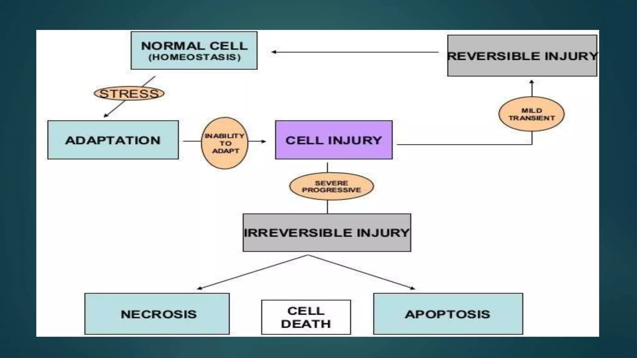
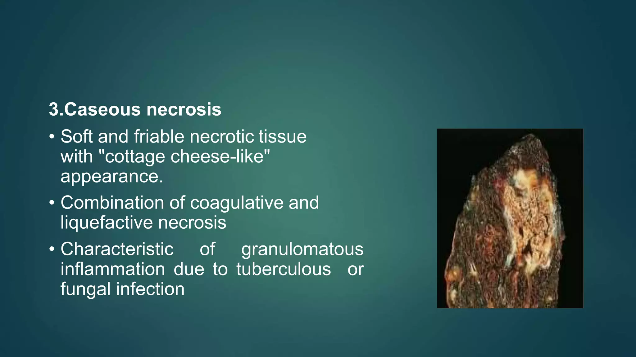
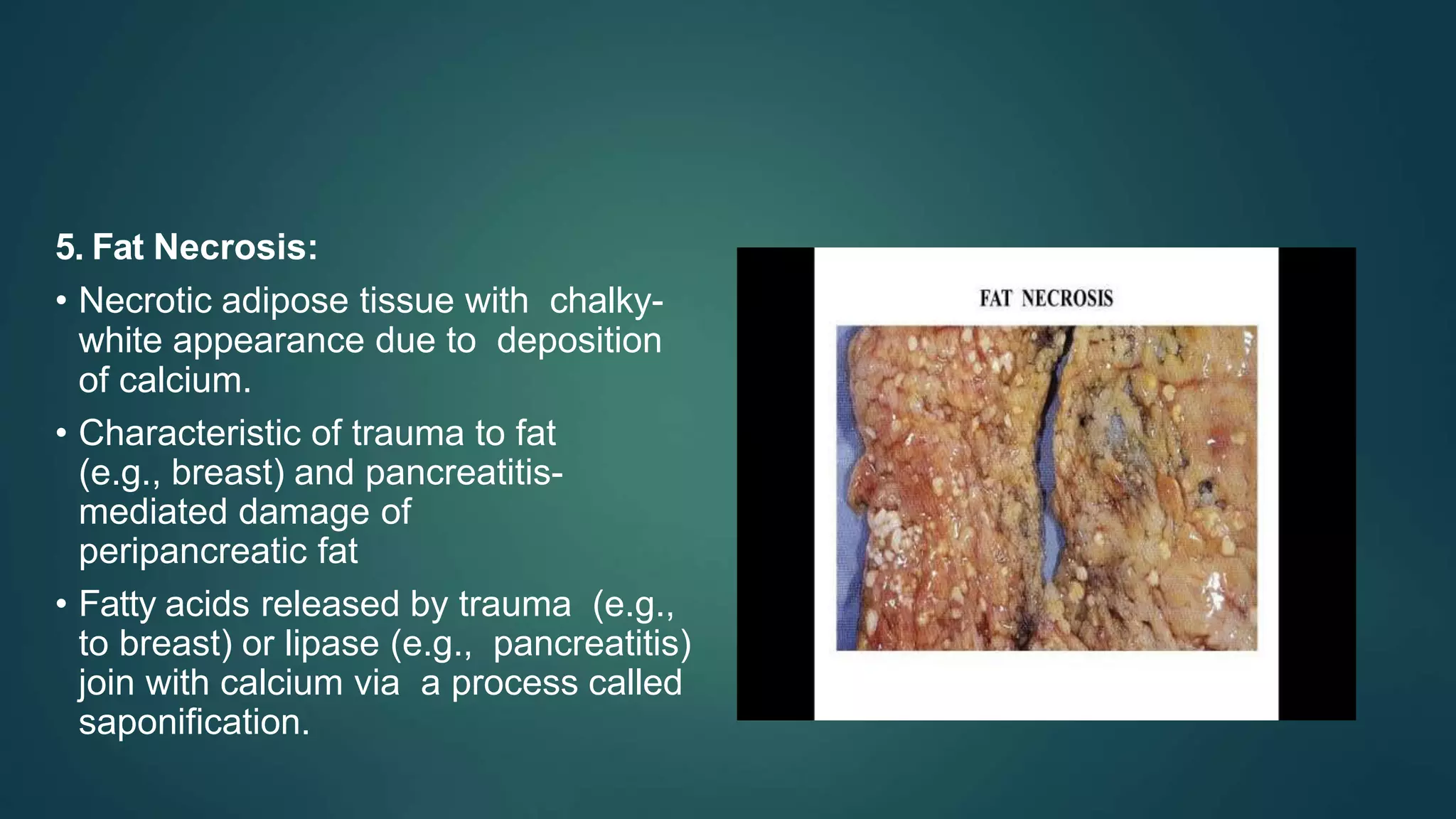
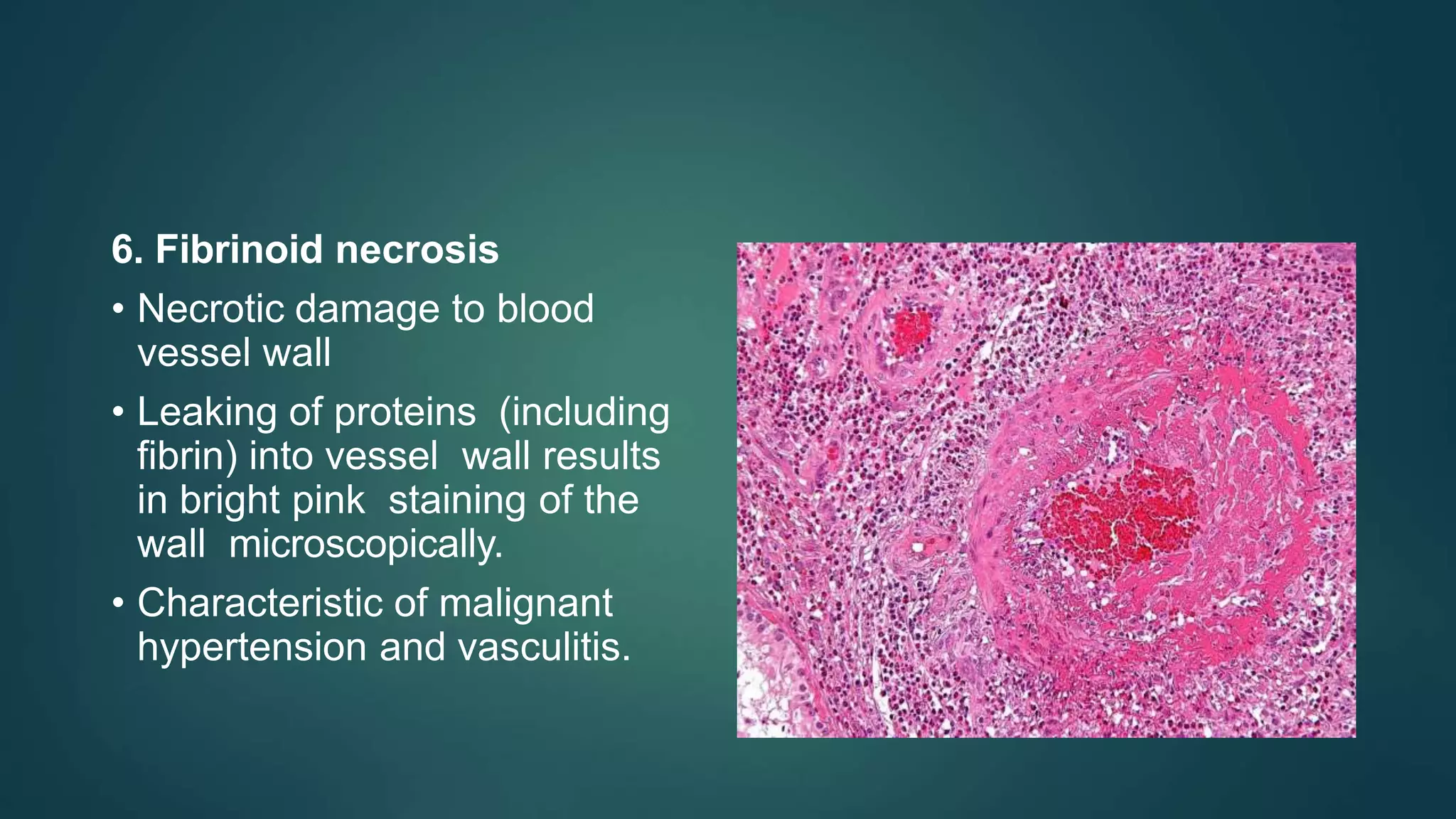
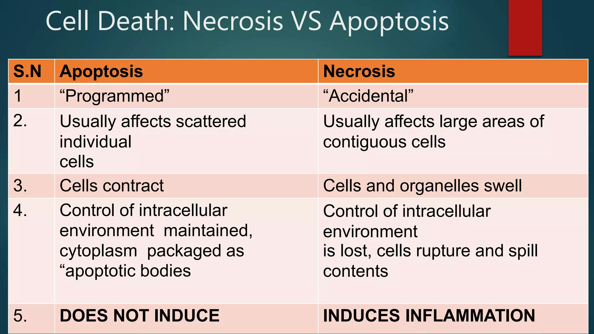
Cell Adaptation and
Cell Injury and Cell
Death
DR. ROSHAN KHATIWADA
Basic Physiology:
 An organ is in homeostasis with the physiologic stress placed on it.
 An increase, decrease, or change in stress on an organ can result in growth
adaptations.
Cell Adapatation
1.Hypertrophy
2.Hyperplasia
3.Metaplasia
4.Dysplasia
5.Atrophy
Hypertrophy and Hyperplasia
• An increase in stress leads to an increase in organ size.
• Occurs via an increase in the size (hypertrophy) and/or the
number (hyperplasia) of cells
• Hypertrophy involves gene activation, protein synthesis, and
production of organelles.
• Hyperplasia involves the production of new cells from stem
cells.
• Hyperplasia and hypertrophy generally occur together
(e.g.uterus during pregnancy).
• Permanent tissues (e.g., cardiac muscle, skeletal muscle,
and nerve), however, cannot make new cells and undergo
hypertrophy only. For example, cardiac myocytes undergo
hypertrophy, not hyperplasia, in response to systemic
hypertension
• Pathologic hyperplasia (e.g. endometrial hyperplasia) can
progress to dysplasia and eventually cancer.
• A notable exception is benign prostatic hyperplasia
(BPH),which does not increase the risk for prostate
cancer.
Atrophy
• A decrease in stress (e.g., decreased hormonal
stimulation, disuse, or decreased nutrients/blood supply)
leads to a decrease in organ size (atrophy).
-- Occurs via a decrease in the size and number of cells
-- Decrease in cell number occurs via apoptosis.
APLASIA AND HYPOPLASIA
• Aplasia is failure of cell production during
embryogenesis (e.g. unilateral renal agenesis).
• Hypoplasia is a decrease in cell production during
embryogenesis, resulting in a relatively small organ (e.g.,
streak ovary in Turner syndrome).
METAPLASIA
• A change in stress on an organ leads to a change in cell type
(metaplasia).
1. Most commonly involves change of one type of surface
epithelium (squamous, columnar, or urothelial) to another
2. Metaplastic cells are better able to handle the new stress.
--Barrett esophagus is a classic example
1. Esophagus is normally lined by non-keratinizing squamous
epithelium (suited to handle friction of a food bolus).
2. Acid reflux from the stomach causes metaplasia to non- ciliated,
mucin-producing columnar cells (better able to handle the stress of
acid).
• Metaplasia occurs via programming of stem cells, which then
produce the new cell type.
• Metaplasia is reversible, in theory, with removal of the driving stressor.
• For example, treatment of gastroesophageal reflux may
reverse Barrett esophagus.
• Under persistent stress, metaplasia can progress to
dysplasia and eventually result in cancer. For example,
Barrett esophagus may progress so adenocarcinoma of
the esophagus.
Dysplasia
• Disordered cellular growth
• Most often refers to proliferation of precancerous cells
• For example, cervical intraepithelial neoplasia (CIN) represents
dysplasia and is a precursor to cervical cancer,
• Often arises from longstanding pathologic hyperplasia (e.g., endometrial
hyperplasia) or metaplasia (e.g., Barrett esophagus)
• Dysplasia is reversible, in theory, with alleviation of inciting stress.
• If stress persists, dysplasia progresses to carcinoma
(irreversible).
CELL INJURY
Cell injury is defined as a variety of stresses a cell encounters as a result of
change in its internal and external environment.
• Cellular injury occurs when a stress exceeds the ability to adapt.
• The likelihood of injury depends on the type of stress, its severity, and the type of
cell affected.
1.Neurons are highly susceptible to ischemic injury; whereas, skeletal muscle is
relatively more resistant.
2.Slowly developing ischemia (eg., renal artery atherosclerosis) resuhs in atrophy,
whereas, acute ischemia (e.g., renal artery embolus) results in injury.
--Common causes of cellular injury include inflammation, nutritional
deficiency or excess, hypoxia, trauma, and genetic mutations.
REVERSIBLE AND IRREVERSIBLE INJURY
• Hypoxia impairs oxidative phosphorylation resulting
in decreased ATP.
• Low ATP disrupts key cellular functions including
1. Na-K pump, resulting in sodium and water buildup in the cell
2. Ca2+ pump, resulting in Calcium buildup in the cytosol of
the cell
3. Aerobic glycolysis, resulting in a switch to anaerobic glycolysis.
Lactic acid buildup results in low pH, which denatures proteins.
• The initial phase of injury is reversible. The hallmark of
reversible injury is cellular swelling.
1. Cytosol swelling results in loss or microvilli and
membrane blebbing.
2. Swelling of the rough endoplasmic reticulum (RER) results
in dissociation of ribosomes and decreased protein synthesis.
• Eventually, the damage becomes irreversible. The hallmark of
irreversible injury is membrane damage.
1.Plasma membrane damage results in
i. Cytosolic enzymes leaking into the serum {e.g., cardiac troponin)
ii. Additional calcium entering into the cell
2.Mitochondrial membrane damage results in
i. Loss of the electron transport chain (inner mitochondrial membrane)
ii. Cytochrome c leaking into cytosol (activates apoptosis)
3.Lysosome membrane damage results in hydrolytic enzymes leaking into the
cytosol, which, in turn, are activated by the high intracellular calcium.
• The end result of irreversible injury is cell death.
Cell Death
BASIC PRINCIPLES
A. The morphologic hallmark of cell death is loss of the
nucleus, which occurs via nuclear condensation
(pyknosis), fragmentation (karyorrhexis), and dissolution
(karyolysis).
B The two mechanisms of cell death are necrosis and
apoptosis.
NECROSIS
• Death of large groups of cells followed by acute
inflammation
• Due to some underlying pathologic process; never physiologic
• Divided into several types based on gross features
1.Coagulative Necrosis :
• Necrotic tissue that remains
firm ; cell shape and organ
structure are preserved by
coagulation of proteins, but
the nucleus disappears.
2. Liquefactive necrosis:
• Necrotic tissue that becomes liquefied;
enzymatic lysis of cells and protein
results in liquefaction.
• Characteristic of
• i. Brain infarction—Proteolytic enzymes
from microglial cells liquefy the brain.
• ii. Abscess—Proteolytic enzymes from
neutrophils liquefy tissue.
• iii. Pancreatitis—Proteolytic enzymes
from pancreas liquefy parenchyma.
3.Caseous necrosis
• Soft and friable necrotic tissue
with "cottage cheese-like"
appearance.
• Combination of coagulative and
liquefactive necrosis
• Characteristic of granulomatous
inflammation due to tuberculous or
fungal infection
4. Gangrenous necrosis
• Coagulative necrosis that
resembles mummified tissue
(dry gangrene)
• Characteristic of ischemia of
lower limb and GI tract
• If superimposed infection of dead
tissues occurs, then liquefactive
necrosis ensues (wet gangrene).
Gangrene: Wet VS Dry
S.N Features Dry Gangrene Wet Gangrene
1. Site Commonly limbs. More common in bowel
2. Mechanisms Arterial occlusion More commonly venous
obstruction, less often arterial occlusion
3. Macroscopy Organ dry, shrunken and
black.
Part moist, soft, swollen and dark.
4. Putrefecatio n Limited due to very
little blood supply.
Marked due to stuffing of organ
with blood
5. Bacteria Bacteria fail to survive. Numerous present.
6. Line of
demarcation
Present but the
junction b/w healthy and gangrenous
part.
No clear line of demarcation
5. Fat Necrosis:
• Necrotic adipose tissue with chalky-
white appearance due to deposition
of calcium.
• Characteristic of trauma to fat
(e.g., breast) and pancreatitis-
mediated damage of
peripancreatic fat
• Fatty acids released by trauma (e.g.,
to breast) or lipase (e.g., pancreatitis)
join with calcium via a process called
saponification.
6. Fibrinoid necrosis
• Necrotic damage to blood
vessel wall
• Leaking of proteins (including
fibrin) into vessel wall results
in bright pink staining of the
wall microscopically.
• Characteristic of malignant
hypertension and vasculitis.
Apoptosis:
• Energy (ATP)-dependent, genetically programmed cell death
involving single cells or small groups of cells. Examples
include
1. Endometrial shedding during menstrual cycle
2. Removal of cells during embryogenesis.
• Apoptosis is mediated by caspases that activate
proteases and endonucleases,
1. Proteases break down the cytoskeleton.
2. Endonucleases break down DNA.
Morphology
1.Dying cell shrinks, leading cytoplasm to become more
eosinophilic (pink)
2.Nucleus condenses (pyknosis) and fragments
(karyorrhexis).
3.Apoptotic bodies fall from the cell and are removed by
macrophages; apoptosis is not followed by inflammation
Cell Death: Necrosis VS Apoptosis
S.N Apoptosis Necrosis
1 “Programmed” “Accidental”
2. Usually affects scattered
individual
cells
Usually affects large areas of
contiguous cells
3. Cells contract Cells and organelles swell
4. Control of intracellular
environment maintained,
cytoplasm packaged as
“apoptotic bodies
Control of intracellular
environment
is lost, cells rupture and spill
contents
5. DOES NOT INDUCE INDUCES INFLAMMATION
Cell adaptation & Cell injury and Cell death

Cell adaptation & Cell injury and Cell death

  • 1.
    Cell Adaptation and CellInjury and Cell Death DR. ROSHAN KHATIWADA
  • 3.
    Basic Physiology:  Anorgan is in homeostasis with the physiologic stress placed on it.  An increase, decrease, or change in stress on an organ can result in growth adaptations.
  • 4.
  • 5.
    Hypertrophy and Hyperplasia •An increase in stress leads to an increase in organ size. • Occurs via an increase in the size (hypertrophy) and/or the number (hyperplasia) of cells • Hypertrophy involves gene activation, protein synthesis, and production of organelles. • Hyperplasia involves the production of new cells from stem cells.
  • 6.
    • Hyperplasia andhypertrophy generally occur together (e.g.uterus during pregnancy). • Permanent tissues (e.g., cardiac muscle, skeletal muscle, and nerve), however, cannot make new cells and undergo hypertrophy only. For example, cardiac myocytes undergo hypertrophy, not hyperplasia, in response to systemic hypertension
  • 7.
    • Pathologic hyperplasia(e.g. endometrial hyperplasia) can progress to dysplasia and eventually cancer. • A notable exception is benign prostatic hyperplasia (BPH),which does not increase the risk for prostate cancer.
  • 8.
    Atrophy • A decreasein stress (e.g., decreased hormonal stimulation, disuse, or decreased nutrients/blood supply) leads to a decrease in organ size (atrophy). -- Occurs via a decrease in the size and number of cells -- Decrease in cell number occurs via apoptosis.
  • 9.
    APLASIA AND HYPOPLASIA •Aplasia is failure of cell production during embryogenesis (e.g. unilateral renal agenesis). • Hypoplasia is a decrease in cell production during embryogenesis, resulting in a relatively small organ (e.g., streak ovary in Turner syndrome).
  • 10.
    METAPLASIA • A changein stress on an organ leads to a change in cell type (metaplasia). 1. Most commonly involves change of one type of surface epithelium (squamous, columnar, or urothelial) to another 2. Metaplastic cells are better able to handle the new stress. --Barrett esophagus is a classic example
  • 11.
    1. Esophagus isnormally lined by non-keratinizing squamous epithelium (suited to handle friction of a food bolus). 2. Acid reflux from the stomach causes metaplasia to non- ciliated, mucin-producing columnar cells (better able to handle the stress of acid). • Metaplasia occurs via programming of stem cells, which then produce the new cell type. • Metaplasia is reversible, in theory, with removal of the driving stressor. • For example, treatment of gastroesophageal reflux may reverse Barrett esophagus.
  • 12.
    • Under persistentstress, metaplasia can progress to dysplasia and eventually result in cancer. For example, Barrett esophagus may progress so adenocarcinoma of the esophagus.
  • 13.
    Dysplasia • Disordered cellulargrowth • Most often refers to proliferation of precancerous cells • For example, cervical intraepithelial neoplasia (CIN) represents dysplasia and is a precursor to cervical cancer, • Often arises from longstanding pathologic hyperplasia (e.g., endometrial hyperplasia) or metaplasia (e.g., Barrett esophagus) • Dysplasia is reversible, in theory, with alleviation of inciting stress. • If stress persists, dysplasia progresses to carcinoma (irreversible).
  • 14.
    CELL INJURY Cell injuryis defined as a variety of stresses a cell encounters as a result of change in its internal and external environment. • Cellular injury occurs when a stress exceeds the ability to adapt. • The likelihood of injury depends on the type of stress, its severity, and the type of cell affected. 1.Neurons are highly susceptible to ischemic injury; whereas, skeletal muscle is relatively more resistant. 2.Slowly developing ischemia (eg., renal artery atherosclerosis) resuhs in atrophy, whereas, acute ischemia (e.g., renal artery embolus) results in injury. --Common causes of cellular injury include inflammation, nutritional deficiency or excess, hypoxia, trauma, and genetic mutations.
  • 15.
    REVERSIBLE AND IRREVERSIBLEINJURY • Hypoxia impairs oxidative phosphorylation resulting in decreased ATP. • Low ATP disrupts key cellular functions including 1. Na-K pump, resulting in sodium and water buildup in the cell 2. Ca2+ pump, resulting in Calcium buildup in the cytosol of the cell 3. Aerobic glycolysis, resulting in a switch to anaerobic glycolysis. Lactic acid buildup results in low pH, which denatures proteins.
  • 16.
    • The initialphase of injury is reversible. The hallmark of reversible injury is cellular swelling. 1. Cytosol swelling results in loss or microvilli and membrane blebbing. 2. Swelling of the rough endoplasmic reticulum (RER) results in dissociation of ribosomes and decreased protein synthesis.
  • 18.
    • Eventually, thedamage becomes irreversible. The hallmark of irreversible injury is membrane damage. 1.Plasma membrane damage results in i. Cytosolic enzymes leaking into the serum {e.g., cardiac troponin) ii. Additional calcium entering into the cell 2.Mitochondrial membrane damage results in i. Loss of the electron transport chain (inner mitochondrial membrane) ii. Cytochrome c leaking into cytosol (activates apoptosis) 3.Lysosome membrane damage results in hydrolytic enzymes leaking into the cytosol, which, in turn, are activated by the high intracellular calcium.
  • 19.
    • The endresult of irreversible injury is cell death.
  • 20.
    Cell Death BASIC PRINCIPLES A.The morphologic hallmark of cell death is loss of the nucleus, which occurs via nuclear condensation (pyknosis), fragmentation (karyorrhexis), and dissolution (karyolysis). B The two mechanisms of cell death are necrosis and apoptosis.
  • 21.
    NECROSIS • Death oflarge groups of cells followed by acute inflammation • Due to some underlying pathologic process; never physiologic • Divided into several types based on gross features
  • 22.
    1.Coagulative Necrosis : •Necrotic tissue that remains firm ; cell shape and organ structure are preserved by coagulation of proteins, but the nucleus disappears.
  • 23.
    2. Liquefactive necrosis: •Necrotic tissue that becomes liquefied; enzymatic lysis of cells and protein results in liquefaction. • Characteristic of • i. Brain infarction—Proteolytic enzymes from microglial cells liquefy the brain. • ii. Abscess—Proteolytic enzymes from neutrophils liquefy tissue. • iii. Pancreatitis—Proteolytic enzymes from pancreas liquefy parenchyma.
  • 24.
    3.Caseous necrosis • Softand friable necrotic tissue with "cottage cheese-like" appearance. • Combination of coagulative and liquefactive necrosis • Characteristic of granulomatous inflammation due to tuberculous or fungal infection
  • 25.
    4. Gangrenous necrosis •Coagulative necrosis that resembles mummified tissue (dry gangrene) • Characteristic of ischemia of lower limb and GI tract • If superimposed infection of dead tissues occurs, then liquefactive necrosis ensues (wet gangrene).
  • 26.
    Gangrene: Wet VSDry S.N Features Dry Gangrene Wet Gangrene 1. Site Commonly limbs. More common in bowel 2. Mechanisms Arterial occlusion More commonly venous obstruction, less often arterial occlusion 3. Macroscopy Organ dry, shrunken and black. Part moist, soft, swollen and dark. 4. Putrefecatio n Limited due to very little blood supply. Marked due to stuffing of organ with blood 5. Bacteria Bacteria fail to survive. Numerous present. 6. Line of demarcation Present but the junction b/w healthy and gangrenous part. No clear line of demarcation
  • 27.
    5. Fat Necrosis: •Necrotic adipose tissue with chalky- white appearance due to deposition of calcium. • Characteristic of trauma to fat (e.g., breast) and pancreatitis- mediated damage of peripancreatic fat • Fatty acids released by trauma (e.g., to breast) or lipase (e.g., pancreatitis) join with calcium via a process called saponification.
  • 28.
    6. Fibrinoid necrosis •Necrotic damage to blood vessel wall • Leaking of proteins (including fibrin) into vessel wall results in bright pink staining of the wall microscopically. • Characteristic of malignant hypertension and vasculitis.
  • 29.
    Apoptosis: • Energy (ATP)-dependent,genetically programmed cell death involving single cells or small groups of cells. Examples include 1. Endometrial shedding during menstrual cycle 2. Removal of cells during embryogenesis. • Apoptosis is mediated by caspases that activate proteases and endonucleases, 1. Proteases break down the cytoskeleton. 2. Endonucleases break down DNA.
  • 30.
    Morphology 1.Dying cell shrinks,leading cytoplasm to become more eosinophilic (pink) 2.Nucleus condenses (pyknosis) and fragments (karyorrhexis). 3.Apoptotic bodies fall from the cell and are removed by macrophages; apoptosis is not followed by inflammation
  • 32.
    Cell Death: NecrosisVS Apoptosis S.N Apoptosis Necrosis 1 “Programmed” “Accidental” 2. Usually affects scattered individual cells Usually affects large areas of contiguous cells 3. Cells contract Cells and organelles swell 4. Control of intracellular environment maintained, cytoplasm packaged as “apoptotic bodies Control of intracellular environment is lost, cells rupture and spill contents 5. DOES NOT INDUCE INDUCES INFLAMMATION