Kohler's disease
•Download as PPTX, PDF•
2 likes•1,073 views

Kohler's Disease Aetiology, Pathogenesis, diagnosis, conservative and operative management from Campbell & Turek's

Recommended
More Related Content
What's hot
What's hot (20)
Similar to Kohler's disease
Similar to Kohler's disease (20)
More from Yash Oza
More from Yash Oza (12)
Recently uploaded
Recently uploaded (20)
Kohler's disease
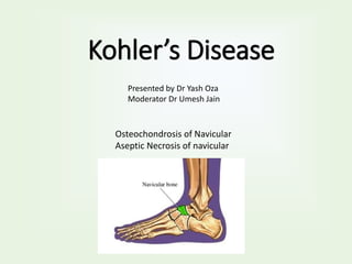
- 1. Kohler’s Disease Osteochondrosis of Navicular Aseptic Necrosis of navicular Presented by Dr Yash Oza Moderator Dr Umesh Jain
- 2. • Osteochondrosis of the tarsal navicular originally was described by Kohler in 1908. • It is characterized by aseptic necrosis of navicular bone, casing a painful limp in a child.
- 3. • Navicular is last bone to ossify. • Ossification centers of the navicular appear between the ages of • 1.5 and 2 years in girls and • 2.5 and 3 years in boys. • Normally, navicular ossification is quite variable & not infrequent to find flattened/dense/fragmented ossific nucleus.
- 4. Pathogenesis • Histological studies shows scattered area of aseptic necrosis and bone absorption. • This suggests vascular origin of the diseases. • In first few years of life, cartilaginous navicular is surrounded by ring of vessels. • From this network, a single artery penetrates to the center of the structure. • Soon it is surrounded by developing ossifications. • And other arteries follow and multiple area of ossification are formed and joined into the large ossific nucleus.
- 5. • Occasionally a single vessel is the sole supply until the age of 4 to 6 years. And ossification is dependent on a single artery. • On weight bearing, the forces compress the navicular constantly and may compromise vascular supply. • The ossification center undergoes aseptic necrosis. • A reactive hyperemia also develops around the bone. Later on, Ingrowth on new vessels leads to resorption & replacement of necrotic bone eventually.
- 6. • Thus, Delayed ossification has been suggested to be the earliest event because the lateness of ossification of the navicular subjects it to more pressure. • 2 types of abnormalities seen 1. Flattened bone with patchy area of increased density 2. Normal shaped bone with overall increased density.
- 7. Clinical Presentation • Age group – 4-10 years • Both saxes affected (M>F) • Child limps with complaint of pain in foot • There is a tender swelling over navicular area. • Local heat may be present due to hyperemia.
- 8. Radiographic finding • In plane radiograph navicular shows increase in density, loss of trabecular structure & alteration in shape and size.
- 9. Diagnosis • Clinical Features + Xray finding = Kohler’s Disease • Asymptomatic + Xray Finding = irregularity of ossification
- 10. Treatment • This is a self-limiting condition , and operative treatment rarely is indicated. • Cast immobilization has been reported to produce quicker resolution of symptoms. But not hasten the restoration process. • Cast worn for several weeks is followed by rigid shank shoe with thomas heel.
- 11. Outcome of conservative treatment Flattened variety with patchy density Normal shape with increased density Over a period of 2 years the size & shape are gradually restored, and the trabecular pattern reappears. Within few months the dense bone is gradually absorbed until a faint ossific shadow remains. At about 2 years after onset, several small ossification make their apperience & bone starts restoring. It taken upto 3 years The navicular is almost always reconstituted to normal
- 12. Surgical Treatment • Pain and disability occasionally develop after osteochondrosis when the navicular becomes distorted and sclerotic, the head of the talus becomes flattened, the articular surfaces of the two bones become fibrillated, and osteophytes form along the margin of the articular surfaces. • Surgery is indicated when disabling symptoms persist.
- 13. • Arthrodesis is the only operation of value, • The midtarsal joints (talonavicular and calcaneocuboid) can be arthrodesed • The calcaneocuboid joint is included because most of its function is lost when the talonavicular joint is fused. • The results of this operation usually are excellent; most patients become symptom free but may notice loss of lateral movements of the foot.
- 14. • When symptoms arise from the naviculocuneiform joints also, these joints should be included in the fusion. • Here arthrodesis is difficult to secure; metallic internal fixation and inlay grafts of autogenous cancellous bone are helpful.