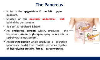
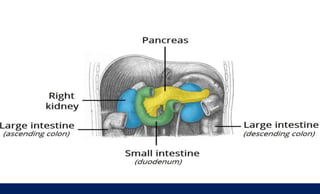
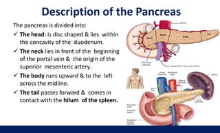
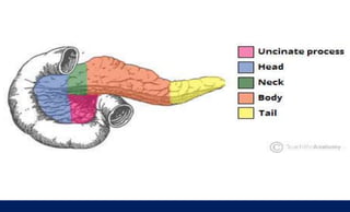
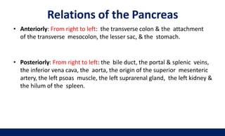
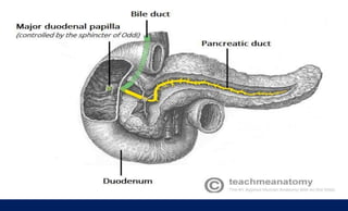
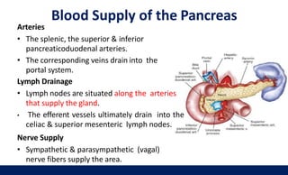
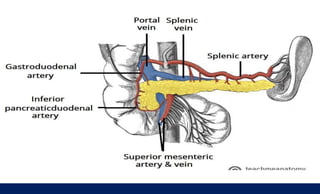
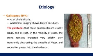
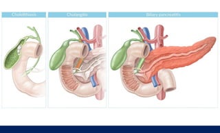
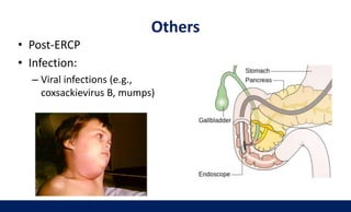
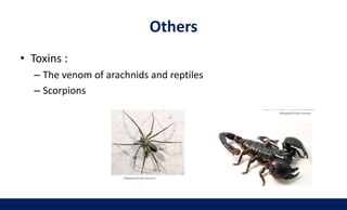
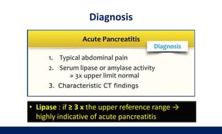
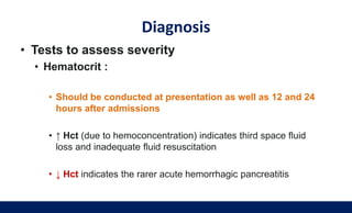
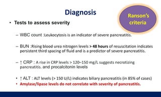
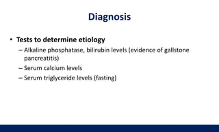
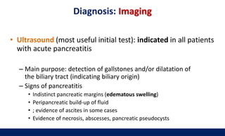
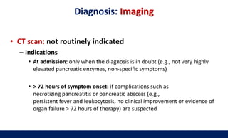
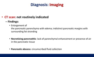
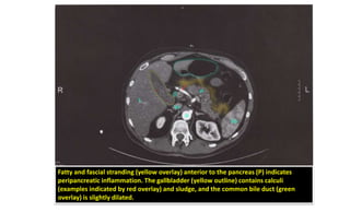
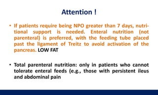
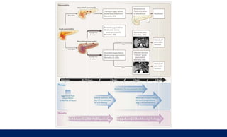
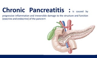
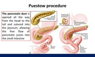
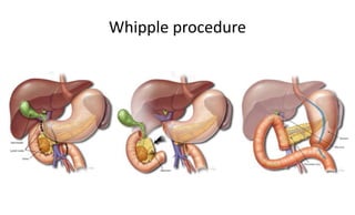
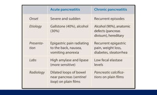
Pancreatitis
Musa Abu Sabha
Outline
Brief Anatomy
Acute Pancreatitis
Chronic Pancreatitis
The Pancreas
• It lies in the epigastrium & the left upper
quadrant.
• Situated on the posterior abdominal wall
behind the peritoneum.
• It is soft & lobulated & have:
 An endocrine portion which, produces the
hormones insulin & glucagon, (play a key role in
carbohydrate metabolism).
 An exocrine portion which produces a secretion
(pancreatic fluids) that contains enzymes capable
of hydrolyzing proteins, fats & carbohydrates.
Description of the Pancreas
The pancreas is divided into:
 The head: is disc shaped & lies within
the concavity of the duodenum.
 The neck lies in front of the beginning
of the portal vein & the origin of the
superior mesenteric artery.
 The body runs upward & to the left
across the midline.
 The tail passes forward & comes in
contact with the hilum of the spleen.
34
Relations of the Pancreas
35
• Anteriorly: From right to left: the transverse colon & the attachment
of the transverse mesocolon, the lesser sac, & the stomach.
• Posteriorly: From right to left: the bile duct, the portal & splenic veins,
the inferior vena cava, the aorta, the origin of the superior mesenteric
artery, the left psoas muscle, the left suprarenal gland, the left kidney &
the hilum of the spleen.
Pancreatic Ducts
36
• The main duct begins in the tail & runs the
length of the gland, receiving numerous
tributaries on the way.
• It opens into the 2nd part of the
duodenum with the bile duct on the
major duodenal papilla.
• The accessory duct of the pancreas, when
present, drains the upper part of the head &
then opens into the duodenum a short
distance above the main duct on the minor
duodenal papilla
Blood Supply of the Pancreas
37
Arteries
• The splenic, the superior & inferior
pancreaticoduodenal arteries.
• The corresponding veins drain into the
portal system.
Lymph Drainage
• Lymph nodes are situated along the arteries
that supply the gland.
• The efferent vessels ultimately drain into the
celiac & superior mesenteric lymph nodes.
Nerve Supply
• Sympathetic & parasympathetic (vagal)
nerve fibers supply the area.
Case Presentation
• A 41-year-old woman presents to the emergency department complaining
of severe and continuous epigastric pain for the past 24-hours. The pain
radiates straight through to her back. She has had progressive nausea with
vomiting. The vomit is bile-stained and without blood. She has had similar
but less severe episodes of abdominal pain in the past, usually after eating
heavy meals, but they always resolved within a few hours. She is gravida
two, para two, with last menses 2 weeks ago, and does not consume any
alcohol. On exam, she is afebrile, heart rate is 115 beats/min, blood
pressure of 128/86 mmHg, and respiratory rate is 18/ min. Her abdomen is
not distended. She has no surgical scars on her abdomen and no obvious
masses visible. She has no bruising around her umbilicus or along her flank.
Bowel sounds are hypoactive. She has marked tenderness to palpation in
her epigastrium, without guarding or rebound. The remainder of her
abdomen is soft and non-tender to palpation. No masses or organomegaly
are appreciated.
Cont.
• Laboratory examination reveals : WBCs count of 17.2K
cells/μL , Amylase of 1545 u/L, Lipase of 1134 u/L, ALT of
245 u/L, AST of 263 u/L , serum glucose of 156 mg/dl and
LDH 180 u/L (0–250 u/L).
• An abdominal series demonstrates gas throughout the
small and large bowel and a focal dilated loop of proximal
small bowel without air fluid levels. There is no free air
under the diaphragm.
What Is the Differential Diagnosis for Epigastric
Abdominal Pain?
• Gastroenteritis
• Acute gastritis
• Acute cholecystitis
• Peptic ulcer disease (PUD)
• Perforated ulcer
• Pancreatitis
• Small bowel obstruction
• Mesenteric ischemia
• Referred pain from MI
Acute Pancreatitis
• Acute inflammation of pancreas
• Inappropriate activation of enzymes → autodigestion
• Epigastric pain, classically radiating to back
• Nausea and vomiting
Etiology
• Most common causes:
– Biliary pancreatitis (e.g., gallstones, constriction of the ampulla
of Vater) ∼ 40% of cases
– Alcohol-induced (∼ 30% of cases)
– Idiopathic (∼ 15%–25% of cases).
• Gallstones 40 % :
– Hx of cholelithiasis.
– Abdominal imaging shows dilated bile ducts.
The gallstones that cause pancreatitis are usually
small, and as such, in the majority of cases, the
stone remains impacted very briefly, only
transiently obstructing the ampulla of Vater, and
soon after passes into the duodenum.
Etiology
• Ethanol 30 %:
– Usually apparent from
history
– AST:ALT <2 .
Etiology
• Hypertriglyceridemia
Elevated triglycerides (> 1000 mg/dl) → acute pancreatitis
Etiology
Etiology
• Hypercalcemia
– Rarely causes pancreatitis
– Calcium may deposit in pancreatic ducts
– Calcium may activate trypsinogen
Don’t Miss Hypercalcemia as the Cause of
Pancreatitis !!
• In the absence of gallstones and alcohol abuse, the
etiology of acute pancreatitis may be unclear.
• consider hypercalcemia as the etiology:
– Pt. on Hydrochlorothiazide for HTN.
– Hyperparathyroidism.
While hypercalcemia can cause pancreatitis, pancreatitis can
cause hypocalcemia Due to Saponification
Low calcium is a poor prognostic indicator
Others
• Post-ERCP
• Infection:
– Viral infections (e.g.,
coxsackievirus B, mumps)
Others
• Toxins :
– The venom of arachnids and reptiles
– Scorpions
Others
• Drugs
• Tumors ( peri-ampullary )
• Autoimmune and
rheumatologically
disorders (e.g., Sjögren
syndrome)
Etiology
• I GET SMASHED
• Idiopathic,
• Gall stones,
• Ethanol,
• Trauma,
• Steroids,
• Mumps,
• Autoimmune
• Scorpion poison
• Hypercalcemia
Hypertriglyceridemia
• ERCP
• Drugs
Clinical presentation
• Constant, severe epigastric pain:
– Classically radiating towards the back
– Worse after meals and when supine
– Improves on leaning forwards
– Nausea, vomiting
• General physical examination:
– Signs of shock Due to distributive shock from cytokine release, and third-spacing of intravascular
fluid: tachycardia, hypotension, oliguria/anuria.
– Possibly Jaundice in patients with biliary pancreatitis
oliguria : urine
output of < 400 mL
per 24 hours in adults
(less than 0.5
mL/kg/h in children).
Anuria:
urine output that is
less than 50 milliliters
per 24 hours.
Clinical presentation
• Abdominal examination
– Abdominal tenderness, distention, guarding
– Ileus
– with reduced bowel sounds and tympany on percussion
– Ascites
– Skin changes (rare)
• Cullen's sign: periumbilical ecchymosis and discoloration (bluish-red)
• Grey Turner's sign: flank ecchymosis with discoloration
• Fox's sign: ecchymosis over the inguinal ligament
Diagnosis
• Lipase : if ≥ 3 x the upper reference range →
highly indicative of acute pancreatitis
Diagnosis
• Tests to assess severity
• Hematocrit :
• Should be conducted at presentation as well as 12 and 24
hours after admissions
• ↑ Hct (due to hemoconcentration) indicates third space fluid
loss and inadequate fluid resuscitation
• ↓ Hct indicates the rarer acute hemorrhagic pancreatitis
Diagnosis
• Tests to assess severity
– WBC count :Leukocytosis is an indicator of severe pancreatitis.
– BUN :Rising blood urea nitrogen levels > 48 hours of resuscitation indicates
persistent third spacing of fluid and is a predictor of severe pancreatitis.
– ↑ CRP : A rise in CRP levels > 120–150 mg/L suggests necrotizing
pancreatitis. and procalcitonin levels
• ↑ ALT : ALT levels (> 150 U/L) indicates biliary pancreatitis (in 85% of cases)
• Amylase/lipase levels do not correlate with severity of pancreatitis.
Ranson’s
criteria
Diagnosis
• Tests to determine etiology
– Alkaline phosphatase, bilirubin levels (evidence of gallstone
pancreatitis)
– Serum calcium levels
– Serum triglyceride levels (fasting)
Diagnosis: Imaging
• Ultrasound (most useful initial test): indicated in all patients
with acute pancreatitis
– Main purpose: detection of gallstones and/or dilatation of
the biliary tract (indicating biliary origin)
– Signs of pancreatitis
• Indistinct pancreatic margins (edematous swelling)
• Peripancreatic build-up of fluid
• ; evidence of ascites in some cases
• Evidence of necrosis, abscesses, pancreatic pseudocysts
Diagnosis: Imaging
• CT scan: not routinely indicated
– Indications
• At admission: only when the diagnosis is in doubt (e.g., not very highly
elevated pancreatic enzymes, non-specific symptoms)
• > 72 hours of symptom onset: if complications such as
necrotizing pancreatitis or pancreatic abscess (e.g.,
persistent fever and leukocytosis, no clinical improvement or evidence of
organ failure > 72 hours of therapy) are suspected
Diagnosis: Imaging
• CT scan: not routinely indicated
– Findings:
• Enlargement of
the pancreatic parenchyma with edema; indistinct pancreatic margins with
surrounding fat stranding
• Necrotizing pancreatitis: lack of parenchymal enhancement or presence of air
in the pancreatic tissue
• Pancreatic abscess: circumscribed fluid collection
Fatty and fascial stranding (yellow overlay) anterior to the pancreas (P) indicates
peripancreatic inflammation. The gallbladder (yellow outline) contains calculi
(examples indicated by red overlay) and sludge, and the common bile duct (green
overlay) is slightly dilated.
Treatment :General measures
• NPO
– No food or liquid
– “Rests” the pancreas (prevents stimulation)
• IV fluids
– Fluid loss due to pancreatic edema
– IV fluids needed to maintain BP and renal perfusion
– Aggressive hydration with crystalloids (e.g., lactated
Ringer's solution , normal saline
• Pain control
– IV opioids (e.g., fentanyl) :Most patients with mild
disease improve in 2-3 days
Treatment : Drugs
• Analgesics: fentanyl or
hydromorphone
• Antibiotics
– Prophylactic antibiotic therapy is not
recommended.
– Antibiotics should only be used in
patients with evidence of infected
necrosis.
• Fenofibrates: in hyperlipidemia-
induced acute pancreatitis
Management : Severity
• Ranson’s criteria are used to predict severity based on
parameters during initial admission and at 48 hours after
Management
algorithm for acute
pancreatitis. (Based
on the practice
guidelines from the
American College of
Gastroenterology)
Biliary pancreatitis
• Urgent ERCP and sphincterotomy (within 24 hours): in
patients with evidence of choledocholithiasis and/or
cholangitis; followed by cholecystectomy
• Cholecystectomy (preferably during same admission once
the patient is stabilized; or within 6 weeks): in all patients
with biliary pancreatitis
There is a 30% risk of recurrent acute pancreatitis within 6
weeks if cholecystectomy is not performed.
The most important therapeutic measure is adequate fluid
replacement minimum of 3–4 liters of crystalloids per day
!!!
• The majority of patients with acute pancreatitis recover in
less than 5 days without any complications. Close to 20% of
patients have a severe presentation with local or systemic
complications (including organ failure)
• Mortality
– In patients without organ failure: < 1%
– In patients with organ failure: ∼ 30%
– Higher mortality in patients with biliary pancreatitis than in patients
with alcoholic pancreatitis
Complications
Present early : MOF
• SIRS, sepsis, DIC
• Pneumonia, respiratory failure, ARDS
• Shock
• Prerenal failure due to volume depletion
• Hypocalcemia
• Pleural effusion, pancreatic ascites
• Paralytic ileus
Complications: Systemic
Local Pancreatic abscess
• Fever and Leukocytosis after 4 Weeks.
• First step : CT scan with contrast looking for necrotic
tissue (i.e., areas that do not enhance) or a pancreatic
abscess.
• Positive CT : aspiration for culture
• Positive culture : start antibiotic and External drainage by
a step-up approach: (1) percutaneous or endoscopic
drainage, (2) video- assisted retroperitoneal debridement,
and (3) open necrosectomy.
6 Weeks Later with Vague Abdominal Pain, a Palpable
Epigastric Mass, and Persistently Elevated Serum Amylase
• pancreatic pseudocyst, a collection of pancreatic fluid sur-
rounded by a wall without epithelium
• The diagnostic test of choice is a CT scan, and most can
be managed conservatively.
• Treatment of a symptomatic, noninfected pseudocyst that
fails to resolve is usually via internal drainage, by creating
a connection between the cyst and the adjacent intestinal
organ, usually the stomach (endoscopic cystogastrostomy)
Attention !
• If patients require being NPO greater than 7 days, nutri-
tional support is needed. Enteral nutrition (not
parenteral) is preferred, with the feeding tube placed
past the ligament of Treitz to avoid activation of the
pancreas. LOW FAT
• Total parenteral nutrition: only in patients who cannot
tolerate enteral feeds (e.g., those with persistent ileus
and abdominal pain
Chronic Pancreatitis : is caused by
progressive inflammation and irreversible damage to the structure and function
(exocrine and endocrine) of the pancreas.
Chronic Pancreatitis : Etiology
• Chronic heavy alcohol use (60–70% of cases, esp. men)
• Pancreatic ductal obstruction (< 10%): strictures (e.g., due to trauma, stones)
• Tobacco use : Dose dependent
• Idiopathic pancreatitis (20–30%)
• Hereditary pancreatitis (∼ 1%)
– PRSS1 gene mutation (autosomal dominant inheritance), SPINK1 gene mutation
– Age of onset < 20 years
– Characterized by a positive family history and the absence of other risk factors
• Autoimmune pancreatitis
• Systemic disease
– Cystic fibrosis: ∼ 2% of cystic fibrosis patients develop chronic pancreatitis.
– Severe hypertriglyceridemia (levels > 1,000 mg/dL)
– Primary hyperparathyroidism (hypercalcemia)
• Tropical pancreatitis
– Most common cause in the tropics (esp. southern India)
– Young age at onset
CT scan: classic finding is calcified pancreas
Clinical Presentation
Chronic abdominal pain
• Epigastric abdominal pain (main symptom)
– Pain radiates to the back, is relieved on bending forward, and is exacerbated after
eating.
– Pain is initially episodic and becomes persistent as the disease progresses.
– Often associated with nausea and vomiting
Features of pancreatic insufficiency: late manifestation (after 90% of
the pancreatic parenchyma is destroyed)
– Steatorrhea (exocrine enzyme deficiency)
• Cramping abdominal pain, bloating, diarrhea
• Can lead to a deficiency of fat-soluble vitamins (A, D, E, and K)
– Malabsorption and weight loss
– Pancreatic diabetes (endocrine hormone deficiency)
In the later stages of chronic
pancreatitis, patients may not
experience any pain.
Approach
• Initial survey: medical history, risk factors, clinical features suggestive of
chronic pancreatitis
• Laboratory testing: helps identify the underlying cause and possible
complications.
• Confirm diagnosis: via imaging and pancreatic function tests.
• Assess disease severity: fecal elastase-1 activity is used to determine the
extent of pancreatic insufficiency.
• Genetic testing: indicated in young patients with idiopathic pancreatitis or
a family history of chronic pancreatitis
Approach :Lab
• Serum pancreatic enzyme levels: Lipase and amylase are
often normal .cannot be used to confirm or rule out the
diagnosis
• Pancreatic function tests:
– Direct tests: Directly measure the levels of pancreatic enzymes
– Indirect tests : Measure the results of pancreatic enzyme
deficiency.
Direct tests : invasive and expensive test
• Cholecystokinin test: Cholecystokinin analog) is
administered IV, which stimulates pancreatic
enzyme and HCO3- secretion. This secretion is collected in a
tube placed in the duodenum during endoscopy and
analyzed.
• Secretin: stimulates pancreatic bicarbonate secretion.
• Cholecystokinin-secretin test: Both of the secretagogues are
administered simultaneously and the collected secretion is
quantitatively analyzed.
Indirect tests : commonly used
• Fecal elastase-1 activity: confirms
that steatorrhea is due to pancreatic
lipase insufficiency.
– FE-1 < 200 μg/g: pancreatic exocrine
insufficiency
– FE-1 < 100 μg/g: severe pancreatic exocrine
insufficiency
• 72-hour quantitative fecal fat estimation
– Patients are asked to consume 100 g of fat per
day for 3 days, after which the fecal fat content
of their stool is measured.
– Fecal fat > 7 g per day is diagnostic
of steatorrhea.
Imaging
• Abdominal CT (plain and contrast-enhanced CT): best initial
imaging modality to screen for CP
– Findings
• Pancreatic ductal dilations and calcifications on plain CT
• “Chain of lakes” appearance of the main pancreatic duct
• Pancreatic atrophy
– Can rule out pancreatic carcinoma or
gastrointestinal malignancy as a possible cause of
epigastric pain and weight loss
Complications
• Pancreatic insufficiency
• Splenic vein thrombosis
• Pseudocyst
• Bile duct obstruction
• Duodenal obstruction
• Increased risk of pancreatic adenocarcinoma
Pancreatic Insufficiency
• Result of chronic pancreatitis
• Fat malabsorption and steatorrhea
– Fat-soluble vitamin deficiencies
• Pancreatic Diabetes (loss of insulin)
• Diagnosis: fecal elastase
– Sensitive and specific test of exocrine pancreas
– High amount if exocrine pancreas normal
– Low fecal elastase in pancreatic insufficiency
Splenic Vain Thrombosis
• Epidemiology: Occurs in 10% of patients with
CP
• Pathophysiology: inflammation of the splenic
vein → thrombus formation → left-sided portal
hypertension → Isolated gastric varices
• Clinical features: can present with upper GI
bleeding, ascites, and splenomegaly
• Diagnostics: ultrasound with doppler, CT/MR
angiography
• Treatment
– Acute: anticoagulation and/or thrombectomy
– Chronic and symptomatic: splenectomy
Management
• Small, low-fat meals
• Avoid alcohol or tobacco
• Pancreatic enzyme supplementation
• Parenteral administration of fat-soluble vitamins (A, D, E,
K) if necessary
• Endocrine insufficiency: insulin administration
Management
• Pain management
– Analgesics: NSAIDs, opioids for severe pain , amitriptyline.
• Intractable pain
– Celiac ganglion block (offers temporary relief)
– Endoscopic papillotomy with ductal dilation, stent placement, and removal of stones if
present
– Extracorporeal shock wave lithotripsy (ESWL): for intraductal stones
• Surgery:
– if pancreatic cancer is suspected or in those with intractable pain
• Procedures
– Pancreaticojejunostomy: if the main pancreatic duct is dilated (> 5 mm)
– Resection of the affected part of the pancreas (distal pancreatectomy, Whipple
procedure)
– Thoracoscopic bilateral splanchnicectomy
Puestow procedure
The pancreatic duct is
opened all the way
from the head to the
tail and sutured into
the jejunum, allowing
the free flow of
pancreatic juices into
the small intestine
Whipple procedure
Thank You !

Disorders of Pancreas

  • 1.
  • 2.
  • 3.
    The Pancreas • Itlies in the epigastrium & the left upper quadrant. • Situated on the posterior abdominal wall behind the peritoneum. • It is soft & lobulated & have:  An endocrine portion which, produces the hormones insulin & glucagon, (play a key role in carbohydrate metabolism).  An exocrine portion which produces a secretion (pancreatic fluids) that contains enzymes capable of hydrolyzing proteins, fats & carbohydrates.
  • 5.
    Description of thePancreas The pancreas is divided into:  The head: is disc shaped & lies within the concavity of the duodenum.  The neck lies in front of the beginning of the portal vein & the origin of the superior mesenteric artery.  The body runs upward & to the left across the midline.  The tail passes forward & comes in contact with the hilum of the spleen. 34
  • 7.
    Relations of thePancreas 35 • Anteriorly: From right to left: the transverse colon & the attachment of the transverse mesocolon, the lesser sac, & the stomach. • Posteriorly: From right to left: the bile duct, the portal & splenic veins, the inferior vena cava, the aorta, the origin of the superior mesenteric artery, the left psoas muscle, the left suprarenal gland, the left kidney & the hilum of the spleen.
  • 8.
    Pancreatic Ducts 36 • Themain duct begins in the tail & runs the length of the gland, receiving numerous tributaries on the way. • It opens into the 2nd part of the duodenum with the bile duct on the major duodenal papilla. • The accessory duct of the pancreas, when present, drains the upper part of the head & then opens into the duodenum a short distance above the main duct on the minor duodenal papilla
  • 10.
    Blood Supply ofthe Pancreas 37 Arteries • The splenic, the superior & inferior pancreaticoduodenal arteries. • The corresponding veins drain into the portal system. Lymph Drainage • Lymph nodes are situated along the arteries that supply the gland. • The efferent vessels ultimately drain into the celiac & superior mesenteric lymph nodes. Nerve Supply • Sympathetic & parasympathetic (vagal) nerve fibers supply the area.
  • 12.
    Case Presentation • A41-year-old woman presents to the emergency department complaining of severe and continuous epigastric pain for the past 24-hours. The pain radiates straight through to her back. She has had progressive nausea with vomiting. The vomit is bile-stained and without blood. She has had similar but less severe episodes of abdominal pain in the past, usually after eating heavy meals, but they always resolved within a few hours. She is gravida two, para two, with last menses 2 weeks ago, and does not consume any alcohol. On exam, she is afebrile, heart rate is 115 beats/min, blood pressure of 128/86 mmHg, and respiratory rate is 18/ min. Her abdomen is not distended. She has no surgical scars on her abdomen and no obvious masses visible. She has no bruising around her umbilicus or along her flank. Bowel sounds are hypoactive. She has marked tenderness to palpation in her epigastrium, without guarding or rebound. The remainder of her abdomen is soft and non-tender to palpation. No masses or organomegaly are appreciated.
  • 13.
    Cont. • Laboratory examinationreveals : WBCs count of 17.2K cells/μL , Amylase of 1545 u/L, Lipase of 1134 u/L, ALT of 245 u/L, AST of 263 u/L , serum glucose of 156 mg/dl and LDH 180 u/L (0–250 u/L). • An abdominal series demonstrates gas throughout the small and large bowel and a focal dilated loop of proximal small bowel without air fluid levels. There is no free air under the diaphragm.
  • 14.
    What Is theDifferential Diagnosis for Epigastric Abdominal Pain? • Gastroenteritis • Acute gastritis • Acute cholecystitis • Peptic ulcer disease (PUD) • Perforated ulcer • Pancreatitis • Small bowel obstruction • Mesenteric ischemia • Referred pain from MI
  • 15.
    Acute Pancreatitis • Acuteinflammation of pancreas • Inappropriate activation of enzymes → autodigestion • Epigastric pain, classically radiating to back • Nausea and vomiting
  • 16.
    Etiology • Most commoncauses: – Biliary pancreatitis (e.g., gallstones, constriction of the ampulla of Vater) ∼ 40% of cases – Alcohol-induced (∼ 30% of cases) – Idiopathic (∼ 15%–25% of cases).
  • 17.
    • Gallstones 40% : – Hx of cholelithiasis. – Abdominal imaging shows dilated bile ducts. The gallstones that cause pancreatitis are usually small, and as such, in the majority of cases, the stone remains impacted very briefly, only transiently obstructing the ampulla of Vater, and soon after passes into the duodenum. Etiology
  • 19.
    • Ethanol 30%: – Usually apparent from history – AST:ALT <2 . Etiology
  • 20.
    • Hypertriglyceridemia Elevated triglycerides(> 1000 mg/dl) → acute pancreatitis Etiology
  • 21.
    Etiology • Hypercalcemia – Rarelycauses pancreatitis – Calcium may deposit in pancreatic ducts – Calcium may activate trypsinogen
  • 22.
    Don’t Miss Hypercalcemiaas the Cause of Pancreatitis !! • In the absence of gallstones and alcohol abuse, the etiology of acute pancreatitis may be unclear. • consider hypercalcemia as the etiology: – Pt. on Hydrochlorothiazide for HTN. – Hyperparathyroidism. While hypercalcemia can cause pancreatitis, pancreatitis can cause hypocalcemia Due to Saponification Low calcium is a poor prognostic indicator
  • 23.
    Others • Post-ERCP • Infection: –Viral infections (e.g., coxsackievirus B, mumps)
  • 24.
    Others • Toxins : –The venom of arachnids and reptiles – Scorpions
  • 25.
    Others • Drugs • Tumors( peri-ampullary ) • Autoimmune and rheumatologically disorders (e.g., Sjögren syndrome)
  • 26.
    Etiology • I GETSMASHED • Idiopathic, • Gall stones, • Ethanol, • Trauma, • Steroids, • Mumps, • Autoimmune • Scorpion poison • Hypercalcemia Hypertriglyceridemia • ERCP • Drugs
  • 27.
    Clinical presentation • Constant,severe epigastric pain: – Classically radiating towards the back – Worse after meals and when supine – Improves on leaning forwards – Nausea, vomiting • General physical examination: – Signs of shock Due to distributive shock from cytokine release, and third-spacing of intravascular fluid: tachycardia, hypotension, oliguria/anuria. – Possibly Jaundice in patients with biliary pancreatitis oliguria : urine output of < 400 mL per 24 hours in adults (less than 0.5 mL/kg/h in children). Anuria: urine output that is less than 50 milliliters per 24 hours.
  • 28.
    Clinical presentation • Abdominalexamination – Abdominal tenderness, distention, guarding – Ileus – with reduced bowel sounds and tympany on percussion – Ascites – Skin changes (rare) • Cullen's sign: periumbilical ecchymosis and discoloration (bluish-red) • Grey Turner's sign: flank ecchymosis with discoloration • Fox's sign: ecchymosis over the inguinal ligament
  • 30.
    Diagnosis • Lipase :if ≥ 3 x the upper reference range → highly indicative of acute pancreatitis
  • 31.
    Diagnosis • Tests toassess severity • Hematocrit : • Should be conducted at presentation as well as 12 and 24 hours after admissions • ↑ Hct (due to hemoconcentration) indicates third space fluid loss and inadequate fluid resuscitation • ↓ Hct indicates the rarer acute hemorrhagic pancreatitis
  • 32.
    Diagnosis • Tests toassess severity – WBC count :Leukocytosis is an indicator of severe pancreatitis. – BUN :Rising blood urea nitrogen levels > 48 hours of resuscitation indicates persistent third spacing of fluid and is a predictor of severe pancreatitis. – ↑ CRP : A rise in CRP levels > 120–150 mg/L suggests necrotizing pancreatitis. and procalcitonin levels • ↑ ALT : ALT levels (> 150 U/L) indicates biliary pancreatitis (in 85% of cases) • Amylase/lipase levels do not correlate with severity of pancreatitis. Ranson’s criteria
  • 33.
    Diagnosis • Tests todetermine etiology – Alkaline phosphatase, bilirubin levels (evidence of gallstone pancreatitis) – Serum calcium levels – Serum triglyceride levels (fasting)
  • 34.
    Diagnosis: Imaging • Ultrasound(most useful initial test): indicated in all patients with acute pancreatitis – Main purpose: detection of gallstones and/or dilatation of the biliary tract (indicating biliary origin) – Signs of pancreatitis • Indistinct pancreatic margins (edematous swelling) • Peripancreatic build-up of fluid • ; evidence of ascites in some cases • Evidence of necrosis, abscesses, pancreatic pseudocysts
  • 35.
    Diagnosis: Imaging • CTscan: not routinely indicated – Indications • At admission: only when the diagnosis is in doubt (e.g., not very highly elevated pancreatic enzymes, non-specific symptoms) • > 72 hours of symptom onset: if complications such as necrotizing pancreatitis or pancreatic abscess (e.g., persistent fever and leukocytosis, no clinical improvement or evidence of organ failure > 72 hours of therapy) are suspected
  • 36.
    Diagnosis: Imaging • CTscan: not routinely indicated – Findings: • Enlargement of the pancreatic parenchyma with edema; indistinct pancreatic margins with surrounding fat stranding • Necrotizing pancreatitis: lack of parenchymal enhancement or presence of air in the pancreatic tissue • Pancreatic abscess: circumscribed fluid collection
  • 37.
    Fatty and fascialstranding (yellow overlay) anterior to the pancreas (P) indicates peripancreatic inflammation. The gallbladder (yellow outline) contains calculi (examples indicated by red overlay) and sludge, and the common bile duct (green overlay) is slightly dilated.
  • 38.
    Treatment :General measures •NPO – No food or liquid – “Rests” the pancreas (prevents stimulation) • IV fluids – Fluid loss due to pancreatic edema – IV fluids needed to maintain BP and renal perfusion – Aggressive hydration with crystalloids (e.g., lactated Ringer's solution , normal saline • Pain control – IV opioids (e.g., fentanyl) :Most patients with mild disease improve in 2-3 days
  • 39.
    Treatment : Drugs •Analgesics: fentanyl or hydromorphone • Antibiotics – Prophylactic antibiotic therapy is not recommended. – Antibiotics should only be used in patients with evidence of infected necrosis. • Fenofibrates: in hyperlipidemia- induced acute pancreatitis
  • 40.
    Management : Severity •Ranson’s criteria are used to predict severity based on parameters during initial admission and at 48 hours after
  • 41.
    Management algorithm for acute pancreatitis.(Based on the practice guidelines from the American College of Gastroenterology)
  • 42.
    Biliary pancreatitis • UrgentERCP and sphincterotomy (within 24 hours): in patients with evidence of choledocholithiasis and/or cholangitis; followed by cholecystectomy • Cholecystectomy (preferably during same admission once the patient is stabilized; or within 6 weeks): in all patients with biliary pancreatitis There is a 30% risk of recurrent acute pancreatitis within 6 weeks if cholecystectomy is not performed.
  • 43.
    The most importanttherapeutic measure is adequate fluid replacement minimum of 3–4 liters of crystalloids per day !!!
  • 44.
    • The majorityof patients with acute pancreatitis recover in less than 5 days without any complications. Close to 20% of patients have a severe presentation with local or systemic complications (including organ failure) • Mortality – In patients without organ failure: < 1% – In patients with organ failure: ∼ 30% – Higher mortality in patients with biliary pancreatitis than in patients with alcoholic pancreatitis Complications
  • 45.
    Present early :MOF • SIRS, sepsis, DIC • Pneumonia, respiratory failure, ARDS • Shock • Prerenal failure due to volume depletion • Hypocalcemia • Pleural effusion, pancreatic ascites • Paralytic ileus Complications: Systemic
  • 46.
    Local Pancreatic abscess •Fever and Leukocytosis after 4 Weeks. • First step : CT scan with contrast looking for necrotic tissue (i.e., areas that do not enhance) or a pancreatic abscess. • Positive CT : aspiration for culture • Positive culture : start antibiotic and External drainage by a step-up approach: (1) percutaneous or endoscopic drainage, (2) video- assisted retroperitoneal debridement, and (3) open necrosectomy.
  • 47.
    6 Weeks Laterwith Vague Abdominal Pain, a Palpable Epigastric Mass, and Persistently Elevated Serum Amylase • pancreatic pseudocyst, a collection of pancreatic fluid sur- rounded by a wall without epithelium • The diagnostic test of choice is a CT scan, and most can be managed conservatively. • Treatment of a symptomatic, noninfected pseudocyst that fails to resolve is usually via internal drainage, by creating a connection between the cyst and the adjacent intestinal organ, usually the stomach (endoscopic cystogastrostomy)
  • 49.
    Attention ! • Ifpatients require being NPO greater than 7 days, nutri- tional support is needed. Enteral nutrition (not parenteral) is preferred, with the feeding tube placed past the ligament of Treitz to avoid activation of the pancreas. LOW FAT • Total parenteral nutrition: only in patients who cannot tolerate enteral feeds (e.g., those with persistent ileus and abdominal pain
  • 51.
    Chronic Pancreatitis :is caused by progressive inflammation and irreversible damage to the structure and function (exocrine and endocrine) of the pancreas.
  • 52.
    Chronic Pancreatitis :Etiology • Chronic heavy alcohol use (60–70% of cases, esp. men) • Pancreatic ductal obstruction (< 10%): strictures (e.g., due to trauma, stones) • Tobacco use : Dose dependent • Idiopathic pancreatitis (20–30%) • Hereditary pancreatitis (∼ 1%) – PRSS1 gene mutation (autosomal dominant inheritance), SPINK1 gene mutation – Age of onset < 20 years – Characterized by a positive family history and the absence of other risk factors • Autoimmune pancreatitis • Systemic disease – Cystic fibrosis: ∼ 2% of cystic fibrosis patients develop chronic pancreatitis. – Severe hypertriglyceridemia (levels > 1,000 mg/dL) – Primary hyperparathyroidism (hypercalcemia) • Tropical pancreatitis – Most common cause in the tropics (esp. southern India) – Young age at onset
  • 53.
    CT scan: classicfinding is calcified pancreas
  • 54.
    Clinical Presentation Chronic abdominalpain • Epigastric abdominal pain (main symptom) – Pain radiates to the back, is relieved on bending forward, and is exacerbated after eating. – Pain is initially episodic and becomes persistent as the disease progresses. – Often associated with nausea and vomiting Features of pancreatic insufficiency: late manifestation (after 90% of the pancreatic parenchyma is destroyed) – Steatorrhea (exocrine enzyme deficiency) • Cramping abdominal pain, bloating, diarrhea • Can lead to a deficiency of fat-soluble vitamins (A, D, E, and K) – Malabsorption and weight loss – Pancreatic diabetes (endocrine hormone deficiency) In the later stages of chronic pancreatitis, patients may not experience any pain.
  • 55.
    Approach • Initial survey:medical history, risk factors, clinical features suggestive of chronic pancreatitis • Laboratory testing: helps identify the underlying cause and possible complications. • Confirm diagnosis: via imaging and pancreatic function tests. • Assess disease severity: fecal elastase-1 activity is used to determine the extent of pancreatic insufficiency. • Genetic testing: indicated in young patients with idiopathic pancreatitis or a family history of chronic pancreatitis
  • 56.
    Approach :Lab • Serumpancreatic enzyme levels: Lipase and amylase are often normal .cannot be used to confirm or rule out the diagnosis • Pancreatic function tests: – Direct tests: Directly measure the levels of pancreatic enzymes – Indirect tests : Measure the results of pancreatic enzyme deficiency.
  • 57.
    Direct tests :invasive and expensive test • Cholecystokinin test: Cholecystokinin analog) is administered IV, which stimulates pancreatic enzyme and HCO3- secretion. This secretion is collected in a tube placed in the duodenum during endoscopy and analyzed. • Secretin: stimulates pancreatic bicarbonate secretion. • Cholecystokinin-secretin test: Both of the secretagogues are administered simultaneously and the collected secretion is quantitatively analyzed.
  • 58.
    Indirect tests :commonly used • Fecal elastase-1 activity: confirms that steatorrhea is due to pancreatic lipase insufficiency. – FE-1 < 200 μg/g: pancreatic exocrine insufficiency – FE-1 < 100 μg/g: severe pancreatic exocrine insufficiency • 72-hour quantitative fecal fat estimation – Patients are asked to consume 100 g of fat per day for 3 days, after which the fecal fat content of their stool is measured. – Fecal fat > 7 g per day is diagnostic of steatorrhea.
  • 59.
    Imaging • Abdominal CT(plain and contrast-enhanced CT): best initial imaging modality to screen for CP – Findings • Pancreatic ductal dilations and calcifications on plain CT • “Chain of lakes” appearance of the main pancreatic duct • Pancreatic atrophy – Can rule out pancreatic carcinoma or gastrointestinal malignancy as a possible cause of epigastric pain and weight loss
  • 60.
    Complications • Pancreatic insufficiency •Splenic vein thrombosis • Pseudocyst • Bile duct obstruction • Duodenal obstruction • Increased risk of pancreatic adenocarcinoma
  • 61.
    Pancreatic Insufficiency • Resultof chronic pancreatitis • Fat malabsorption and steatorrhea – Fat-soluble vitamin deficiencies • Pancreatic Diabetes (loss of insulin) • Diagnosis: fecal elastase – Sensitive and specific test of exocrine pancreas – High amount if exocrine pancreas normal – Low fecal elastase in pancreatic insufficiency
  • 62.
    Splenic Vain Thrombosis •Epidemiology: Occurs in 10% of patients with CP • Pathophysiology: inflammation of the splenic vein → thrombus formation → left-sided portal hypertension → Isolated gastric varices • Clinical features: can present with upper GI bleeding, ascites, and splenomegaly • Diagnostics: ultrasound with doppler, CT/MR angiography • Treatment – Acute: anticoagulation and/or thrombectomy – Chronic and symptomatic: splenectomy
  • 63.
    Management • Small, low-fatmeals • Avoid alcohol or tobacco • Pancreatic enzyme supplementation • Parenteral administration of fat-soluble vitamins (A, D, E, K) if necessary • Endocrine insufficiency: insulin administration
  • 64.
    Management • Pain management –Analgesics: NSAIDs, opioids for severe pain , amitriptyline. • Intractable pain – Celiac ganglion block (offers temporary relief) – Endoscopic papillotomy with ductal dilation, stent placement, and removal of stones if present – Extracorporeal shock wave lithotripsy (ESWL): for intraductal stones • Surgery: – if pancreatic cancer is suspected or in those with intractable pain • Procedures – Pancreaticojejunostomy: if the main pancreatic duct is dilated (> 5 mm) – Resection of the affected part of the pancreas (distal pancreatectomy, Whipple procedure) – Thoracoscopic bilateral splanchnicectomy
  • 65.
    Puestow procedure The pancreaticduct is opened all the way from the head to the tail and sutured into the jejunum, allowing the free flow of pancreatic juices into the small intestine
  • 66.
  • 68.

Editor's Notes

  • #28 Inflammation and cytokine storm cause endothelial injury and increased permeability in the peripancreatic vascula- ture, leading to fluid leaking into the retroperitoneal space (known as third spacing). The cytokine storm also causes massive vasodilation, which along with a shrunken intravas- cular volume can cause severe hypotension
  • #31 Acute pancreatitis is diagnosed based on a typical clinical presentation, with abdominal pain radiating to the back, and either detection of highly elevated pancreatic enzymes or characteristic findings on imaging. Serum hematocrit is an easy test that should be conducted to help quickly predict disease severity
  • #32 1-There is no advantage in serially trending the enzymes for monitoring the clinical progress of the patient. The enzymes also have no role in determining the etiology or severity of acute pancreatitis. 2-The enzyme levels are not directly proportional to severity or prognosis! 
  • #33 1-There is no advantage in serially trending the enzymes for monitoring the clinical progress of the patient. The enzymes also have no role in determining the etiology or severity of acute pancreatitis. 2-The enzyme levels are not directly proportional to severity or prognosis! 
  • #38 MRCP and ERCP Indications: suspected biliary or pancreatic duct obstructions MRCP is noninvasive but less sensitive than ERCP ERCP can be combined with sphincterotomy and stone extraction; but may worsen pancreatitis. Conventional x-ray Sentinel loop sign: dilatation of a loop of small intestine in the upper abdomen (duodenum/jejunum) Colon cut off sign: gaseous distention of the ascending and transverse colon that abruptly terminates at the splenic flexure Evidence of possible complications: pleural effusions, pancreatic calcium stones; helps rule out intestinal perforation with free air
  • #39 Lactated Ringer's solution may be preferred to normal saline solution because studies showed it reduces the risk of patient's developing SIRS compared to normal saline. Also, large quanitities of normal saline infusion can cause non-anion gap, hyperchloremic acidosis.
  • #40 Lactated Ringer's solution may be preferred to normal saline solution because studies showed it reduces the risk of patient's developing SIRS compared to normal saline. Also, large quanitities of normal saline infusion can cause non-anion gap, hyperchloremic acidosis.
  • #67 Cancer of the duodenum, cholangiocarcinoma, and ampullary carcinoma