Download as PDF, PPTX


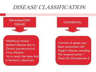
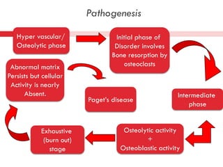
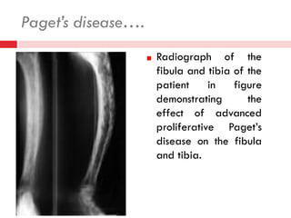
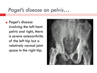
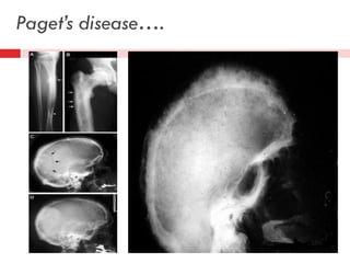


This document provides information on Paget's disease, including: - It is a chronic bone disorder characterized by abnormal bone remodeling that can cause bone deformities and fractures. - The cause is unknown but may involve viruses or genetic factors. It most commonly affects older adults and bones like the pelvis and spine. - Symptoms can include bone pain, stiffness, fractures, and hearing loss. Lab tests show elevated alkaline phosphatase levels. Imaging like x-rays are used for diagnosis. - The disease involves abnormal bone breakdown and formation seen on imaging as thickened and misshapen bones. While often asymptomatic, treatment with medications may be used for painful symptoms.


