
Recommended
More Related Content
What's hot
What's hot (20)
Similar to Anatomy of female reproductive system
Similar to Anatomy of female reproductive system (20)
Recently uploaded
Recently uploaded (20)
Anatomy of female reproductive system
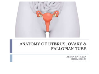
- 1. ANATOMY OF UTERUS, OVARY & FALLOPIAN TUBE ASWIN SATHYAN ROLL NO: 31
- 2. INTERNAL GENITAL ORGANS VAGINA CERVIX UTERUS FALLOPIAN TUBES OVARIES
- 4. UTERUS • Hollow pyriform muscular organ • Situation – between bladder in front and rectum behind • Position- anteversion and anteflexion • Usually inclines to right ( dextrorotation) & cervix is directed to the left (levorotation) • 8 x 5 x 1.25 cm • Weight – 50-80g • Three parts 1) Body 2) Isthmus 3) Cervix
- 5. BODY OR CORPUS Body is further divided into fundus and cervix FUNDUS- the part which lies above the openings of the uterine tubes. BODY-is triangular and lies between the openingsof the tubes and the isthmus CORNUA-superolateral angles of the body of the uterus project outwards from the junction of the fundus and body ISTHMUS-the isthmus is a constricted part measuring about 0.5cm situated between the body and the cervix
- 6. CERVIX The cervix is the lowermost part of the uterus. It extends from the histological internal os and ends at external os which opens into the vagina. Cylindrical in shape Measures about 2.5cm in length and diameter. It is divided into a supravaginal part and a vaginal part which lies within the vagina each measuring 1.25cm NULLIPAROUS- vaginal part of the cervix is conical with external os looking circular. PAROUS- is cylindrical with the external os having bilateral slits.
- 7. UTERINE LIGAMENTS FALSE LIGAMENTS TRUE LIGAMENTS (all paired) Broad ligament(paired) Round ligaments Rectouterine folds(paired) Transverse cervical ligament Uterovesical fold(anterior ligament –unpaired) Uterosacral ligament Rectovaginal fold(posterior ligament-unpaired) Pubocervical ligament
- 8. RELATIONS Anteriorly- bladder Posteriorly-rectum Peritoneum covers the body of the uterus and the superavaginal portion of the cervix posteriorly. Pouch of Douglas- reflected peritoneum over the rectum. Anteriorly peritoneum covers the body.And reflected on to bladder creating uterovesical fold. Laterally-broad ligament.
- 9. BLOOD SUPPLY The uterine artery (a branch of the internal iliac artery) LYMPHATIC DRAINAGE Lymphatics from the fundus accompany the ovarian artery and drain into the para-aortic nodes. lymphatics from the body and cervix drain to the internal and external iliac lymph node NERVE SUPPLY Sensory fibres in the T11 and T12 roots carry pain sensation from the uterus Sensory from the cervix and upper vagina pass through the pelvic nerves to the 2,3,4th sacral nerves
- 10. OVARIES Ovaries are paired bodies situated on either side of the uterus in close approximation within the lateral pelvic wall. Ovaries are located in the fossa of waldeyer. Measure-4*3*2cm in size It is the only intraabdominal structure not covered by peritoneum.
- 11. ATTACHMENT Medial pole is attached to the uterine cornua by the ovarian ligament. Laterally it is attached by infundibulopelvic ligament to the lateral pelvic wall. The fimbrial end of the tube is in close proximity to the ovary and connected to it by the fimbria ovarica.
- 12. STRUCTURE Ovary has two parts. Outer cortex and inner medulla. CORTEX Outer portion of the cortex is the tunica albuginea. On the surface there is a single layer of cuboidal epithelium-germinal epithelium of waldeyer. MEDULLA It is composed of loose connective tissue and is vascular.
- 13. DEVELOPMENT Triple orgin From coelomic epithelium of genital ridge Underlying mesoderm Primordial germ cells
- 14. BLOOD SUPPLY Ovarian artery(branch of abdominal aorta) VENOUS DRAINAGE To inferior vena cava on the right ,renal vain on the left LYMPHATIC DRAINAGE- to the parla- aortic node. NERVE SUPPLY Sympathetic-ovarian plexus Parasympathetic-inferior hypogastric plexus.
- 15. FALLOPIAN TUBE •7-12 cm long •Lying in the superior border of the broad ligament •Medial end opens into the uterus at the cornua. •Lateral extremity is free from broad lig. and in close association with ovary •Parts of tube--- infundibulum-comprising fimbriae ampulla-widest part isthmus-narrowest part Interstitial part-inside myometrium •Fertilisation normally takes place in the ampulla.
- 16. Clinical significance Implantation in ampulla-ectopic pregnancy Structure •Outer serosal layer •Middle muscular layer •Inner ciliated epithelium Development Nonfused portion of mullerian duct Blood supply,lymphatic drainage and nerve supply Arterial supply----Ovarian and uterine artery Venous drainage----ovarian veins via pampiniform plexus Lymphatic drainage----paraaortic nodes
- 17. THANK YOU