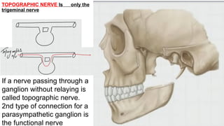
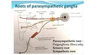
TOPOGRAPHIC NERVE Isonly the
trigeminal nerve
If a nerve passing through a
ganglion without relaying is
called topographic nerve.
2nd type of connection for a
parasympathetic ganglion is
the functional nerve
4.
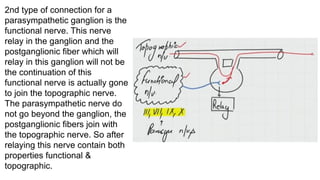
2nd type ofconnection for a
parasympathetic ganglion is the
functional nerve. This nerve
relay in the ganglion and the
postganglionic fiber which will
relay in this ganglion will not be
the continuation of this
functional nerve is actually gone
to join the topographic nerve.
The parasympathetic nerve do
not go beyond the ganglion, the
postganglionic fibers join with
the topographic nerve. So after
relaying this nerve contain both
properties functional &
topographic.
5.
So only eithera branch of ophthalmic, maxillary or
mandibular nerve come out of these ganglions after
relaying. So every postganglionic fiber is a branch of
trigeminal nerve. For Example the glossopharyngeal
nerve goes to supply the parotid gland but after
relaying in ganglion it does not go to supply this
gland no branch of glossopharyngeal nerve enter
into this gland , the glossopharyngeal nerve will
reach in ganglion like otic ganglion so from ganglion
to the parotid gland it has to be supply by trigeminal
nerve.
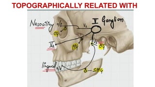
6.
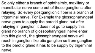
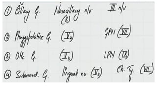
So trigeminal
ganglion inmeckle’s
cave and then its
three branches i.e
ophthalmic nerve
( enter into orbit by
superior ophthalmic
fissure one of its
branch concern with
ganglion is
Nasocillary nerve
which enter in orbit
this nerve
tropographically
related with ciliary
ganglion ) ,
7.
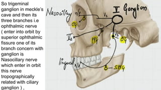
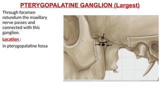
maxillary nerve (Leave the
cranial cavity by foramen rotundum
and come in pterygopalatine fossa
then it goes inside the maxilla is
topographically related with
pterygopalatine ganglion) PPG &
mandibular( Coming out of
through foramen ovale , its trunk is
topographically related to otic
ganglion (OG) , then divides into
anterior & posterior divisions, one
of its branch which is concern
there is called lingual nerve & that
lingual nerve will be
topographically related with the
submandibular ganglion)
8.
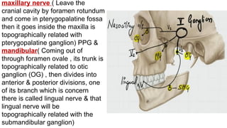
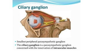
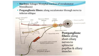
CILIARY
GANGLION
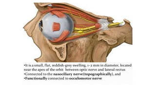
Location Between theoptic nerve
& lateral rectus muscle but close
to the apex of the orbit.
The third cranial nerve that is
oculomotor nerve present in
midbrain at level of superior
colliculus has two nuclei one is 3rd
nucleus itself and 2nd
edenger
westphal nucleus, this third nerve
goes through the lateral wall of
cavernous sinus and devides into
upper and lower division , fibers
from lower division supplying to
inferior oblique muscle
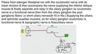
9.
Fibers from edengerWestphal run with the oculomotor nerve with its
lower division & then accompany the nerve supplying the inferior oblique
muscle & finally separate and relay in the ciliary ganglion so oculomotor
nerve is a functional nerve then from the ciliary ganglion the post
ganglionic fibers i.e short ciliary nerves(8-10 in No:) Supplying the ciliaris
and sphincter pupillae muscles, so for ciliary ganglion oculomotor is
functional nerve & topographic nerve is Nasociliary nerve
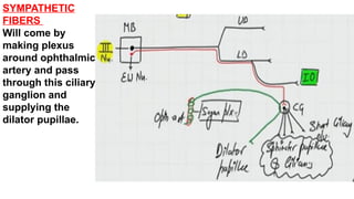
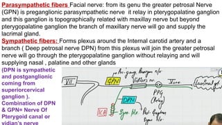
Parasympathetic fibers Facialnerve: from its genu the greater petrosal Nerve
(GPN) is preganglionic parasympathetic nerve it relay in pterygopalatine ganglion
and this ganglion is topographically related with maxillay nerve but beyond
pterygopalatine ganglion the branch of maxillary nerve will go and supply the
lacrimal gland.
Sympathetic fibers: Forms plexus around the Internal carotid artery and a
branch ( Deep petrosal nerve DPN) from this plexus will join the greater petrosal
nerve will go through the pterygopalatine ganglion without relaying and will
supplying nasal , palatine and other glands
(DPN is sympathetic
and postganglionic
coming from
superiorcervical
ganglion ).
Combination of DPN
& GPN= Nerve Of
Pterygoid canal or
17.
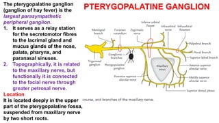
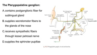
PTERYGOPALATINE GANGLION
The pterygopalatineganglion
(ganglion of hay fever) is the
largest parasympathetic
peripheral ganglion.
1. It serves as a relay station
for the secretomotor fibres
to the lacrimal gland and
mucus glands of the nose,
palate, pharynx, and
paranasal sinuses.
2. Topographically, it is related
to the maxillary nerve, but
functionally it is connected
to the facial nerve through
greater petrosal nerve.
Location
It is located deeply in the upper
part of the pterygopalatine fossa,
suspended from maxillary nerve
by two short roots.
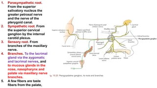
18.
1. Parasympathetic root.
Fromthe superior
salivatory nucleus the
greater petrosal nerve
and the nerve of the
pterygoid canal.
2. Sympathetic root. From
the superior cervical
ganglion by the internal
carotid plexus.
3. Sensory root. From
branches of the maxillary
nerve.
4. Branches. To the lacrimal
gland via the zygomatic
and lacrimal nerves, and
to mucous glands in the
nose, nasopharynx and
palate via maxillary nerve
branches.
5. A few fibers are taste
fibers from the palate,
19.
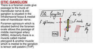
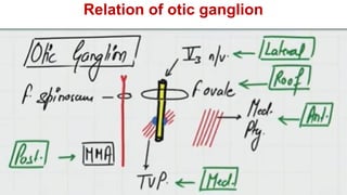
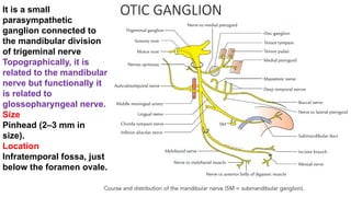
OTIC GANGLION
There isa foramen ovale give
passage to the trunk of
mandibular nerve & otic
ganglion is situated in the
infratemporal fossa & medial
side of mandibular nerve.
Foramen spinosum which is
situated behind the foramen
ovale allows the passage of
middle meningeal artery
(MMA), Anteriorly there is a
muscle called medial
pterygoid & another muscle
which is medial to the ganglion
is tensor velli palatini (TVP)
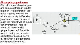
Glossopharyngeal nerve
Starts frommedulla oblongata
and come out through jugular
foramen (JF) a branch of this
nerve is going towards the
middle ear cavity (ME) called
jacobson;’s nerve, this nerve
reach the medial wall of middle
ear (Promontory) here its
forms the plexus called
tympanic plexus & from this
plexus coming out nerve is
called lesser petrosal nerve
(LPN) which is preganglionic
parasympathetic nerve. .
23.
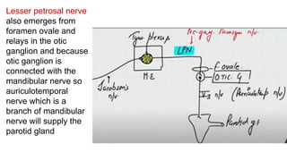
Lesser petrosal nerve
alsoemerges from
foramen ovale and
relays in the otic
ganglion and because
otic ganglion is
connected with the
mandibular nerve so
auriculotemporal
nerve which is a
branch of mandibular
nerve will supply the
parotid gland
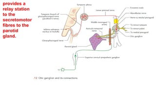
OTIC GANGLION
It isa small
parasympathetic
ganglion connected to
the mandibular division
of trigeminal nerve
Topographically, it is
related to the mandibular
nerve but functionally it
is related to
glossopharyngeal nerve.
Size
Pinhead (2–3 mm in
size).
Location
Infratemporal fossa, just
below the foramen ovale.
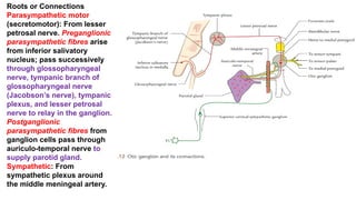
Roots or Connections
Parasympatheticmotor
(secretomotor): From lesser
petrosal nerve. Preganglionic
parasympathetic fibres arise
from inferior salivatory
nucleus; pass successively
through glossopharyngeal
nerve, tympanic branch of
glossopharyngeal nerve
(Jacobson’s nerve), tympanic
plexus, and lesser petrosal
nerve to relay in the ganglion.
Postganglionic
parasympathetic fibres from
ganglion cells pass through
auriculo-temporal nerve to
supply parotid gland.
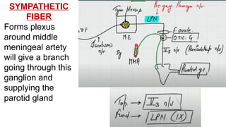
Sympathetic: From
sympathetic plexus around
the middle meningeal artery.
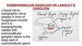
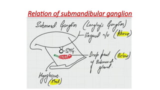
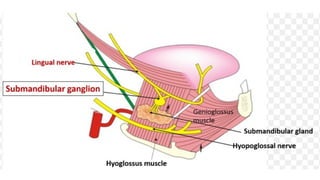
SUBMANDIBULAR GANGLION ORLANGLEY’S
GANGLION
Lingual nerve
topographic nerve
passes in front of
hyoglossus muscle
and it is
topographically
related with
submandibular
ganglion below is the
deep part of
submandibular gland
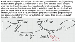
Facial nerve frompons and also you can see the lingual nerve which is topographically
related with this ganglion . Another branch of facial nerve called as chorda tympani
which join the lingual nerve and then reach the submandibular ganglion . In this case
chorda tympani is a functional nerve but it is not able to reach the ganglion itself so it
joins the lingual nerve in the infra temporal fossa and by using the lingual nerve the
chorda tympani will reach the submandibular ganglion and then this ganglion will give
rise postganglionic branch in two ways, the first way supply direct branches to supply
the submandibular gland
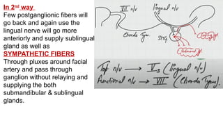
32.
In 2nd
way
Few postganglionicfibers will
go back and again use the
lingual nerve will go more
anteriorly and supply sublingual
gland as well as
SYMPATHETIC FIBERS
Through pluxes around facial
artery and pass through
ganglion without relaying and
supplying the both
submandibular & sublingual
glands.
35.
. Loss ofsensation from the
temporal region and loss of
secretory function of the
parotid gland would be
caused by interruption of
which one of the following
nerve.
A.Facial
B.Chorda tympani
C.Auriculotemporal
D.Greater auricular
36.
The Pterygopalatine ganglion:
A.containspostganglionic fiber for
sublingual gland
B.supplies secretomotor fibers to
the glands of the nose
C.receives sympathetic fibers
through lesser petrosal nerve
D.supplies the sphincter pupillae
37.
Which one ofthe following ganglion is present in
infratemporal fossa:
A.Ciliary
B.Otic
C.Pterygopalatine
D.Submandibular
38.
The chorda tympanicontains
which component before it
joins the lingual nerve?
A. Preganglionic sympathetic
B. Postganglionic sympathetic
C. Postganglionic
parasympathetic
D. Preganglionic
parasympathetic
39.
The absence oftears is due to
lesion in which of the following
ganglion?
A.Ciliary
B.Sphenopalatine
C.Superior cervical sympathetic
D.Trigeminal