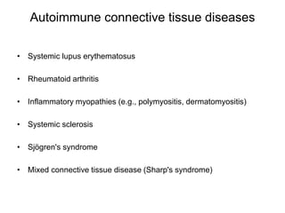
Connective Tissue Disorder
Dr. Deepesh Sharma(PT)
MPT(Cardio-Pulmonary)
Assistant Professor
JNU College Of Physiotherapy ,Jaipur
Introduction
• Connective tissue is an important biological tissue composed of
an extracellular matrix that binds, anchors, and supports organs.
• Over 200 conditions, which may be inherited or autoimmune,
affect connective tissue and they are collectively known
as connective tissue diseases (CTDs).
• Inherited CTDs are caused by mutations that affect one of the two
fibers (collagen and fibrin). Autoimmune CTDs have no clear
etiology, but the incidence is higher in women and among
genetically predisposed individuals.
• As the name suggests, in autoimmune CTDs, the immune
system develops antibodies against components of connective
tissue. Individual conditions can affect a vast range of bodily
structures (including cartilage, blood vessels, bone, tendons, and
organs) and thus present with a wide array of clinical findings.
Types
1. Autoimmune connective tissue diseases
2. Inherited connective tissue diseases
Autoimmune connective tissue diseases
• Systemic lupus erythematosus
• Rheumatoid arthritis
• Inflammatory myopathies (e.g., polymyositis, dermatomyositis)
• Systemic sclerosis
• Sjögren's syndrome
• Mixed connective tissue disease (Sharp's syndrome)
Inherited connective tissue diseases
• Ehlers-Danlos syndrome
• Marfan syndrome
• Osteogenesis imperfecta
• Alport syndrome
• Loeys-Dietz syndrome [1]
– Autosomal dominant inheritance
– Features shared with Marfan syndrome: marfanoid
habitus, increased risk of ascending aortic
aneurysms and aortic dissection
– Distinct features: hypertelorism, bifid uvula, cleft palate,
easy bruising and keloids, arterial tortuosity (winding
course of arteries)
Clinical manifestations of connective
tissue disorders
• Clinical features vary greatly among individual diseases. The table
below provides a general overview of the more common features.
Marfan syndrome (MFS)
• It is an autosomal dominant connective tissue
disorder affecting the microfibrils
and elastin in connective tissue throughout the body.
• MFS is associated with pathological manifestations in
the cardiovascular system (e.g., mitral valve
prolapse, aortic aneurysm, and dissection),
the musculoskeletal system (e.g., tall stature with
disproportionately long extremities, joint hypermobility),
and the eyes (e.g., subluxation of the lens of the eye).
Etiology & Pathophysiology
• Autosomal dominant disease caused
by mutation of fibrillin-1 gene (FBN1)
on chromosome 15
• Defective fibrillin → defective elastin →
defective connective tissue throughout the body
Clinical features
• Cardiovascular Features
– Aortic necrosis (cystic medial degeneration), characterized by
histopathologic findings such as:
• Loss, thinning, disorganization, and fragmentation of elastic
fibers
• Accumulation of mucoid extracellular matrix
• Loss of smooth muscle nuclei
– Aortic regurgitation
– Aortic aneurysm
• Thoracic or abdominal aortic aneurysm
• Aortic root dilation: aneurysm of the proximal thoracic aorta
– Aortic dissection
• Features of mitral valve prolapse
• Berry aneurysms, which can rupture → subarachnoid hemorrhage
Clinical features
Musculoskeletal features
• Tall stature with rapid linear growth and long extremities
• Joint hypermobility
• High-arched palate
• Arachnodactyly (achromachia): abnormally long, slender
fingers and toes
• Pectus deformity: pectus carinatum (sternal kyphosis; more
specific for Marfan syndrome) or pectus excavatum
• Pes planus (flat foot) or hindfoot valgus
• Spinal deformities (scoliosis, hyperkyphosis)
Clinical features
Skin
• Loss of skin elasticity/striae (stretch marks)
Eyes
• Visual impairment: ectopia lentis (lens
dislocation) → lens subluxation superiorly and
temporally
• Severe myopia
Ehlers-Danlos syndrome (EDS)
• It is a heterogeneous group of six connective tissue
disorders with variable inheritance in which the synthesis
and processing of collagen are defective.
• Patients present with varying degrees of hyperelastic
skin, joint hypermobility, and tissue fragility (including
that of vasculature).
Etiology & Pathophysiology
• Etiology
• Heterogeneous group of six connective tissue disorders
with variable inheritance (autosomal
recessive or dominant)
• Mutation in genes controlling synthesis of collagen
• Classic type: mutations in COL5A1, COL5A2 → type
V collagen defect
• Vascular type: type III procollagen defect
• Pathophysiology
– Defective collagen cross-linking and fibril synthesis
Clinical Features
Cardiovascular Features
• heart valve defects (particularly mitral valve prolapse)
• Features of aneurysms/dissections of the iliac, splenic, renal
arterties, or the aorta
• Berry/saccular aneurysms of the cerebral arteries → features
of subarachnoid hemorrhage
Musculoskeletal
• Joint hypermobility with tendency to dislocate
• Skeletal abnormalitis (e.g., scoliosis)
Skin
• Tendency to bruise easily
• Skin hyperextensibility
• Frequent skin lacerations and poor skin healing (e.g., following
surgery)
Other
• Hernias
• Features of organ rupture (e.g., shock, local pain), especially in
vascular EDS
Prognosis
• Marfan syndrome: patients can expect a normal
lifespan, if the disorder is diagnosed early and
complications are managed appropriately
– Aortic root disease is the most common cause
of mortality
• Ehlers-Danlos syndrome: Life expectancy is typically
normal with the exception of vascular EDS, which
has a reduced life expectancy of ∼ 50 years.
Osteogenesis imperfecta (brittle bone
disease)
• A genetic disorder characterized by defective synthesis
of type 1 collagen, which is important in bone formation.
• Patients present with signs that are sometimes mistaken
for child abuse (e.g., easy bruising, predisposition to
bony fractures).
Etiology & Pathophysiology
• Etiology :
• Autosomal
dominant mutation in COL1A1 or COL1A2 genes
• Pathophysiology:
• ↓ formation of hydrogen and disulfide
bonds between type I preprocollagen molecules → ↓
triple helix formation → ↓ type
I collagen synthesis → impaired bone matrix formation
(osteogenesis)
Clinical features
• Osteogenesis imperfecta type I (the mildest and the most common
form)
– Growth delay
– Skeletal deformities, brittle bones
– Bowing of bones and saber shins
– Fractures during childbirth and recurrently from minimal trauma
thereafter
– Blue sclerae
– Progressive hearing loss
– Brittle, opalescent teeth (dentinogenesis imperfecta; due to a
lack of dentin)
– Ligamentous laxity and joint hypermobility
• Osteogenesis imperfecta type II
– Most severe form; lethal prenatally or within the first year
– Multiple intrauterine and/or preinatal fractures
– Underdeveloped lungs and subsequent respiratory problems
Scurvy
• Clinical manifestation of vitamin C deficiency,
which leads to impaired collagen
synthesis and easily damaged connective
tissue.
Clinical features
• Swollen gums
• Mucosal bleeding
• Poor wound healing
• Curly body hair
• Follicular hyperkeratosis
• Hemarthrosis
• Generalized weakness/fatigue
Systemic lupus erythematosus (SLE)
• It is a multisystem autoimmune disease that
predominantly affects women of childbearing
age.
• The exact cause is still unknown, but hormonal
and immunological influences as well as genetic
predisposition are considered likely etiological
factors.
• The presentation of the disease is variable and
may range from mild localized symptoms to life-
threatening systemic disease.
Epidemiology
• Sex: ♀ >> ♂ (10:1)
• Peak incidence: women aged 20–40 years; no
particular age of manifestation in men
Etiology
• The exact etiology is unknown, but several predisposing
factors have been identified:
• Genetic predisposition:
– HLA-DR2 and HLA-DR3 are commonly present in individuals
with SLE
– Genetic deficiency of classical
pathway complement proteins (C1q, C2, C4)
• Hormonal factors: studies suggest
that hyperestrogenic states (e.g., due to oral
contraceptive use, postmenopausal hormonal
therapy, endometriosis) are associated with an increased risk
of SLE.
• Environmental factors: UV light, stimulation of immune cells
through infection with bacteria and viruses, medications.
Pathophysiology
• Possible mechanisms for the development
of autoantibodies
– Deficiency of classical complement proteins (C1q, C4, C2)
→ failure of macrophages to phagocytose immune
complexes and apoptotic cell material (i.e., plasma and nuclear
antigens) → dysregulated, intolerant lymphocytes begin
targeting normally protected intracellular antigens
→ autoantibody production (e.g., anti-ds DNA)
• Mechanism of tissue damage
– Type III hypersensitivity→ antibody-antigen complex formation
in microvasculature → complement activation
and inflammation → damage to skin, kidneys, joints, small
vessels
– Type II hypersensitivity → IgG and IgM antibodies directed
against antigens on cells (e.g., red blood cells) → cytopenia
Clinical features
• Skin (> 70% of cases)
– Malar rash (butterfly rash) with sparing of the nasolabial
folds
– Photosensitivity
– Discoid rash
– Oral ulcers
– Alopecia (nonscarring)
• Joints
– Arthritis and arthralgia (> 90% of cases)
– Mostly nonerosive polyarthritis (normal x-ray)
• Fever (> 50% of cases), fatigue (> 80% of cases), weight
loss
Other signs and symptoms
• Musculoskeletal: myalgia and lymphadenopathy
• Serositis: pleuritis and pericarditis; effusions and chest pain may
occur
• Kidneys: nephritis with proteinuria
• Heart: involvement of the myocardium, pericardium, valves,
and coronary arteries
• Lungs: pneumonitis, interstitial lung disease, pulmonary
hypertension
• Gastrointestinal: esophagitis, hepatitis, pancreatitis
• Vascular: Raynaud phenomenon, vasculitis, thromboembolism
• Neurologic: e.g., seizures, psychosis, personality changes,
aseptic meningitis, polyneuropathy, myasthenia gravis
• Hematologic: hemolytic anemia, thrombocytopenia, leukopenia
• Eyes: keratoconjunctivitis sicca
Sjogren syndrome
• Sjogren syndrome is a chronic inflammatory autoimmune disease
that occurs mainly in middle-aged women.
• The cause of primary Sjogren syndrome is unknown, whereas
secondary Sjogren syndrome is associated with underlying
autoimmune diseases (e.g., rheumatoid arthritis).
• As the immune system mainly attacks lacrimal and salivary glands,
patients typically present with xerophthalmia (dry eyes)
and xerostomia (dry mouth), the combination of which is also known
as sicca syndrome.
• The disease may also involve the skin, joints, internal organs, and
nervous system .
Epidemiology
• Sex: ♀ > ♂ (∼ 9:1)
• Age of onset: typically 40–60 years
Etiology
• Primary Sjogren syndrome: idiopathic (association
with HLA-DR52 estimated in 87% of cases)
• Secondary Sjogren syndrome
– Autoimmune connective tissue diseases: rheumatoid
arthritis, systemic lupus erythematosus, systemic
sclerosis, polymyositis
– Primary biliary cirrhosis (PBC)
Clinical features
Glandular symptoms
• Inflammation of the salivary glands: decreased production of saliva
→ xerostomia (dry mouth)
– Dysphagia
– Increased formation of dental caries and tendency to oral infections
– Parotitis
• Inflammation of the lacrimal glands (chronic dacryoadenitis): decreased
secretion of tears → xerophthalmia (dry eyes) → keratoconjunctivitis sicca
– Redness, itching, burning of eyes
– Sensation of sand or foreign body in the eyes
– Blurred vision
• Sicca syndrome: combination of dry mouth and dry eyes
• Nasal dryness → chronic rhinitis, nosebleeds
• Pharyngeal, tracheal, and bronchial dryness → persistent, dry cough
• Vaginal dryness → dyspareunia (painful intercourse) and increased risk of
infections
Extraglandular symptoms
• General symptoms: fatigue and arthralgias (∼ 70% of cases)
• Skin manifestations: xerosis ; Raynaud's phenomenon (∼
15–30% of cases)
• Vasculitis (∼ 10% of cases)
– Palpable purpura of the legs
– Recurrent urticaria, skin ulcerations
– Glomerulonephritis
• Neurological and psychiatric manifestations
– Depression
– Variety of focal and/or diffuse findings (e.g., impaired gross
motor control, paresis, seizures, peripheral neuropathy)
• Gastrointestinal manifestations: e.g., dyspepsia,
reflux esophagitis
Mixed connective tissue
disease (Sharp's syndrome)
• Definition: overlapping symptoms of
systemic sclerosis, systemic lupus erythematosus (SLE),
and polymyositis
• Clinical presentation
– Myositis
– Arthritis
– Acrosclerosis
– Raynaud's phenomenon
– Pulmonary hypertension
– The course is usually milder than that of other connective
tissue diseases (CTD), but may progress into another CTD.

Connective tissue disorder

  • 1.
    Connective Tissue Disorder Dr.Deepesh Sharma(PT) MPT(Cardio-Pulmonary) Assistant Professor JNU College Of Physiotherapy ,Jaipur
  • 2.
    Introduction • Connective tissueis an important biological tissue composed of an extracellular matrix that binds, anchors, and supports organs. • Over 200 conditions, which may be inherited or autoimmune, affect connective tissue and they are collectively known as connective tissue diseases (CTDs). • Inherited CTDs are caused by mutations that affect one of the two fibers (collagen and fibrin). Autoimmune CTDs have no clear etiology, but the incidence is higher in women and among genetically predisposed individuals. • As the name suggests, in autoimmune CTDs, the immune system develops antibodies against components of connective tissue. Individual conditions can affect a vast range of bodily structures (including cartilage, blood vessels, bone, tendons, and organs) and thus present with a wide array of clinical findings.
  • 3.
    Types 1. Autoimmune connectivetissue diseases 2. Inherited connective tissue diseases
  • 4.
    Autoimmune connective tissuediseases • Systemic lupus erythematosus • Rheumatoid arthritis • Inflammatory myopathies (e.g., polymyositis, dermatomyositis) • Systemic sclerosis • Sjögren's syndrome • Mixed connective tissue disease (Sharp's syndrome)
  • 5.
    Inherited connective tissuediseases • Ehlers-Danlos syndrome • Marfan syndrome • Osteogenesis imperfecta • Alport syndrome • Loeys-Dietz syndrome [1] – Autosomal dominant inheritance – Features shared with Marfan syndrome: marfanoid habitus, increased risk of ascending aortic aneurysms and aortic dissection – Distinct features: hypertelorism, bifid uvula, cleft palate, easy bruising and keloids, arterial tortuosity (winding course of arteries)
  • 6.
    Clinical manifestations ofconnective tissue disorders • Clinical features vary greatly among individual diseases. The table below provides a general overview of the more common features.
  • 7.
    Marfan syndrome (MFS) •It is an autosomal dominant connective tissue disorder affecting the microfibrils and elastin in connective tissue throughout the body. • MFS is associated with pathological manifestations in the cardiovascular system (e.g., mitral valve prolapse, aortic aneurysm, and dissection), the musculoskeletal system (e.g., tall stature with disproportionately long extremities, joint hypermobility), and the eyes (e.g., subluxation of the lens of the eye).
  • 8.
    Etiology & Pathophysiology •Autosomal dominant disease caused by mutation of fibrillin-1 gene (FBN1) on chromosome 15 • Defective fibrillin → defective elastin → defective connective tissue throughout the body
  • 9.
    Clinical features • CardiovascularFeatures – Aortic necrosis (cystic medial degeneration), characterized by histopathologic findings such as: • Loss, thinning, disorganization, and fragmentation of elastic fibers • Accumulation of mucoid extracellular matrix • Loss of smooth muscle nuclei – Aortic regurgitation – Aortic aneurysm • Thoracic or abdominal aortic aneurysm • Aortic root dilation: aneurysm of the proximal thoracic aorta – Aortic dissection • Features of mitral valve prolapse • Berry aneurysms, which can rupture → subarachnoid hemorrhage
  • 10.
    Clinical features Musculoskeletal features •Tall stature with rapid linear growth and long extremities • Joint hypermobility • High-arched palate • Arachnodactyly (achromachia): abnormally long, slender fingers and toes • Pectus deformity: pectus carinatum (sternal kyphosis; more specific for Marfan syndrome) or pectus excavatum • Pes planus (flat foot) or hindfoot valgus • Spinal deformities (scoliosis, hyperkyphosis)
  • 11.
    Clinical features Skin • Lossof skin elasticity/striae (stretch marks) Eyes • Visual impairment: ectopia lentis (lens dislocation) → lens subluxation superiorly and temporally • Severe myopia
  • 13.
    Ehlers-Danlos syndrome (EDS) •It is a heterogeneous group of six connective tissue disorders with variable inheritance in which the synthesis and processing of collagen are defective. • Patients present with varying degrees of hyperelastic skin, joint hypermobility, and tissue fragility (including that of vasculature).
  • 14.
    Etiology & Pathophysiology •Etiology • Heterogeneous group of six connective tissue disorders with variable inheritance (autosomal recessive or dominant) • Mutation in genes controlling synthesis of collagen • Classic type: mutations in COL5A1, COL5A2 → type V collagen defect • Vascular type: type III procollagen defect • Pathophysiology – Defective collagen cross-linking and fibril synthesis
  • 15.
    Clinical Features Cardiovascular Features •heart valve defects (particularly mitral valve prolapse) • Features of aneurysms/dissections of the iliac, splenic, renal arterties, or the aorta • Berry/saccular aneurysms of the cerebral arteries → features of subarachnoid hemorrhage Musculoskeletal • Joint hypermobility with tendency to dislocate • Skeletal abnormalitis (e.g., scoliosis) Skin • Tendency to bruise easily • Skin hyperextensibility • Frequent skin lacerations and poor skin healing (e.g., following surgery) Other • Hernias • Features of organ rupture (e.g., shock, local pain), especially in vascular EDS
  • 17.
    Prognosis • Marfan syndrome:patients can expect a normal lifespan, if the disorder is diagnosed early and complications are managed appropriately – Aortic root disease is the most common cause of mortality • Ehlers-Danlos syndrome: Life expectancy is typically normal with the exception of vascular EDS, which has a reduced life expectancy of ∼ 50 years.
  • 18.
    Osteogenesis imperfecta (brittlebone disease) • A genetic disorder characterized by defective synthesis of type 1 collagen, which is important in bone formation. • Patients present with signs that are sometimes mistaken for child abuse (e.g., easy bruising, predisposition to bony fractures).
  • 19.
    Etiology & Pathophysiology •Etiology : • Autosomal dominant mutation in COL1A1 or COL1A2 genes • Pathophysiology: • ↓ formation of hydrogen and disulfide bonds between type I preprocollagen molecules → ↓ triple helix formation → ↓ type I collagen synthesis → impaired bone matrix formation (osteogenesis)
  • 20.
    Clinical features • Osteogenesisimperfecta type I (the mildest and the most common form) – Growth delay – Skeletal deformities, brittle bones – Bowing of bones and saber shins – Fractures during childbirth and recurrently from minimal trauma thereafter – Blue sclerae – Progressive hearing loss – Brittle, opalescent teeth (dentinogenesis imperfecta; due to a lack of dentin) – Ligamentous laxity and joint hypermobility • Osteogenesis imperfecta type II – Most severe form; lethal prenatally or within the first year – Multiple intrauterine and/or preinatal fractures – Underdeveloped lungs and subsequent respiratory problems
  • 22.
    Scurvy • Clinical manifestationof vitamin C deficiency, which leads to impaired collagen synthesis and easily damaged connective tissue.
  • 23.
    Clinical features • Swollengums • Mucosal bleeding • Poor wound healing • Curly body hair • Follicular hyperkeratosis • Hemarthrosis • Generalized weakness/fatigue
  • 24.
    Systemic lupus erythematosus(SLE) • It is a multisystem autoimmune disease that predominantly affects women of childbearing age. • The exact cause is still unknown, but hormonal and immunological influences as well as genetic predisposition are considered likely etiological factors. • The presentation of the disease is variable and may range from mild localized symptoms to life- threatening systemic disease.
  • 25.
    Epidemiology • Sex: ♀>> ♂ (10:1) • Peak incidence: women aged 20–40 years; no particular age of manifestation in men
  • 26.
    Etiology • The exactetiology is unknown, but several predisposing factors have been identified: • Genetic predisposition: – HLA-DR2 and HLA-DR3 are commonly present in individuals with SLE – Genetic deficiency of classical pathway complement proteins (C1q, C2, C4) • Hormonal factors: studies suggest that hyperestrogenic states (e.g., due to oral contraceptive use, postmenopausal hormonal therapy, endometriosis) are associated with an increased risk of SLE. • Environmental factors: UV light, stimulation of immune cells through infection with bacteria and viruses, medications.
  • 27.
    Pathophysiology • Possible mechanismsfor the development of autoantibodies – Deficiency of classical complement proteins (C1q, C4, C2) → failure of macrophages to phagocytose immune complexes and apoptotic cell material (i.e., plasma and nuclear antigens) → dysregulated, intolerant lymphocytes begin targeting normally protected intracellular antigens → autoantibody production (e.g., anti-ds DNA) • Mechanism of tissue damage – Type III hypersensitivity→ antibody-antigen complex formation in microvasculature → complement activation and inflammation → damage to skin, kidneys, joints, small vessels – Type II hypersensitivity → IgG and IgM antibodies directed against antigens on cells (e.g., red blood cells) → cytopenia
  • 28.
    Clinical features • Skin(> 70% of cases) – Malar rash (butterfly rash) with sparing of the nasolabial folds – Photosensitivity – Discoid rash – Oral ulcers – Alopecia (nonscarring) • Joints – Arthritis and arthralgia (> 90% of cases) – Mostly nonerosive polyarthritis (normal x-ray) • Fever (> 50% of cases), fatigue (> 80% of cases), weight loss
  • 29.
    Other signs andsymptoms • Musculoskeletal: myalgia and lymphadenopathy • Serositis: pleuritis and pericarditis; effusions and chest pain may occur • Kidneys: nephritis with proteinuria • Heart: involvement of the myocardium, pericardium, valves, and coronary arteries • Lungs: pneumonitis, interstitial lung disease, pulmonary hypertension • Gastrointestinal: esophagitis, hepatitis, pancreatitis • Vascular: Raynaud phenomenon, vasculitis, thromboembolism • Neurologic: e.g., seizures, psychosis, personality changes, aseptic meningitis, polyneuropathy, myasthenia gravis • Hematologic: hemolytic anemia, thrombocytopenia, leukopenia • Eyes: keratoconjunctivitis sicca
  • 31.
    Sjogren syndrome • Sjogrensyndrome is a chronic inflammatory autoimmune disease that occurs mainly in middle-aged women. • The cause of primary Sjogren syndrome is unknown, whereas secondary Sjogren syndrome is associated with underlying autoimmune diseases (e.g., rheumatoid arthritis). • As the immune system mainly attacks lacrimal and salivary glands, patients typically present with xerophthalmia (dry eyes) and xerostomia (dry mouth), the combination of which is also known as sicca syndrome. • The disease may also involve the skin, joints, internal organs, and nervous system .
  • 32.
    Epidemiology • Sex: ♀> ♂ (∼ 9:1) • Age of onset: typically 40–60 years
  • 33.
    Etiology • Primary Sjogrensyndrome: idiopathic (association with HLA-DR52 estimated in 87% of cases) • Secondary Sjogren syndrome – Autoimmune connective tissue diseases: rheumatoid arthritis, systemic lupus erythematosus, systemic sclerosis, polymyositis – Primary biliary cirrhosis (PBC)
  • 34.
    Clinical features Glandular symptoms •Inflammation of the salivary glands: decreased production of saliva → xerostomia (dry mouth) – Dysphagia – Increased formation of dental caries and tendency to oral infections – Parotitis • Inflammation of the lacrimal glands (chronic dacryoadenitis): decreased secretion of tears → xerophthalmia (dry eyes) → keratoconjunctivitis sicca – Redness, itching, burning of eyes – Sensation of sand or foreign body in the eyes – Blurred vision • Sicca syndrome: combination of dry mouth and dry eyes • Nasal dryness → chronic rhinitis, nosebleeds • Pharyngeal, tracheal, and bronchial dryness → persistent, dry cough • Vaginal dryness → dyspareunia (painful intercourse) and increased risk of infections
  • 35.
    Extraglandular symptoms • Generalsymptoms: fatigue and arthralgias (∼ 70% of cases) • Skin manifestations: xerosis ; Raynaud's phenomenon (∼ 15–30% of cases) • Vasculitis (∼ 10% of cases) – Palpable purpura of the legs – Recurrent urticaria, skin ulcerations – Glomerulonephritis • Neurological and psychiatric manifestations – Depression – Variety of focal and/or diffuse findings (e.g., impaired gross motor control, paresis, seizures, peripheral neuropathy) • Gastrointestinal manifestations: e.g., dyspepsia, reflux esophagitis
  • 36.
    Mixed connective tissue disease(Sharp's syndrome) • Definition: overlapping symptoms of systemic sclerosis, systemic lupus erythematosus (SLE), and polymyositis • Clinical presentation – Myositis – Arthritis – Acrosclerosis – Raynaud's phenomenon – Pulmonary hypertension – The course is usually milder than that of other connective tissue diseases (CTD), but may progress into another CTD.