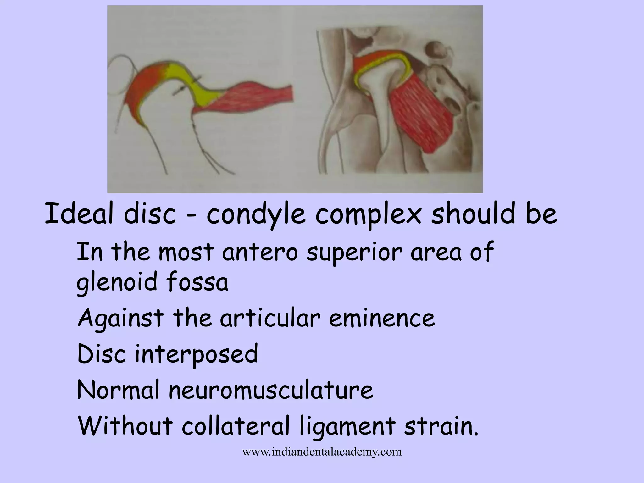
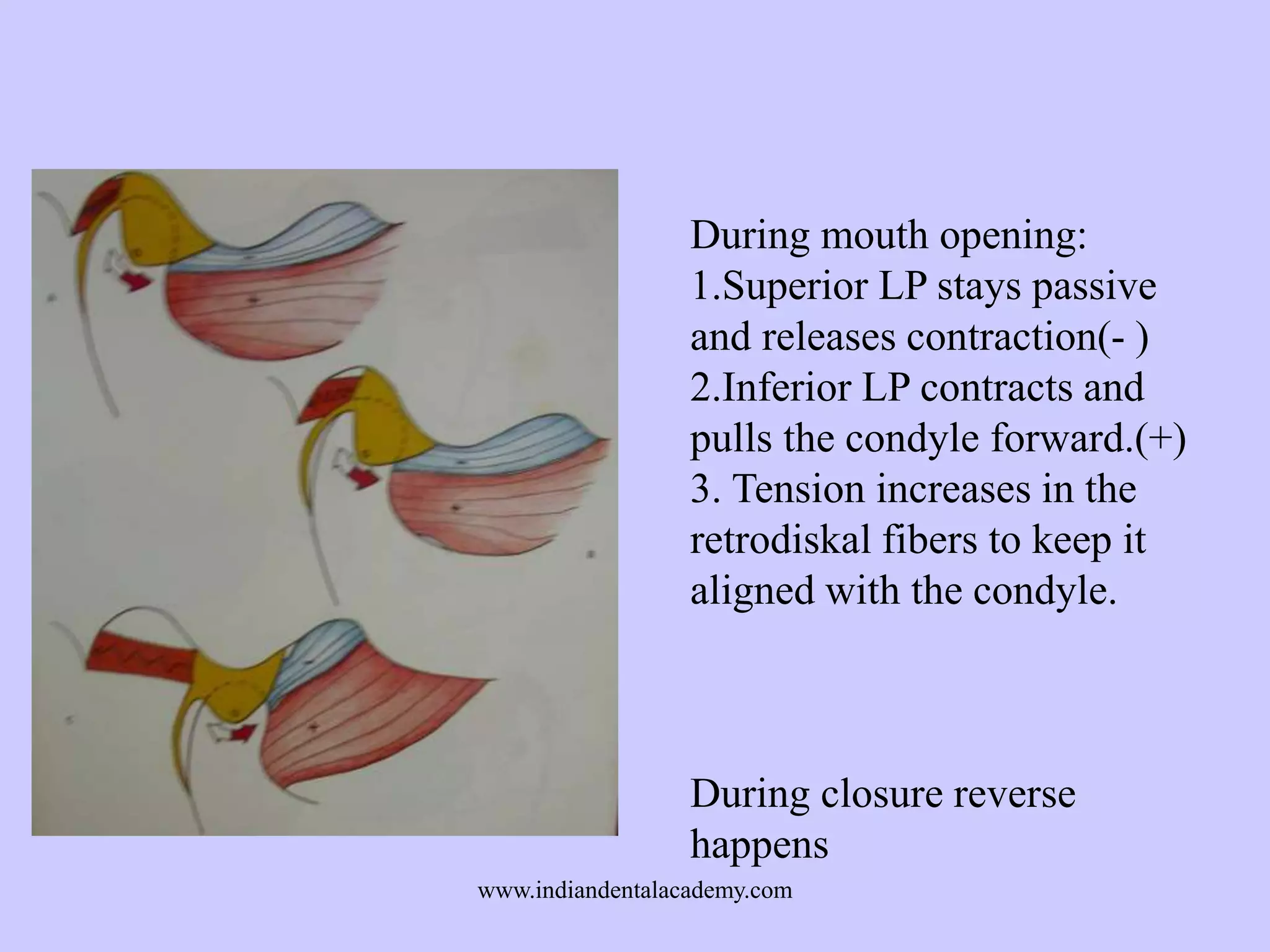
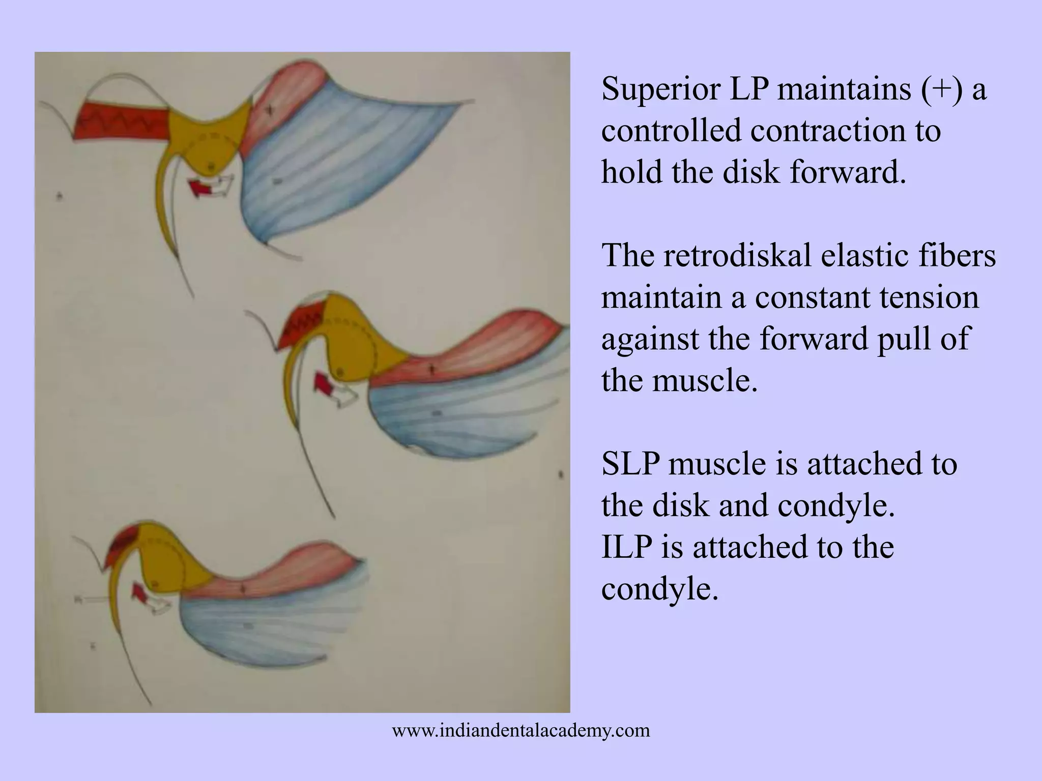
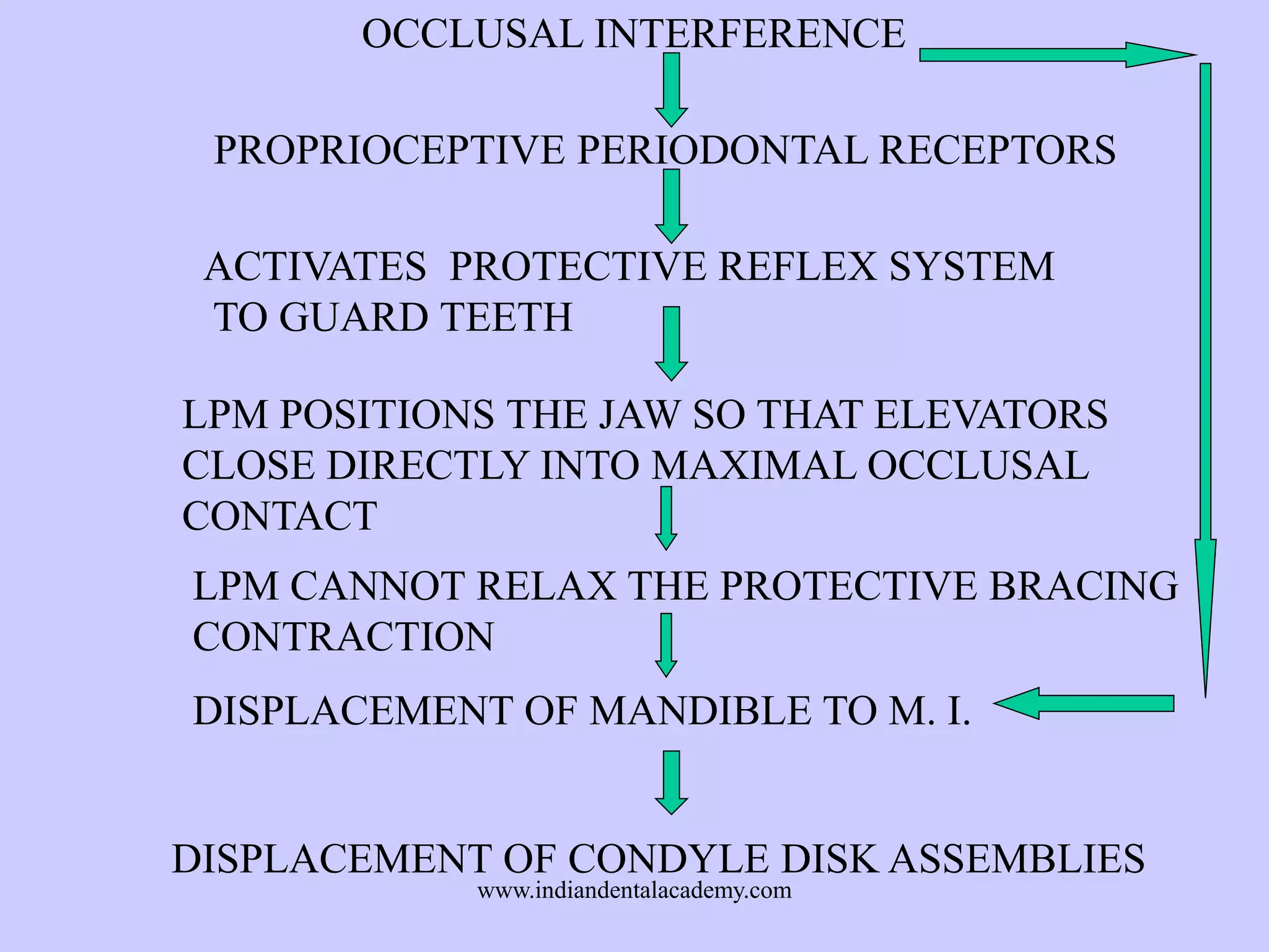
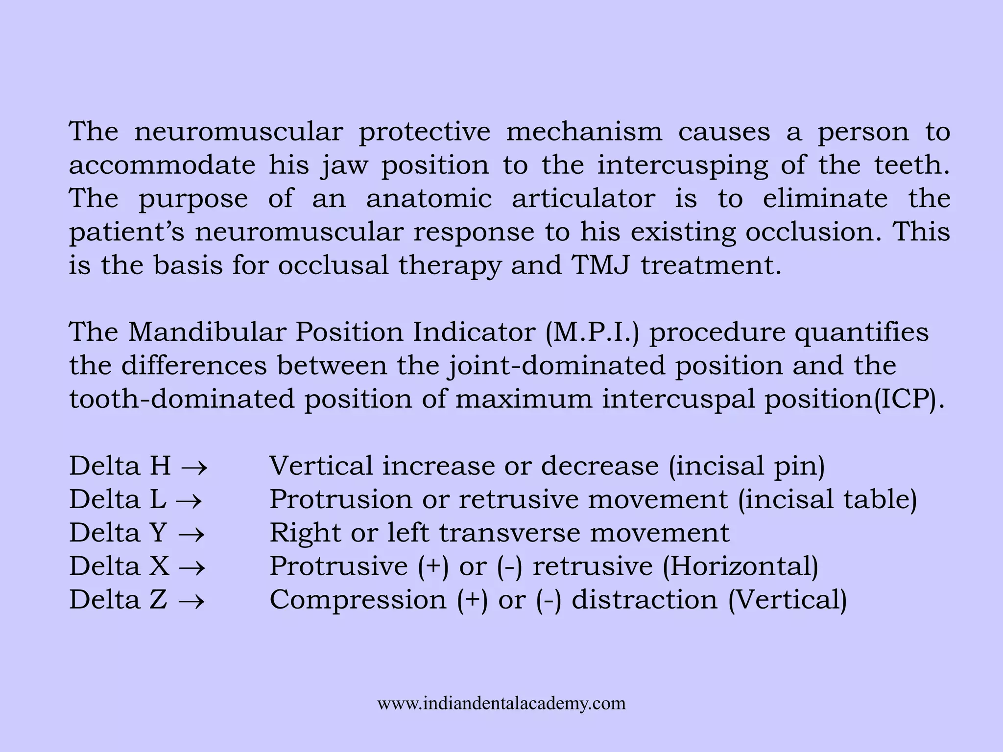
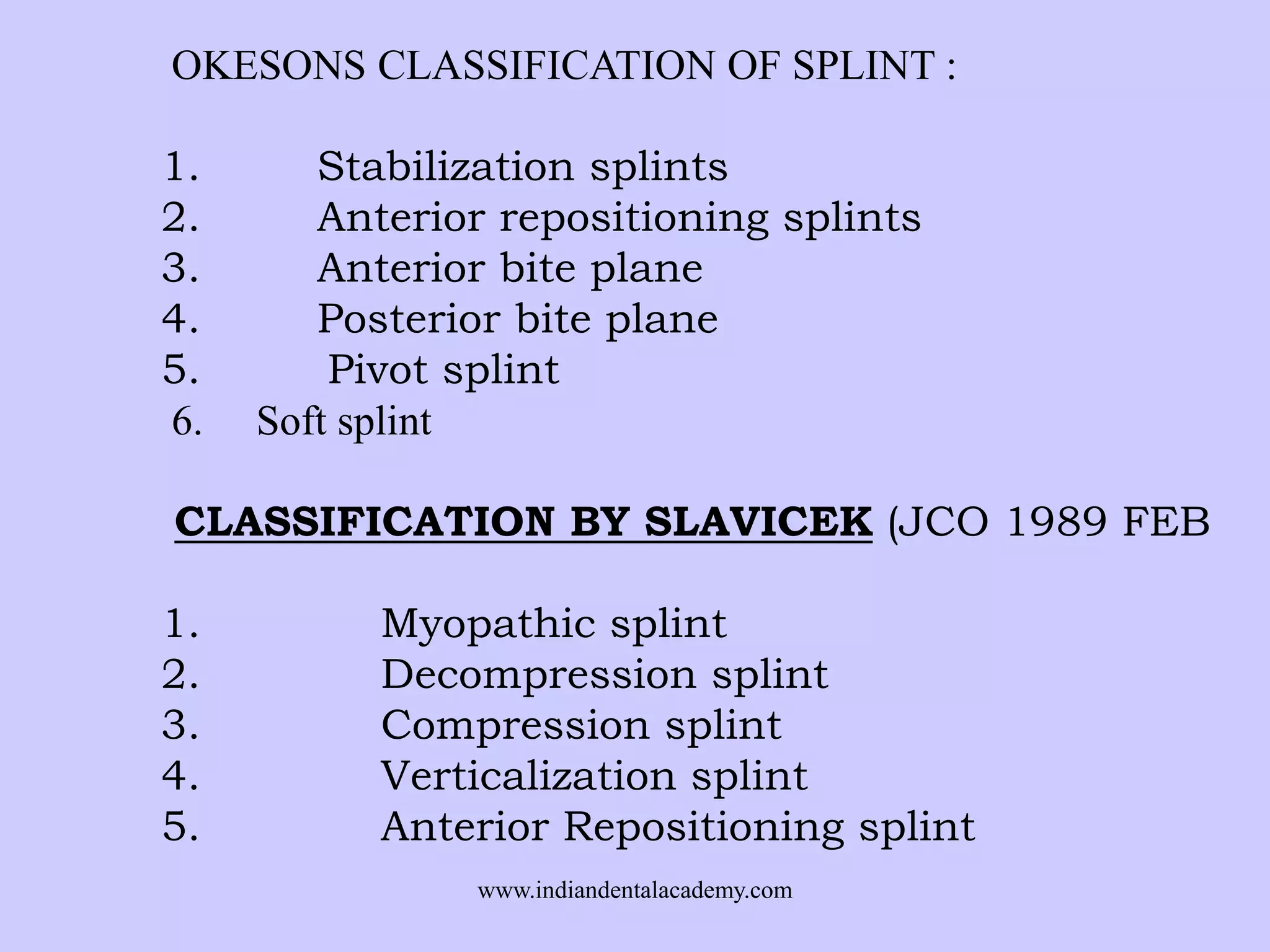
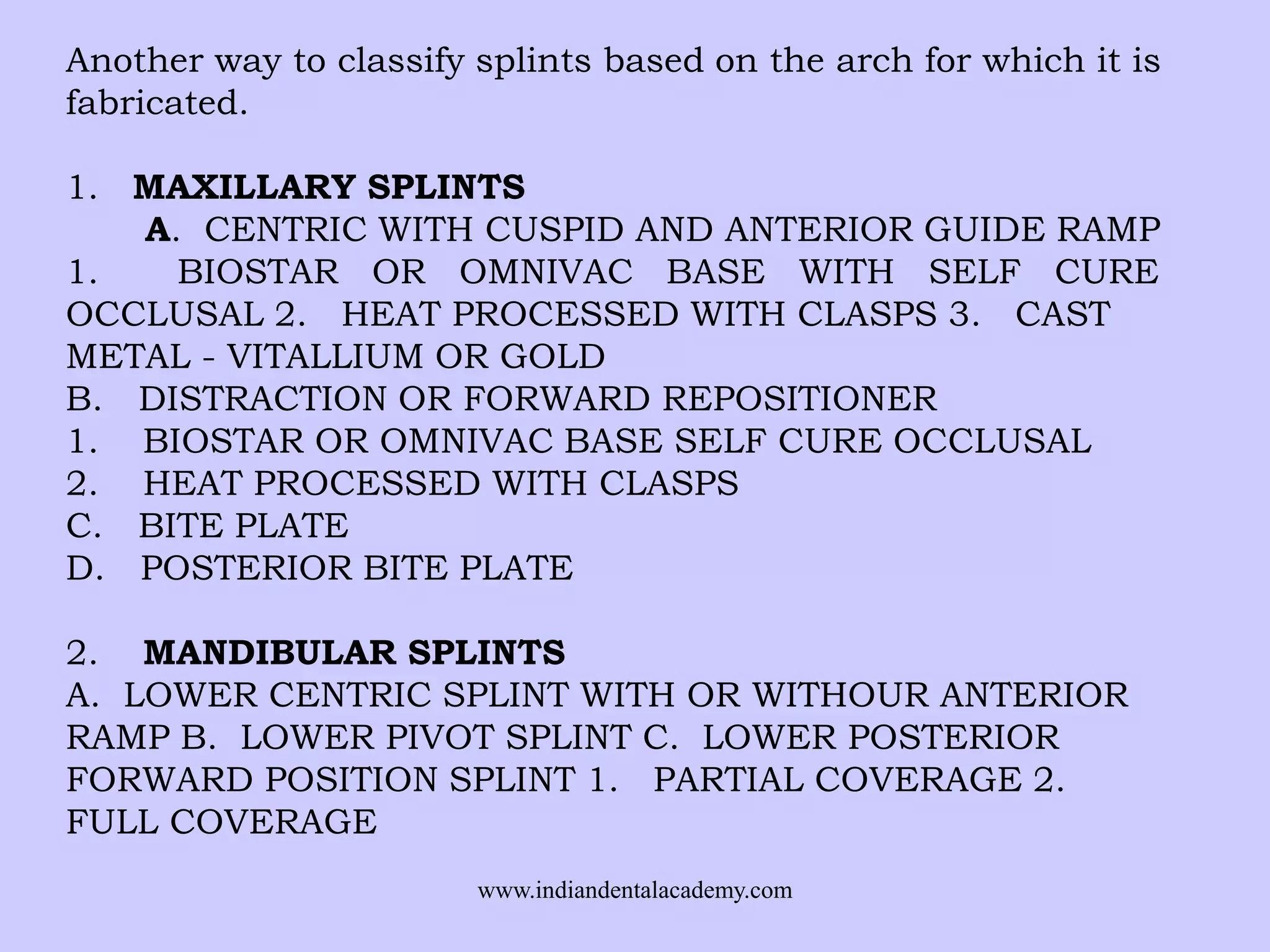
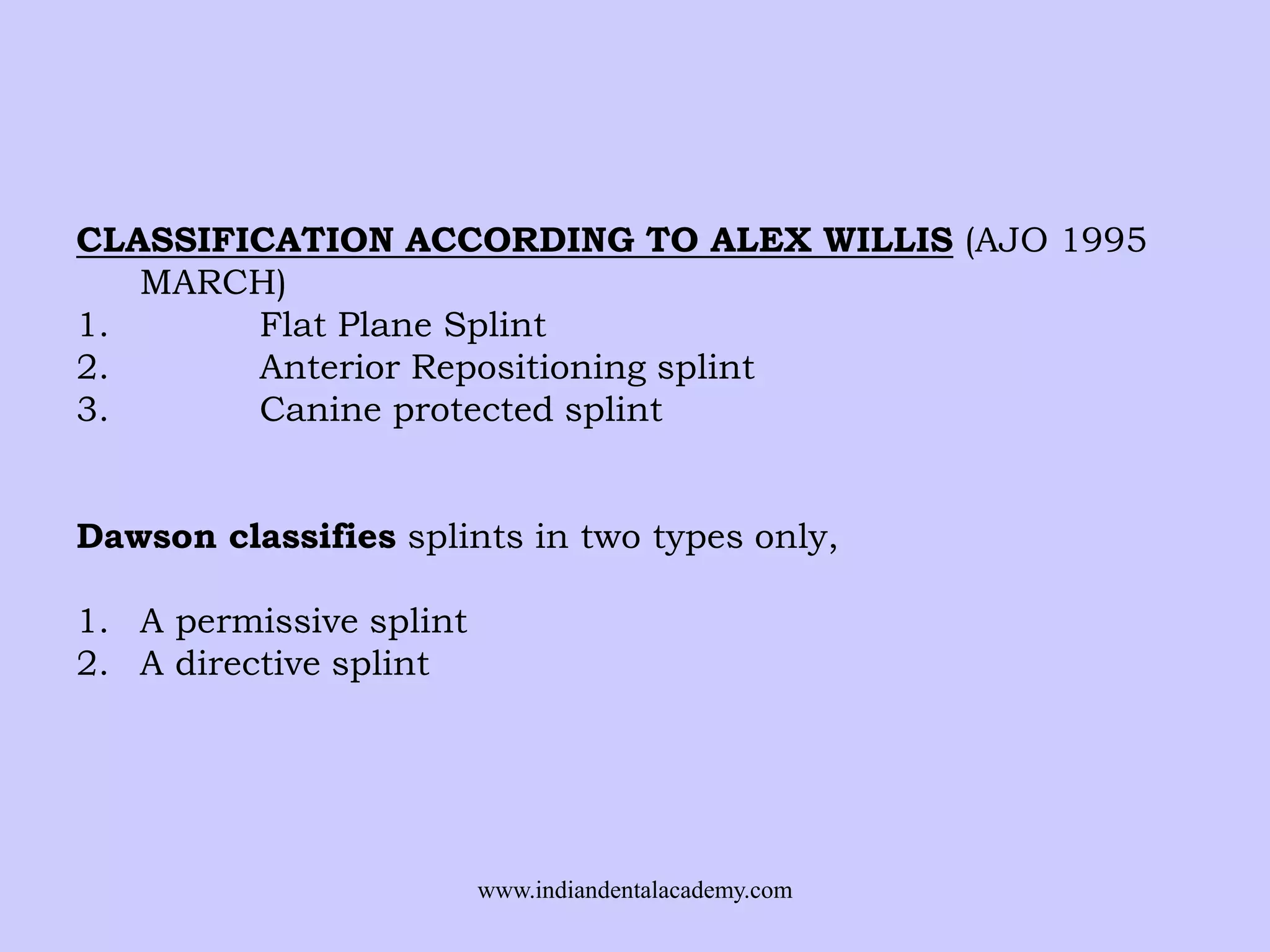
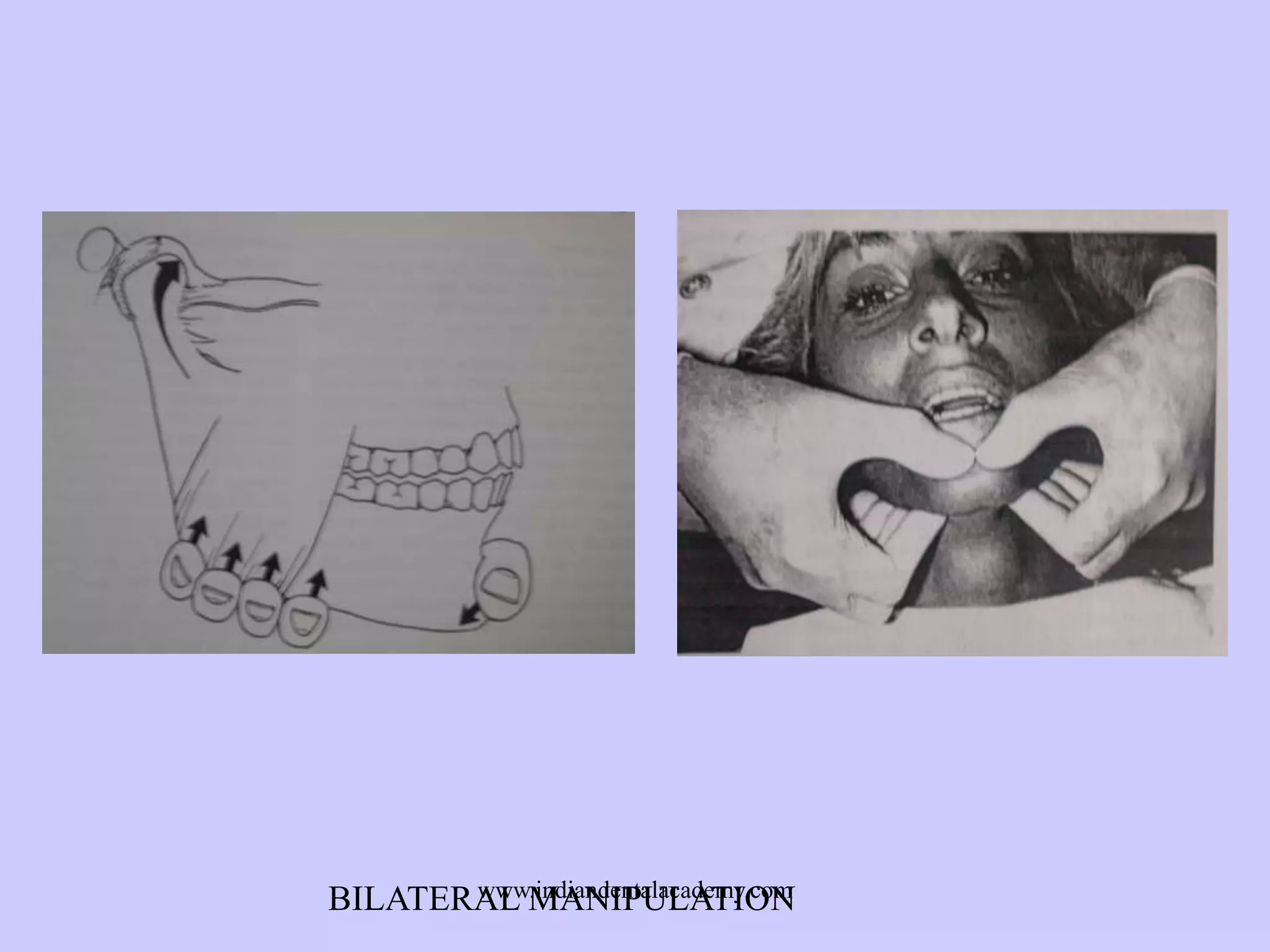
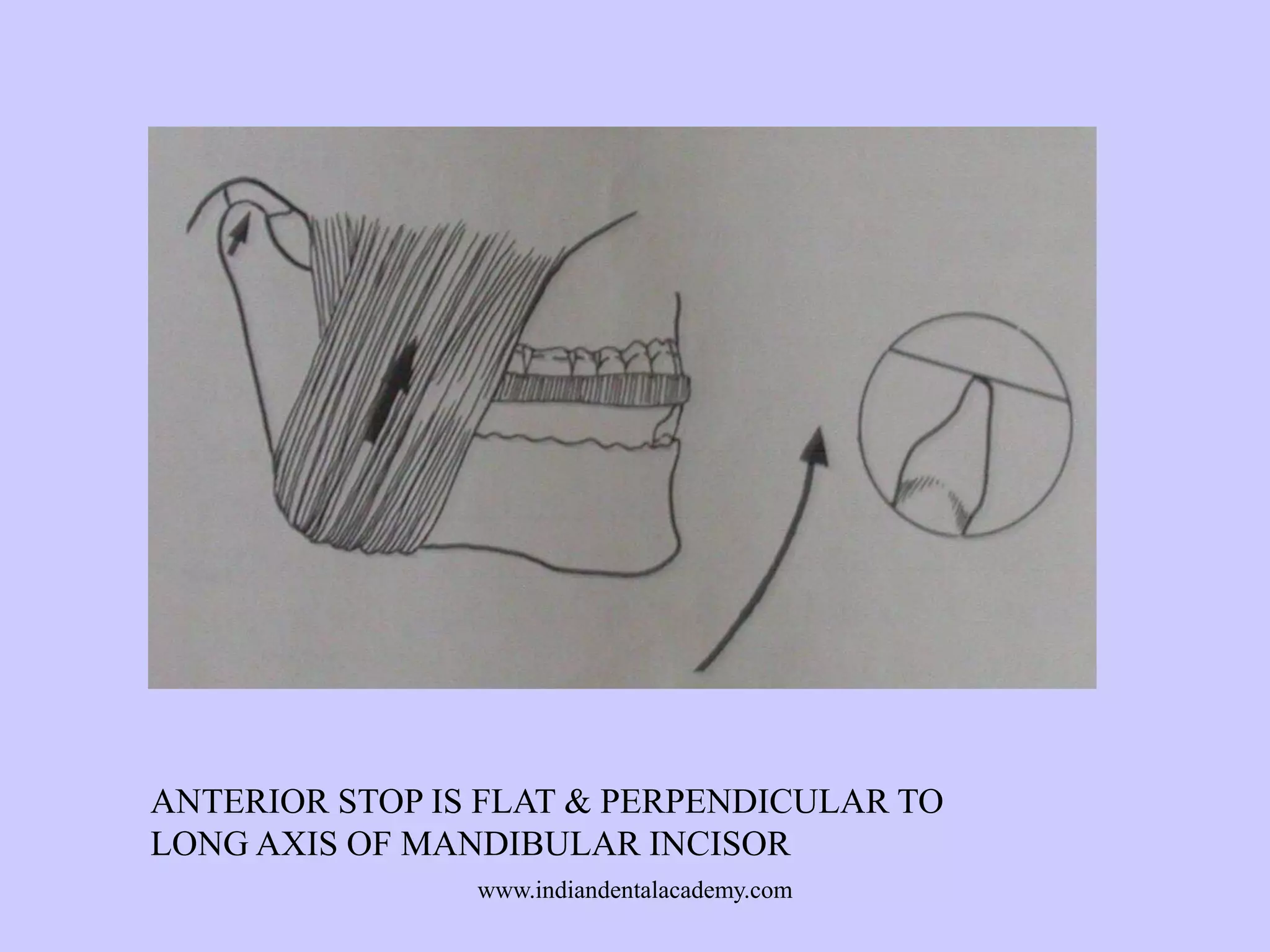
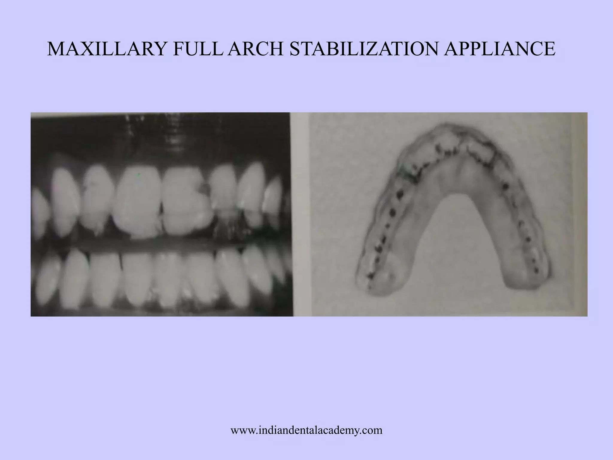
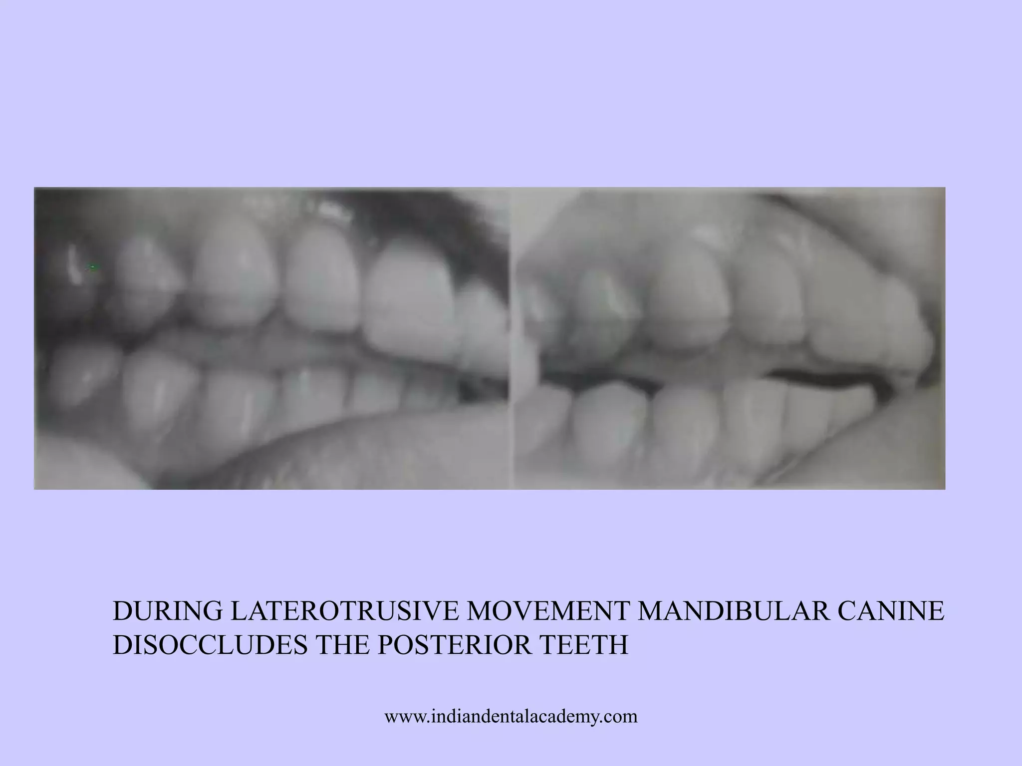
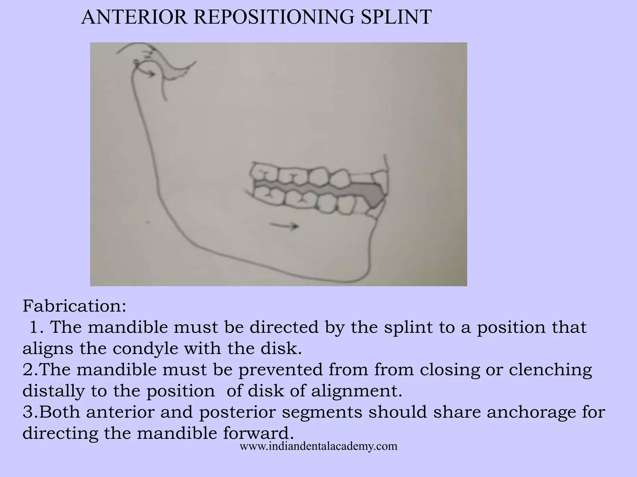
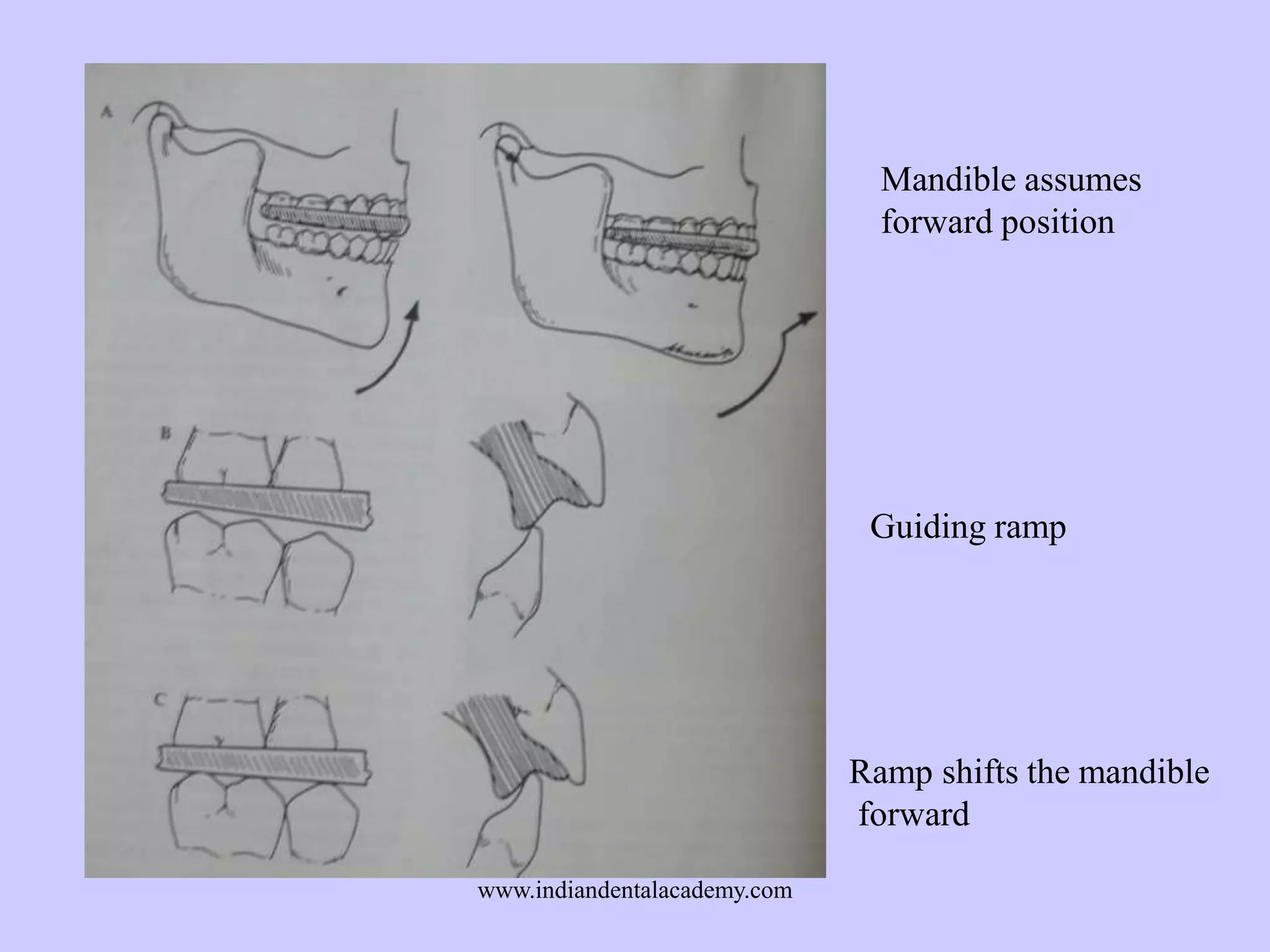
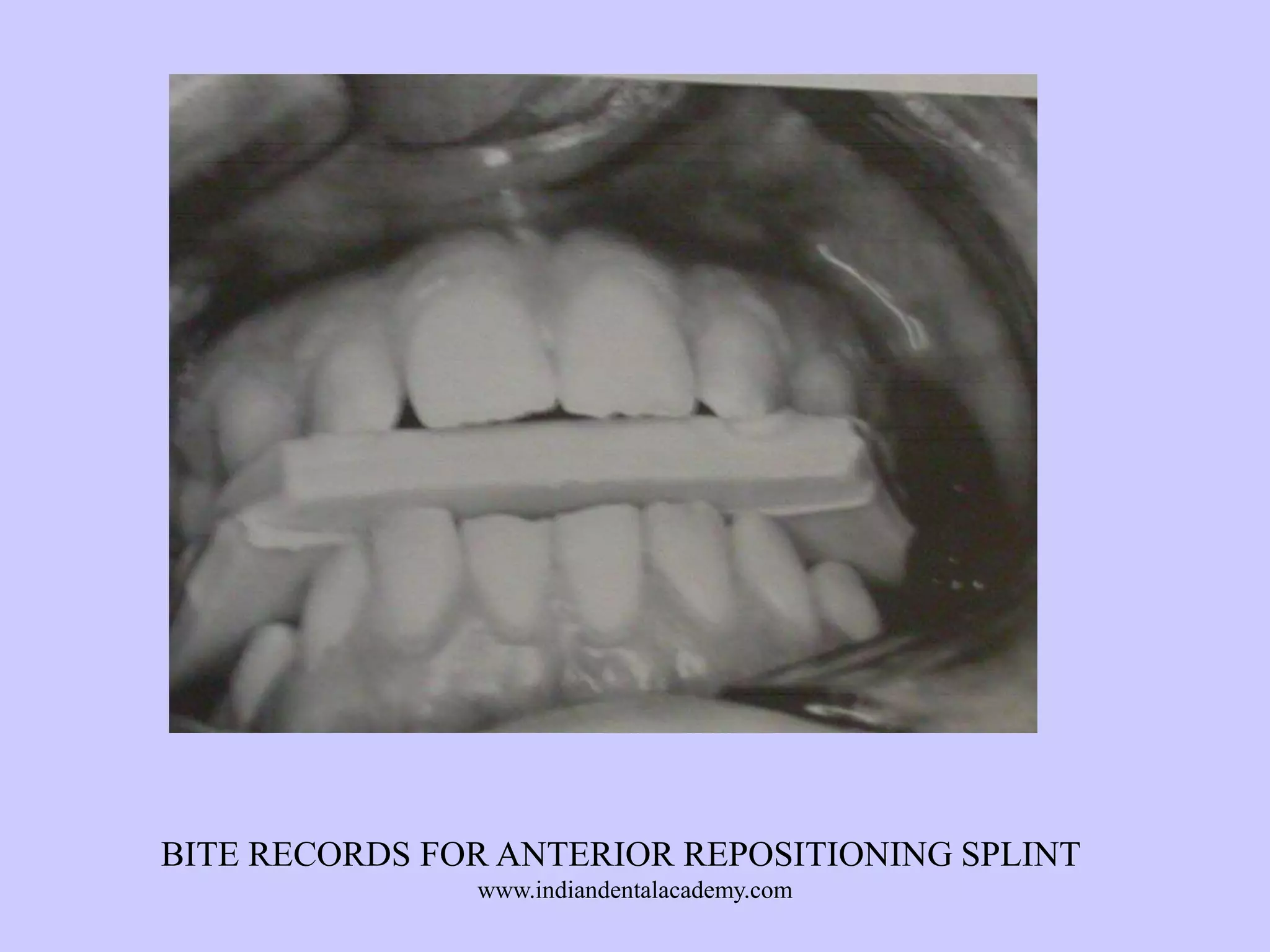
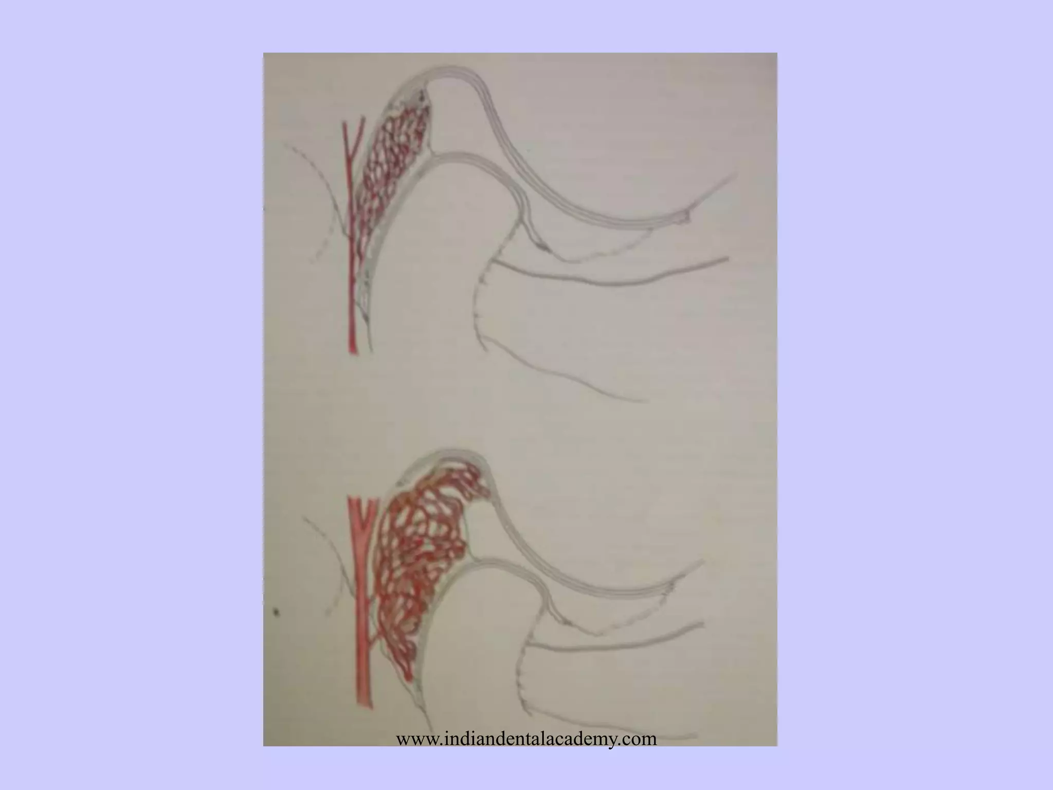
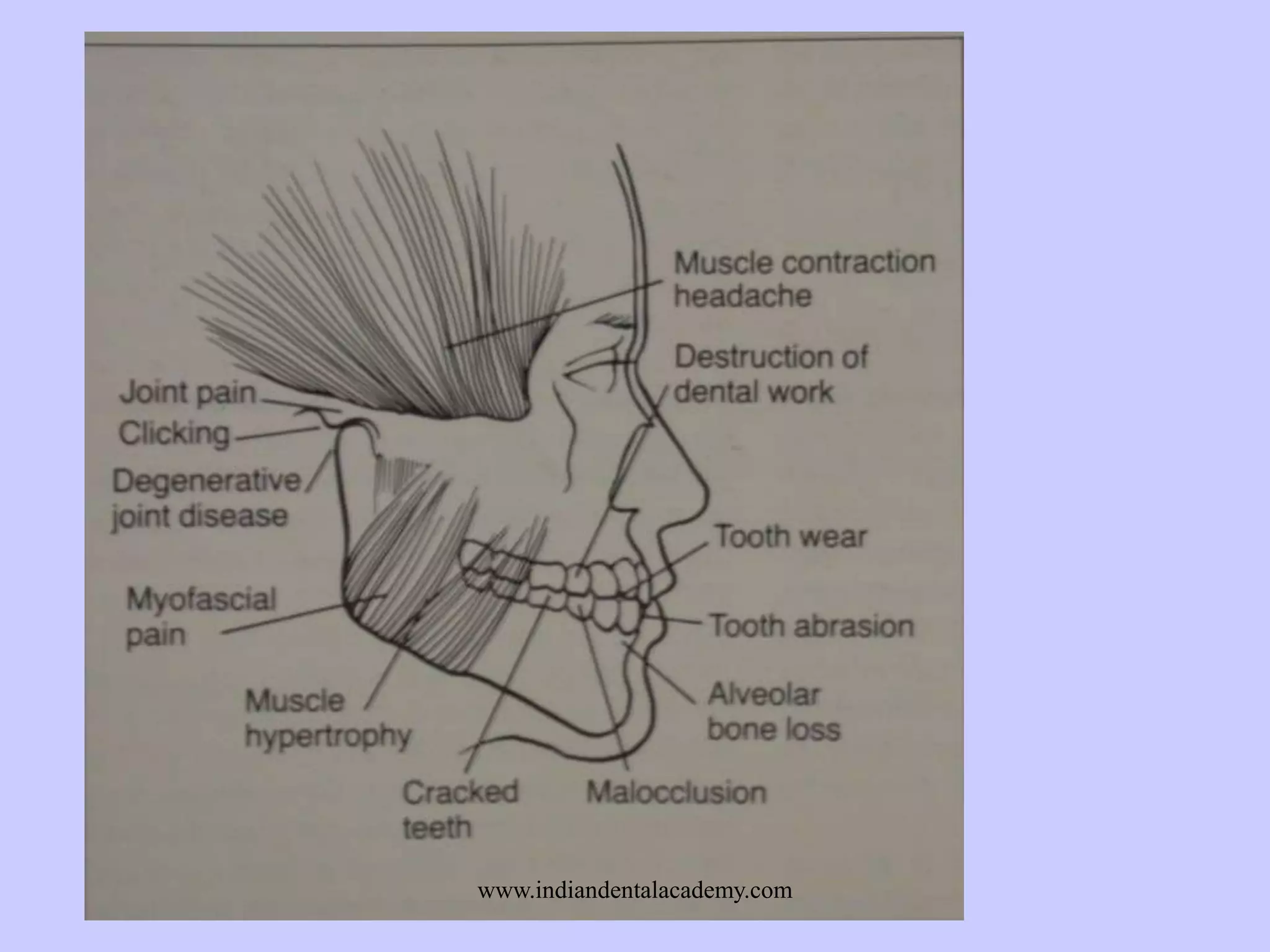
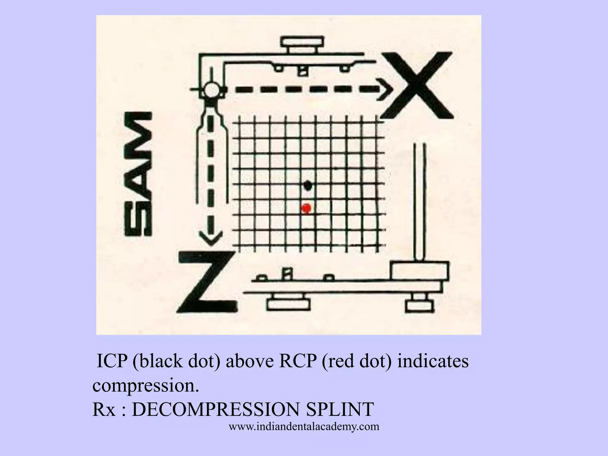
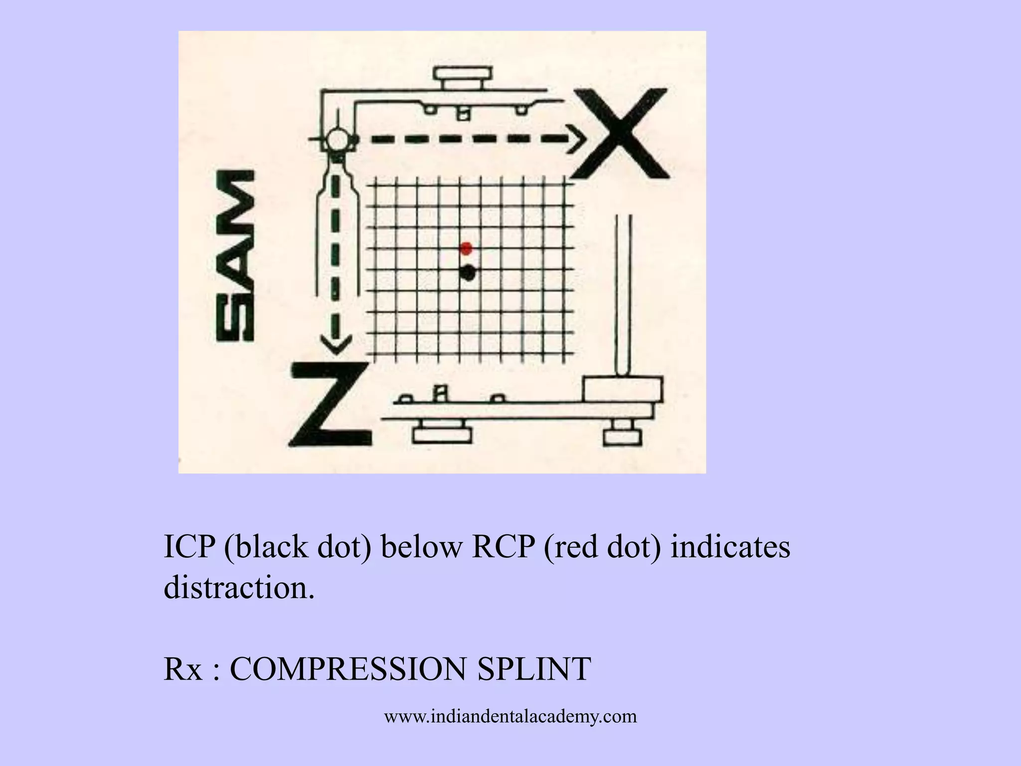
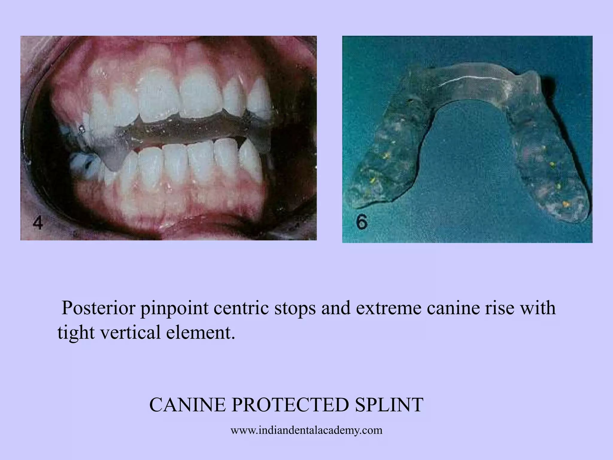
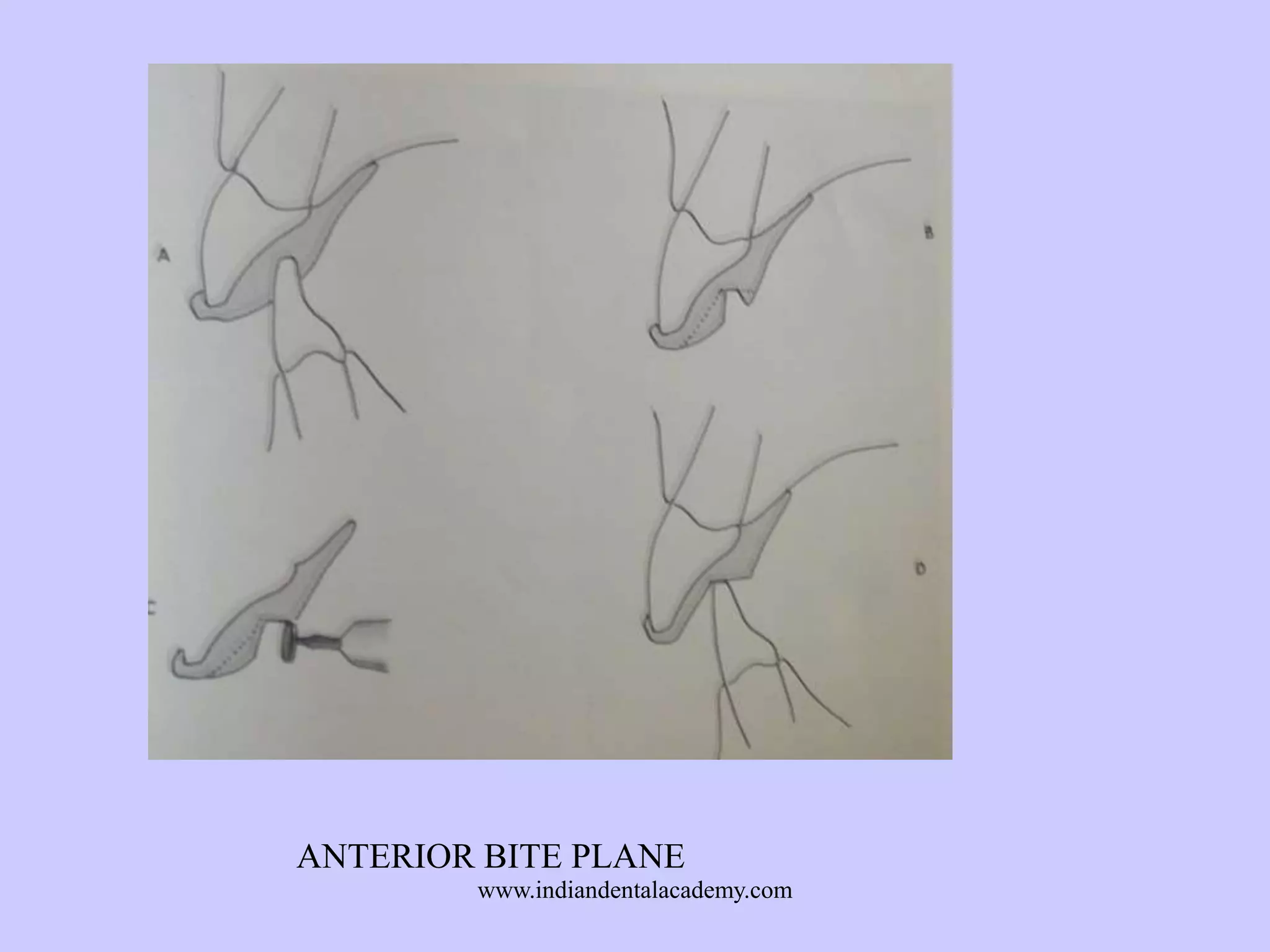
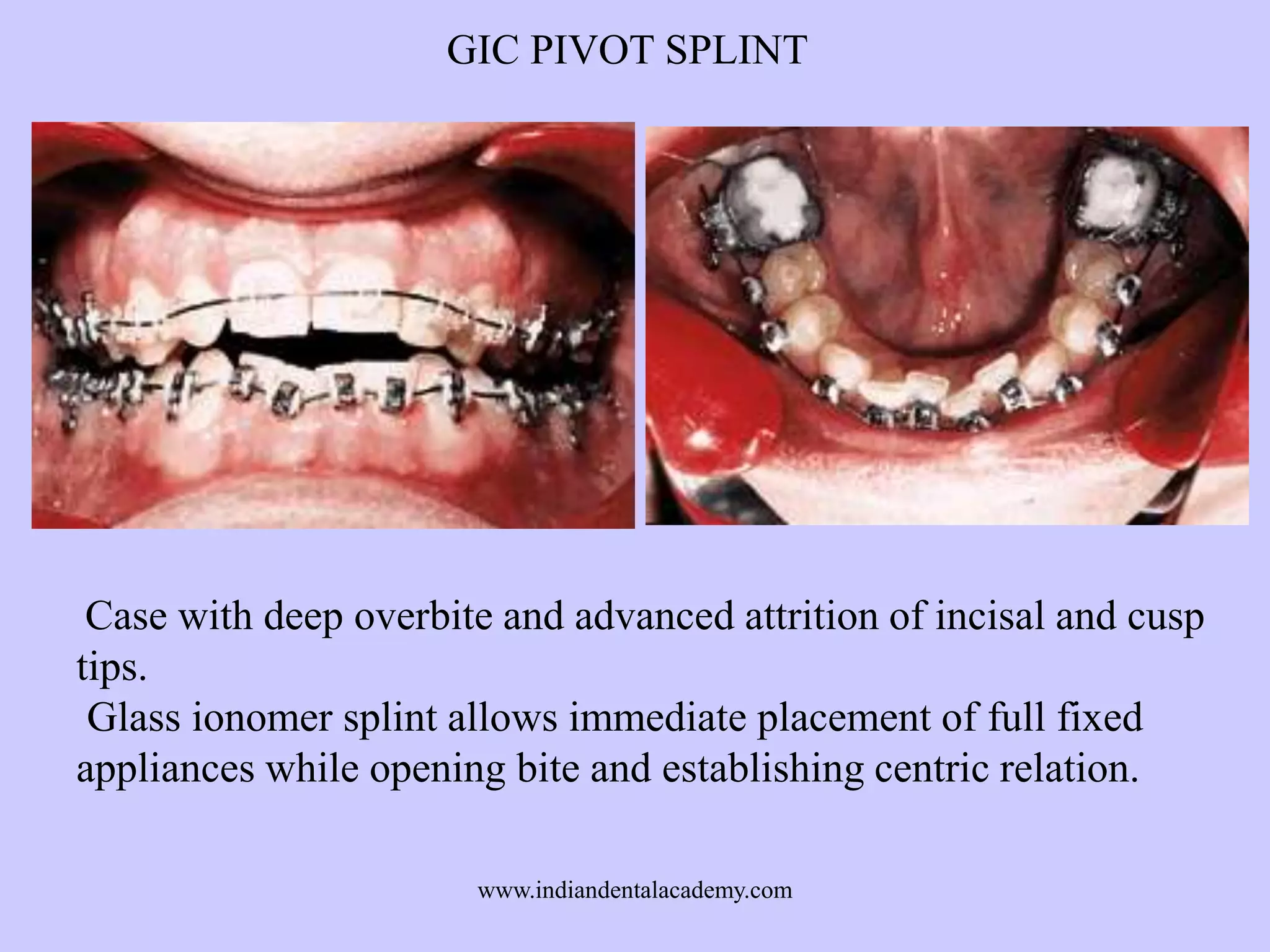
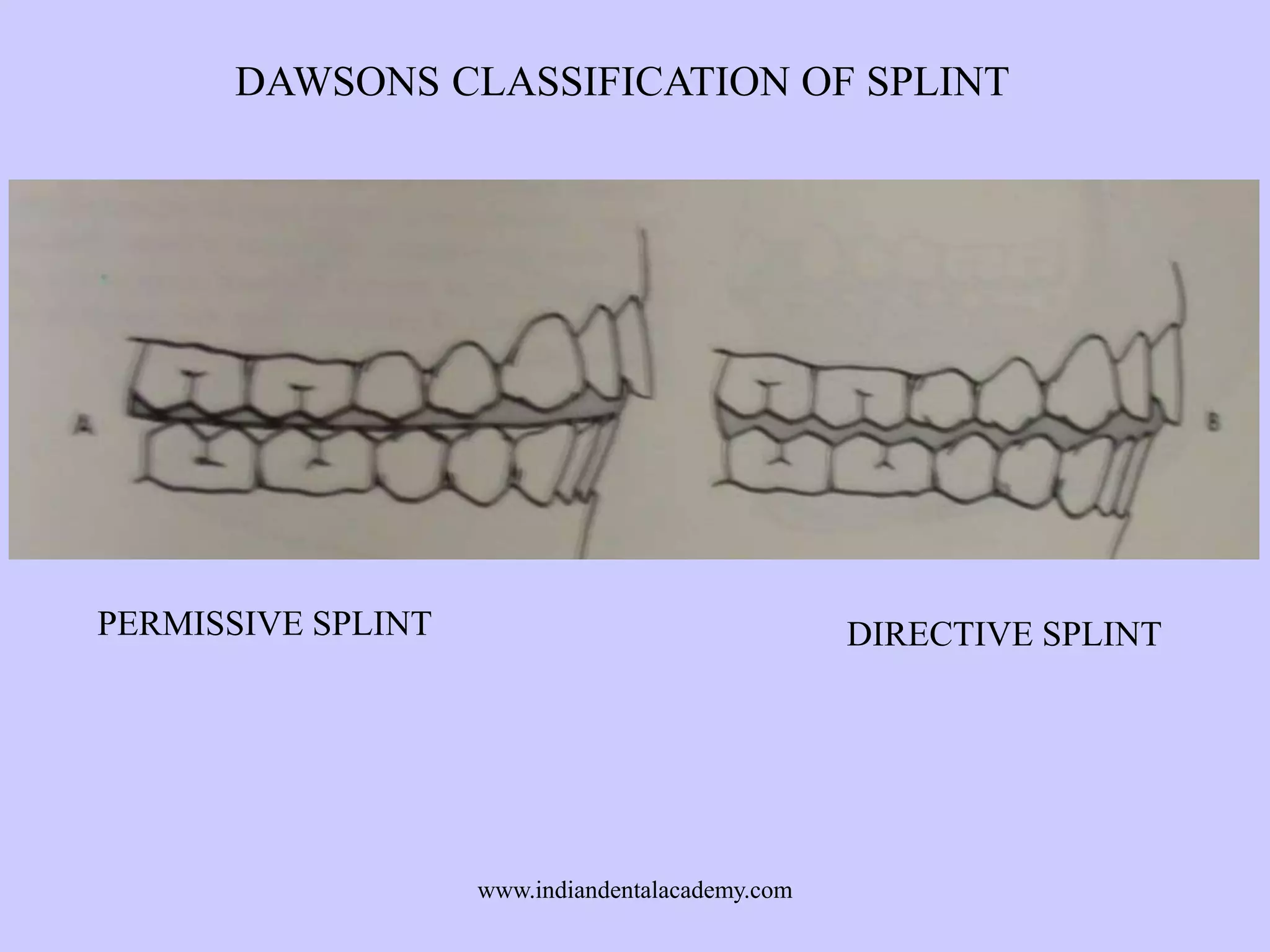
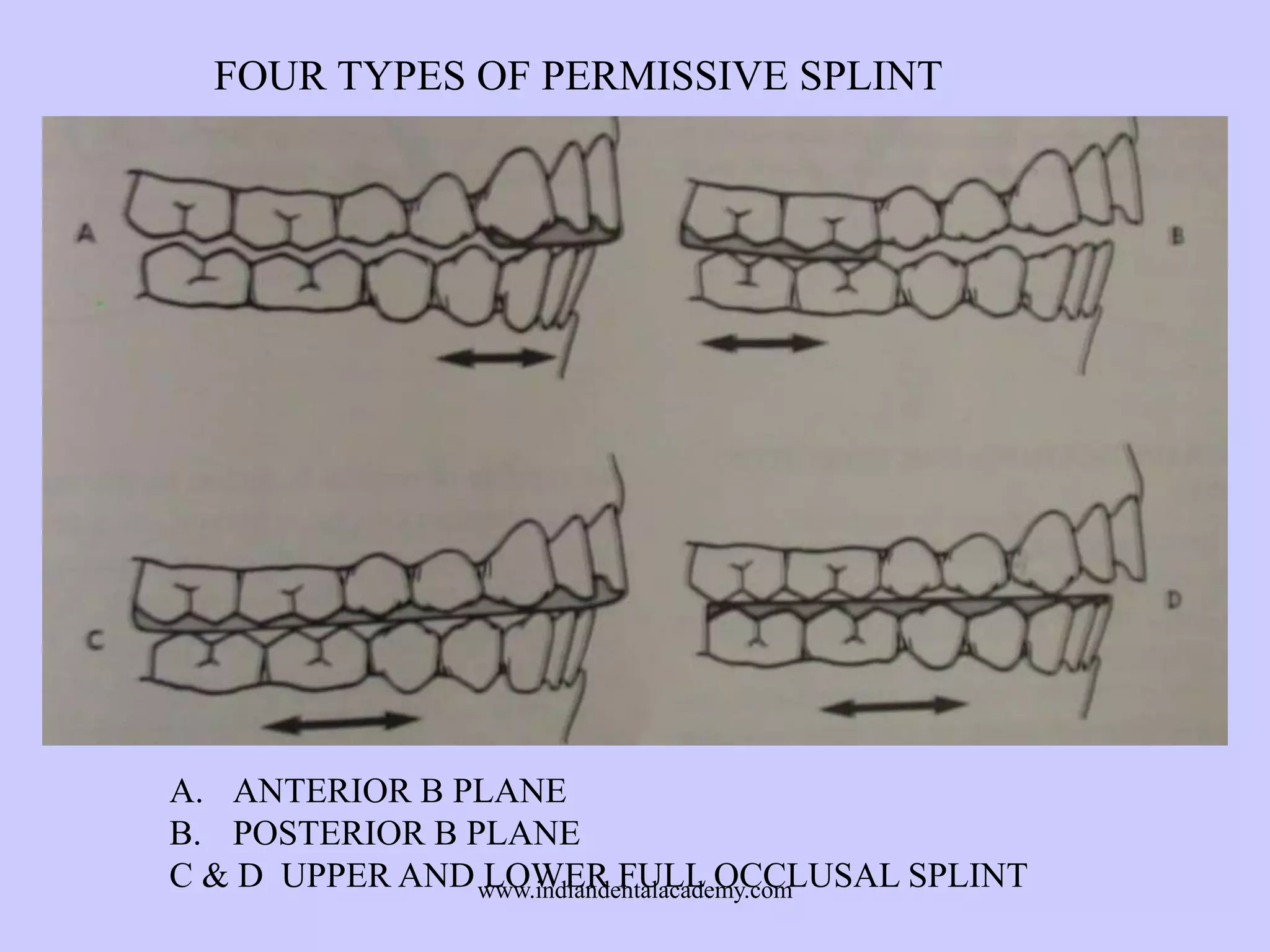
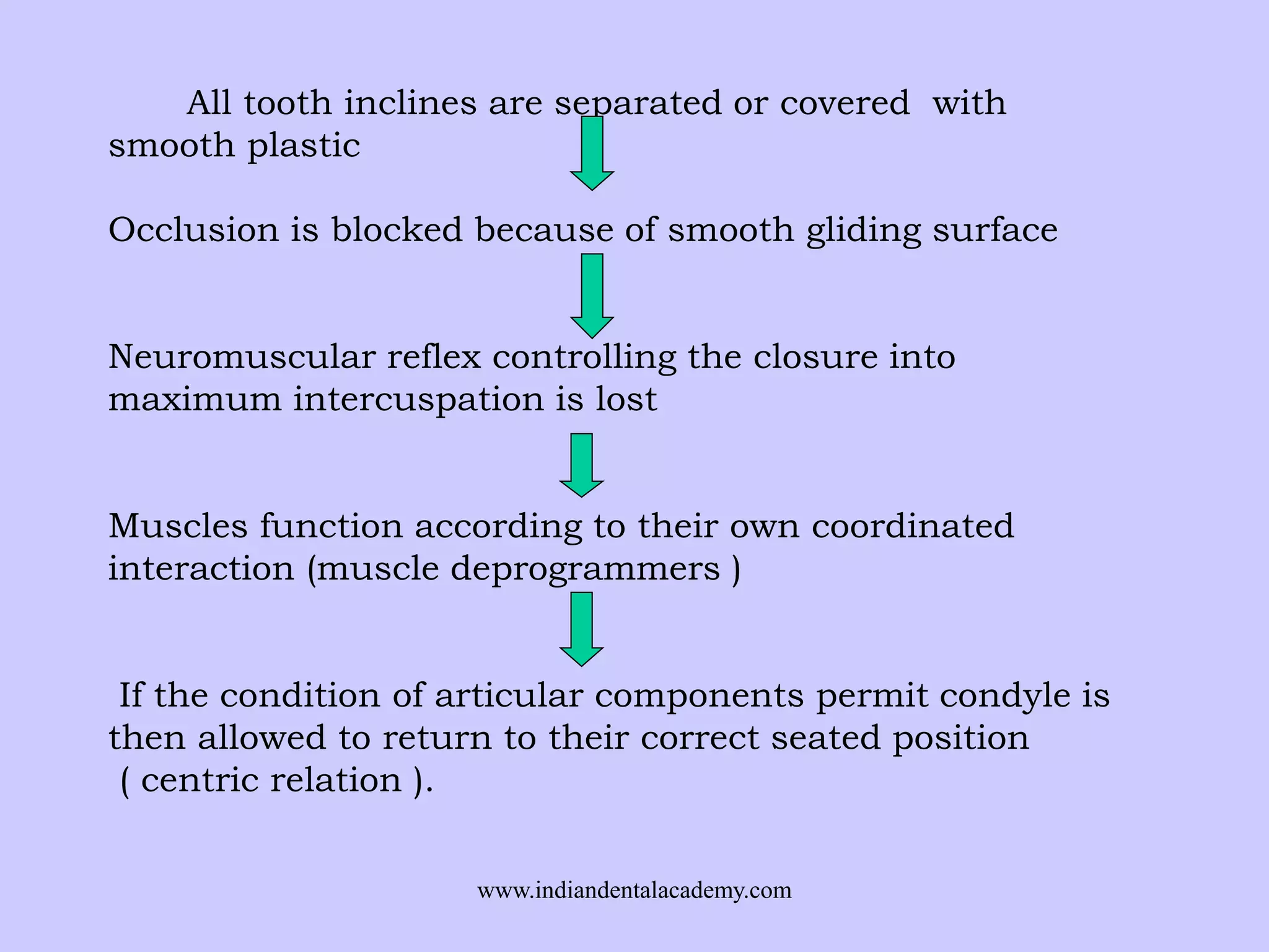
INDIAN DENTAL ACADEMY
Leader in continuing dental education
www.indiandentalacademy.com

www.indiandentalacademy.com
• An occlusal appliance, called a splint,
is a removable device, usually made
of hard acrylic that fits over the
occlusal and incisal surfaces of the
teeth in one arch, creating precise
occlusal contact with the teeth of the
opposing arch. It is commonly
referred to as a bite guard, night
guard, interocclusal appliance, or
orthopedic device.
www.indiandentalacademy.com
Ideal disc - condyle complex should be
In the most antero superior area of
glenoid fossa
Against the articular eminence
Disc interposed
Normal neuromusculature
Without collateral ligament strain.
www.indiandentalacademy.com
During mouth opening:
1.Superior LP stays passive
and releases contraction(- )
2.Inferior LP contracts and
pulls the condyle forward.(+)
3. Tension increases in the
retrodiskal fibers to keep it
aligned with the condyle.

During closure reverse
happens
www.indiandentalacademy.com
Superior LP maintains (+) a
controlled contraction to
hold the disk forward.
The retrodiskal elastic fibers
maintain a constant tension
against the forward pull of
the muscle.
SLP muscle is attached to
the disk and condyle.
ILP is attached to the
condyle.

www.indiandentalacademy.com
OCCLUSAL INTERFERENCE
PROPRIOCEPTIVE PERIODONTAL RECEPTORS

ACTIVATES PROTECTIVE REFLEX SYSTEM
TO GUARD TEETH
LPM POSITIONS THE JAW SO THAT ELEVATORS
CLOSE DIRECTLY INTO MAXIMAL OCCLUSAL
CONTACT
LPM CANNOT RELAX THE PROTECTIVE BRACING
CONTRACTION

DISPLACEMENT OF MANDIBLE TO M. I.

DISPLACEMENT OF CONDYLE DISK ASSEMBLIES
www.indiandentalacademy.com
WILLIAMSONS EXPERIMENT (JPD 1983)
WILLIAMSON DEMONSTRATED THE PRECISE EFFECT
OF OCCLUSAL INTERFERNCE ON MUSCLE
COORDINATION AND NORMAL MUSCLE ACTIVITY
Using EMG procedure, he showed that interfering contacts on
the posterior teeth in any eccentric position caused hyperactivity
of the elevator muscles.
But if anterior guidance was allowed to disclude all posterior
teeth from any contact other than CR, the elevator muscles either
stopped or reduced active contraction the moment posterior teeth
were discluded.
www.indiandentalacademy.com
HOW SPLINT WORKS ?

Five distinct theories explain it:
1.

Occlusal disengagement theory

2.

Vertical dimension theory

3.
Maxillomandibular realignment
theory
4.
TMJ repositioning theory
5.

Cognitive awareness theory
www.indiandentalacademy.com
The neuromuscular protective mechanism causes a person to
accommodate his jaw position to the intercusping of the teeth.
The purpose of an anatomic articulator is to eliminate the
patient’s neuromuscular response to his existing occlusion. This
is the basis for occlusal therapy and TMJ treatment.
The Mandibular Position Indicator (M.P.I.) procedure quantifies
the differences between the joint-dominated position and the
tooth-dominated position of maximum intercuspal position(ICP).
Delta
Delta
Delta
Delta
Delta

H
L
Y
X
Z

Vertical increase or decrease (incisal pin)
Protrusion or retrusive movement (incisal table)
Right or left transverse movement
Protrusive (+) or (-) retrusive (Horizontal)
Compression (+) or (-) distraction (Vertical)

www.indiandentalacademy.com
OKESONS CLASSIFICATION OF SPLINT :
1.
2.
3.
4.
5.
6.

Stabilization splints
Anterior repositioning splints
Anterior bite plane
Posterior bite plane
Pivot splint
Soft splint

CLASSIFICATION BY SLAVICEK (JCO 1989 FEB
1.
2.
3.
4.
5.

Myopathic splint
Decompression splint
Compression splint
Verticalization splint
Anterior Repositioning splint
www.indiandentalacademy.com
Another way to classify splints based on the arch for which it is
fabricated.
1. MAXILLARY SPLINTS
A. CENTRIC WITH CUSPID AND ANTERIOR GUIDE RAMP
1.
BIOSTAR OR OMNIVAC BASE WITH SELF CURE
OCCLUSAL 2. HEAT PROCESSED WITH CLASPS 3. CAST
METAL - VITALLIUM OR GOLD
B. DISTRACTION OR FORWARD REPOSITIONER
1. BIOSTAR OR OMNIVAC BASE SELF CURE OCCLUSAL
2. HEAT PROCESSED WITH CLASPS
C. BITE PLATE
D. POSTERIOR BITE PLATE
2. MANDIBULAR SPLINTS
A. LOWER CENTRIC SPLINT WITH OR WITHOUR ANTERIOR
RAMP B. LOWER PIVOT SPLINT C. LOWER POSTERIOR
FORWARD POSITION SPLINT 1. PARTIAL COVERAGE 2.
FULL COVERAGE
www.indiandentalacademy.com
CLASSIFICATION ACCORDING TO ALEX WILLIS (AJO 1995
MARCH)
1.
Flat Plane Splint
2.
Anterior Repositioning splint
3.
Canine protected splint
Dawson classifies splints in two types only,
1. A permissive splint
2. A directive splint

www.indiandentalacademy.com
BILATERAL MANIPULATION
www.indiandentalacademy.com
LEAF GAUGE
www.indiandentalacademy.com
MESIAL INCLINE

DISTAL INCLINE

www.indiandentalacademy.com
ANTERIOR STOP IS FLAT & PERPENDICULAR TO
LONG AXIS OF MANDIBULAR INCISOR
www.indiandentalacademy.com
MAXILLARY FULL ARCH STABILIZATION APPLIANCE

www.indiandentalacademy.com
DURING LATEROTRUSIVE MOVEMENT MANDIBULAR CANINE
DISOCCLUDES THE POSTERIOR TEETH
www.indiandentalacademy.com
DESIGN :
1.In centric relation all posterior mandibular
buccal cusps must contact on flat surfaces
with even force.
2. During protrusive movement, the
mandibular canines must contact the
appliance with even force.
3.In any lateral movement only the mandibular
canines should exhibit laterotrusive contact on
the appliance.
4.The mandibular posterior teeth must contact
the appliance only in centric relation closure.
5.In the alert feeding position, the posterior
teeth must contact the appliance more
www.indiandentalacademy.com
prominently than the anterior teeth.
ANTERIOR REPOSITIONING SPLINT

Fabrication:
1. The mandible must be directed by the splint to a position that
aligns the condyle with the disk.
2.The mandible must be prevented from from closing or clenching
distally to the position of disk of alignment.
3.Both anterior and posterior segments should share anchorage for
directing the mandible forward.
www.indiandentalacademy.com
Mandible assumes
forward position

Guiding ramp

Ramp shifts the mandible
forward
www.indiandentalacademy.com
BITE RECORDS FOR ANTERIOR REPOSITIONING SPLINT
www.indiandentalacademy.com
S SPLINT

www.indiandentalacademy.com
ICP (black dot) above RCP (red dot) indicates
compression.
Rx : DECOMPRESSION SPLINT
www.indiandentalacademy.com
ICP (black dot) below RCP (red dot) indicates
distraction.
Rx : COMPRESSION SPLINT
www.indiandentalacademy.com
Posterior pinpoint centric stops and extreme canine rise with
tight vertical element.

CANINE PROTECTED SPLINT
www.indiandentalacademy.com
ANTERIOR BITE PLANE
www.indiandentalacademy.com
1.
2.
3.
4.

POSTERIORS ARE INTRUDED BY ELEVATOR MUSCLES
UPPER INCISORS STARTS TO MOVE LINGUALLY
LOWER INCISORS MIGHT MOVE LABIALLY & SUPRAERUPT
LINGUALIZED UPPER INCISOR INTERFERE WITH ARC OF
CLOSURE & FORCE THE CONDYLE TO DISTALIZE
www.indiandentalacademy.com
POSTERIOR BITE PLANE & EFFECT AFTER 1 YEAR WEAR
www.indiandentalacademy.com
Bite Plane anchored in headgear tubes.
www.indiandentalacademy.com
Full Arch Maxillary Removable Bite Splint
www.indiandentalacademy.com
I. Inclined plane for the alignment of the upper front teeth.
II. Biting splint for the nivellisation of the compensating curve.
www.indiandentalacademy.com
POSTERIOR PIVOTING APPLIANCE LOADS THE CONDYLE
IT DOESNOT DISTRACT THE CONDYLE
www.indiandentalacademy.com
GIC PIVOT SPLIT

Four balanced points of maxillomandibular contact in centric relation:
two articular disc assemblies and
two glass ionomer padswww.indiandentalacademy.com
on lower first molars.
GIC PIVOT SPLINT

Case with deep overbite and advanced attrition of incisal and cusp
tips.
Glass ionomer splint allows immediate placement of full fixed
appliances while opening bite and establishing centric relation.
www.indiandentalacademy.com
DAWSONS CLASSIFICATION OF SPLINT

PERMISSIVE SPLINT

DIRECTIVE SPLINT

www.indiandentalacademy.com
FOUR TYPES OF PERMISSIVE SPLINT

A. ANTERIOR B PLANE
B. POSTERIOR B PLANE
C & D UPPER AND www.indiandentalacademy.com
LOWER FULL OCCLUSAL SPLINT
All tooth inclines are separated or covered with
smooth plastic
Occlusion is blocked because of smooth gliding surface
Neuromuscular reflex controlling the closure into
maximum intercuspation is lost
Muscles function according to their own coordinated
interaction (muscle deprogrammers )
If the condition of articular components permit condyle is
then allowed to return to their correct seated position
( centric relation ).
www.indiandentalacademy.com
Thank you
For more details please visit
www.indiandentalacademy.com

www.indiandentalacademy.com

Orthodontic splints /certified fixed orthodontic courses by Indian dental academy

  • 1.
    INDIAN DENTAL ACADEMY Leaderin continuing dental education www.indiandentalacademy.com www.indiandentalacademy.com
  • 2.
    • An occlusalappliance, called a splint, is a removable device, usually made of hard acrylic that fits over the occlusal and incisal surfaces of the teeth in one arch, creating precise occlusal contact with the teeth of the opposing arch. It is commonly referred to as a bite guard, night guard, interocclusal appliance, or orthopedic device. www.indiandentalacademy.com
  • 3.
    Ideal disc -condyle complex should be In the most antero superior area of glenoid fossa Against the articular eminence Disc interposed Normal neuromusculature Without collateral ligament strain. www.indiandentalacademy.com
  • 4.
    During mouth opening: 1.SuperiorLP stays passive and releases contraction(- ) 2.Inferior LP contracts and pulls the condyle forward.(+) 3. Tension increases in the retrodiskal fibers to keep it aligned with the condyle. During closure reverse happens www.indiandentalacademy.com
  • 5.
    Superior LP maintains(+) a controlled contraction to hold the disk forward. The retrodiskal elastic fibers maintain a constant tension against the forward pull of the muscle. SLP muscle is attached to the disk and condyle. ILP is attached to the condyle. www.indiandentalacademy.com
  • 6.
    OCCLUSAL INTERFERENCE PROPRIOCEPTIVE PERIODONTALRECEPTORS ACTIVATES PROTECTIVE REFLEX SYSTEM TO GUARD TEETH LPM POSITIONS THE JAW SO THAT ELEVATORS CLOSE DIRECTLY INTO MAXIMAL OCCLUSAL CONTACT LPM CANNOT RELAX THE PROTECTIVE BRACING CONTRACTION DISPLACEMENT OF MANDIBLE TO M. I. DISPLACEMENT OF CONDYLE DISK ASSEMBLIES www.indiandentalacademy.com
  • 7.
    WILLIAMSONS EXPERIMENT (JPD1983) WILLIAMSON DEMONSTRATED THE PRECISE EFFECT OF OCCLUSAL INTERFERNCE ON MUSCLE COORDINATION AND NORMAL MUSCLE ACTIVITY Using EMG procedure, he showed that interfering contacts on the posterior teeth in any eccentric position caused hyperactivity of the elevator muscles. But if anterior guidance was allowed to disclude all posterior teeth from any contact other than CR, the elevator muscles either stopped or reduced active contraction the moment posterior teeth were discluded. www.indiandentalacademy.com
  • 8.
    HOW SPLINT WORKS? Five distinct theories explain it: 1. Occlusal disengagement theory 2. Vertical dimension theory 3. Maxillomandibular realignment theory 4. TMJ repositioning theory 5. Cognitive awareness theory www.indiandentalacademy.com
  • 9.
    The neuromuscular protectivemechanism causes a person to accommodate his jaw position to the intercusping of the teeth. The purpose of an anatomic articulator is to eliminate the patient’s neuromuscular response to his existing occlusion. This is the basis for occlusal therapy and TMJ treatment. The Mandibular Position Indicator (M.P.I.) procedure quantifies the differences between the joint-dominated position and the tooth-dominated position of maximum intercuspal position(ICP). Delta Delta Delta Delta Delta H L Y X Z Vertical increase or decrease (incisal pin) Protrusion or retrusive movement (incisal table) Right or left transverse movement Protrusive (+) or (-) retrusive (Horizontal) Compression (+) or (-) distraction (Vertical) www.indiandentalacademy.com
  • 10.
    OKESONS CLASSIFICATION OFSPLINT : 1. 2. 3. 4. 5. 6. Stabilization splints Anterior repositioning splints Anterior bite plane Posterior bite plane Pivot splint Soft splint CLASSIFICATION BY SLAVICEK (JCO 1989 FEB 1. 2. 3. 4. 5. Myopathic splint Decompression splint Compression splint Verticalization splint Anterior Repositioning splint www.indiandentalacademy.com
  • 11.
    Another way toclassify splints based on the arch for which it is fabricated. 1. MAXILLARY SPLINTS A. CENTRIC WITH CUSPID AND ANTERIOR GUIDE RAMP 1. BIOSTAR OR OMNIVAC BASE WITH SELF CURE OCCLUSAL 2. HEAT PROCESSED WITH CLASPS 3. CAST METAL - VITALLIUM OR GOLD B. DISTRACTION OR FORWARD REPOSITIONER 1. BIOSTAR OR OMNIVAC BASE SELF CURE OCCLUSAL 2. HEAT PROCESSED WITH CLASPS C. BITE PLATE D. POSTERIOR BITE PLATE 2. MANDIBULAR SPLINTS A. LOWER CENTRIC SPLINT WITH OR WITHOUR ANTERIOR RAMP B. LOWER PIVOT SPLINT C. LOWER POSTERIOR FORWARD POSITION SPLINT 1. PARTIAL COVERAGE 2. FULL COVERAGE www.indiandentalacademy.com
  • 12.
    CLASSIFICATION ACCORDING TOALEX WILLIS (AJO 1995 MARCH) 1. Flat Plane Splint 2. Anterior Repositioning splint 3. Canine protected splint Dawson classifies splints in two types only, 1. A permissive splint 2. A directive splint www.indiandentalacademy.com
  • 13.
  • 14.
  • 15.
  • 16.
  • 17.
    ANTERIOR STOP ISFLAT & PERPENDICULAR TO LONG AXIS OF MANDIBULAR INCISOR www.indiandentalacademy.com
  • 18.
    MAXILLARY FULL ARCHSTABILIZATION APPLIANCE www.indiandentalacademy.com
  • 19.
    DURING LATEROTRUSIVE MOVEMENTMANDIBULAR CANINE DISOCCLUDES THE POSTERIOR TEETH www.indiandentalacademy.com
  • 20.
    DESIGN : 1.In centricrelation all posterior mandibular buccal cusps must contact on flat surfaces with even force. 2. During protrusive movement, the mandibular canines must contact the appliance with even force. 3.In any lateral movement only the mandibular canines should exhibit laterotrusive contact on the appliance. 4.The mandibular posterior teeth must contact the appliance only in centric relation closure. 5.In the alert feeding position, the posterior teeth must contact the appliance more www.indiandentalacademy.com prominently than the anterior teeth.
  • 21.
    ANTERIOR REPOSITIONING SPLINT Fabrication: 1.The mandible must be directed by the splint to a position that aligns the condyle with the disk. 2.The mandible must be prevented from from closing or clenching distally to the position of disk of alignment. 3.Both anterior and posterior segments should share anchorage for directing the mandible forward. www.indiandentalacademy.com
  • 22.
    Mandible assumes forward position Guidingramp Ramp shifts the mandible forward www.indiandentalacademy.com
  • 23.
    BITE RECORDS FORANTERIOR REPOSITIONING SPLINT www.indiandentalacademy.com
  • 24.
  • 25.
  • 26.
  • 27.
  • 28.
  • 29.
    ICP (black dot)above RCP (red dot) indicates compression. Rx : DECOMPRESSION SPLINT www.indiandentalacademy.com
  • 30.
    ICP (black dot)below RCP (red dot) indicates distraction. Rx : COMPRESSION SPLINT www.indiandentalacademy.com
  • 31.
    Posterior pinpoint centricstops and extreme canine rise with tight vertical element. CANINE PROTECTED SPLINT www.indiandentalacademy.com
  • 32.
  • 33.
  • 34.
    1. 2. 3. 4. POSTERIORS ARE INTRUDEDBY ELEVATOR MUSCLES UPPER INCISORS STARTS TO MOVE LINGUALLY LOWER INCISORS MIGHT MOVE LABIALLY & SUPRAERUPT LINGUALIZED UPPER INCISOR INTERFERE WITH ARC OF CLOSURE & FORCE THE CONDYLE TO DISTALIZE www.indiandentalacademy.com
  • 35.
    POSTERIOR BITE PLANE& EFFECT AFTER 1 YEAR WEAR www.indiandentalacademy.com
  • 36.
    Bite Plane anchoredin headgear tubes. www.indiandentalacademy.com
  • 37.
    Full Arch MaxillaryRemovable Bite Splint www.indiandentalacademy.com
  • 38.
    I. Inclined planefor the alignment of the upper front teeth. II. Biting splint for the nivellisation of the compensating curve. www.indiandentalacademy.com
  • 39.
    POSTERIOR PIVOTING APPLIANCELOADS THE CONDYLE IT DOESNOT DISTRACT THE CONDYLE www.indiandentalacademy.com
  • 40.
    GIC PIVOT SPLIT Fourbalanced points of maxillomandibular contact in centric relation: two articular disc assemblies and two glass ionomer padswww.indiandentalacademy.com on lower first molars.
  • 41.
    GIC PIVOT SPLINT Casewith deep overbite and advanced attrition of incisal and cusp tips. Glass ionomer splint allows immediate placement of full fixed appliances while opening bite and establishing centric relation. www.indiandentalacademy.com
  • 42.
    DAWSONS CLASSIFICATION OFSPLINT PERMISSIVE SPLINT DIRECTIVE SPLINT www.indiandentalacademy.com
  • 43.
    FOUR TYPES OFPERMISSIVE SPLINT A. ANTERIOR B PLANE B. POSTERIOR B PLANE C & D UPPER AND www.indiandentalacademy.com LOWER FULL OCCLUSAL SPLINT
  • 44.
    All tooth inclinesare separated or covered with smooth plastic Occlusion is blocked because of smooth gliding surface Neuromuscular reflex controlling the closure into maximum intercuspation is lost Muscles function according to their own coordinated interaction (muscle deprogrammers ) If the condition of articular components permit condyle is then allowed to return to their correct seated position ( centric relation ). www.indiandentalacademy.com
  • 45.
  • 46.
  • 47.
  • 48.
  • 49.
  • 50.
  • 51.
  • 52.
  • 53.
  • 54.
  • 55.
  • 56.
  • 57.
  • 58.
  • 59.
  • 60.
  • 61.
    Thank you For moredetails please visit www.indiandentalacademy.com www.indiandentalacademy.com
  • 62.