Download to read offline
![Orthop Rheumatol Open Access J 1(2): OROAJ.MS.ID.555557(2015) 001
and extends down the shaft of the femur for about 10 cm. The
gluteal fascia and the ilio-tibial tract are exposed; the deep fascia
Introduction
The Posterior Approach is the most commonly used
approaches to the Hip Joint for Endo prosthesis, total Joint
replacement or Revision Hip Surgery wherein it gives excellent
visibility to the entire joint, when compared to other approaches
to the Hip Joint [4]. The author’s original paper [5] written 35
years ago presented an original technique designed after taking
into consideration the surgical anatomy of the Hip Joint, where
by the posterior overhanging part of the greater trochanter was
osteotomised to include the short lateral rotators along with the
posterior one-thirds of the gluteus medius and the capsule of the
HipJoint, which was then turnedback as a single flap to exposethe
acetabulum in detail along with a bloodless exposure. This was
also confirmed before any clinical application of this approach
on cadavers, which concluded greater stability as compared with
routine suture or reattachment of the short lateral rotators
Technique and Introduction
In all, many approaches to the Hip Joint are described in
literature. The author’s original technique was implemented
after a detailed cadaveric study (Figures 1-3), where the forces
required to dislocate the Hip Joint was considerably more when
compared to the routine suture or reattachment of the short
lateral rotators.
Clinical Technique
The patient is placed on the sound side. The skin incision
extends from just distal and lateral to the posterior superior
iliac spine towards the lateral edge of the greater trochanter,
with a curve in the direction of the fibres of gluteus maximus,
K Mohan lyer*
Orthopaedic Surgeon, India
Submission: September 26, 2015; Published: October 08, 2015
*Corresponding author: K Mohan lyer, Senior Consultant, Orthopaedic Surgeon, Flat No: 120/H-2K, First Floor, Kailash Apartments, 8th Main
Road, Malleswaram, Bangalore, 560003, Karnataka State, India, Tel: +919632683264; Email:
Modified Posterior Approach to the Hip
Joint
Mini Review
Volume 1 Issue 2 - 2015
Orthop Rheumatol Open Access J
Copyright © All rights are reserved by K Mohan lyer
Abstract
The Modification offers greater visibility and decreased blood loss to the Hip Joint, there by conferring greater stability posteriorly as
compared with the conventional Posterior Approach as described by Austin Moore in 1957.This Modification was devised at a time when
the cause of dislocation was being blamed on the Posterior Approach to the Hip Joint [1,2] In this Approach, since bone is attached to bone, it
confers greater stability than an ordinary suture through soft tissues and hence reduces dislocation of the Hip Joint [3].
Keywords: Trochanteric Osteotomy; Dislocation.
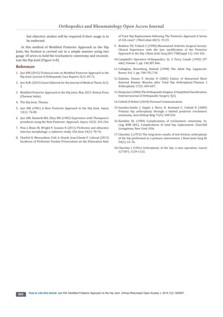
Figure 1: Device used to test stability of the hip joint showing pelvis fixed and protactors to
measure the angle of flexion/extension, adduction/abduction and internal/external rotations
(Courtesy: Photograph reproduced with the kind permission of Injury/Elsevier).
Figure 2: Device used to test stability of the hip joint showing pelvis fixed and protactors to
measure the angle of flexion/extension, adduction/abduction and internal/external rotations
(Courtesy: Photograph reproduced with the kind permission of Injury/Elsevier).](https://image.slidesharecdn.com/oroaj-151012120047-lva1-app6891/85/Oroaj-ms-id-555557-1-1-320.jpg)
![Orthop Rheumatol Open Access J 1(2): OROAJ.MS.ID.555557(2015) 001
and extends down the shaft of the femur for about 10 cm. The
gluteal fascia and the ilio-tibial tract are exposed; the deep fascia
Introduction
The Posterior Approach is the most commonly used
approaches to the Hip Joint for Endo prosthesis, total Joint
replacement or Revision Hip Surgery wherein it gives excellent
visibility to the entire joint, when compared to other approaches
to the Hip Joint [4]. The author’s original paper [5] written 35
years ago presented an original technique designed after taking
into consideration the surgical anatomy of the Hip Joint, where
by the posterior overhanging part of the greater trochanter was
osteotomised to include the short lateral rotators along with the
posterior one-thirds of the gluteus medius and the capsule of the
HipJoint, which was then turnedback as a single flap to exposethe
acetabulum in detail along with a bloodless exposure. This was
also confirmed before any clinical application of this approach
on cadavers, which concluded greater stability as compared with
routine suture or reattachment of the short lateral rotators
Technique and Introduction
In all, many approaches to the Hip Joint are described in
literature. The author’s original technique was implemented
after a detailed cadaveric study (Figures 1-3), where the forces
required to dislocate the Hip Joint was considerably more when
compared to the routine suture or reattachment of the short
lateral rotators.
Clinical Technique
The patient is placed on the sound side. The skin incision
extends from just distal and lateral to the posterior superior
iliac spine towards the lateral edge of the greater trochanter,
with a curve in the direction of the fibres of gluteus maximus,
K Mohan lyer*
Orthopaedic Surgeon, India
Submission: September 26, 2015; Published: October 08, 2015
*Corresponding author: K Mohan lyer, Senior Consultant, Orthopaedic Surgeon, Flat No: 120/H-2K, First Floor, Kailash Apartments, 8th Main
Road, Malleswaram, Bangalore, 560003, Karnataka State, India, Tel: +919632683264; Email:
Modified Posterior Approach to the Hip
Joint
Mini Review
Volume 1 Issue 2 - 2015
Orthop Rheumatol Open Access J
Copyright © All rights are reserved by K Mohan lyer
Abstract
The Modification offers greater visibility and decreased blood loss to the Hip Joint, there by conferring greater stability posteriorly as
compared with the conventional Posterior Approach as described by Austin Moore in 1957.This Modification was devised at a time when
the cause of dislocation was being blamed on the Posterior Approach to the Hip Joint [1,2] In this Approach, since bone is attached to bone, it
confers greater stability than an ordinary suture through soft tissues and hence reduces dislocation of the Hip Joint [3].
Keywords: Trochanteric Osteotomy; Dislocation.
Figure 1: Device used to test stability of the hip joint showing pelvis fixed and protactors to
measure the angle of flexion/extension, adduction/abduction and internal/external rotations
(Courtesy: Photograph reproduced with the kind permission of Injury/Elsevier).
Figure 2: Device used to test stability of the hip joint showing pelvis fixed and protactors to
measure the angle of flexion/extension, adduction/abduction and internal/external rotations
(Courtesy: Photograph reproduced with the kind permission of Injury/Elsevier).](https://image.slidesharecdn.com/oroaj-151012120047-lva1-app6891/75/Oroaj-ms-id-555557-1-1-2048.jpg)
![Orthopedics and Rheumatology Open Access Journal
How to cite this article: lyer KM. Modified Posterior Approach to the Hip Joint. Orthop Rheumatol Open Access J. 2015;1(2): 555557.
002
incised vertically in the lower part of the incision and the incision
is curved upwards through the middle of the fibres of gluteus
maximus.
The muscles now seen converging on the greater trochanter
from above downwards are gluteus medius; piriformis; obturator
internus, flanked by the superior and inferior gaemelli; quatratus
femoris, and the upper edge of the adductor magnus. All these
muscles lie edge to edge, with the sciatic nerve well away from
the insertion of the short lateral rotators (Figure 4).
The posterior border of the gluteus medius in the upper part
and the quadrate tubercle with the lower border of the quadrate
femoris in the lower part is then identified.
The greater trochanter is cut through so that the
detached part includes the insertion of the following
structures. From below upwards these are quatratus
femoris, obturator internus with the inferior and
superior gaemelli, piriformis and the posterior third of the fibres
of the gluteus medius. The osteotomy extends from the junction
of the posterior third and anterior two-thirds of the lateral border
of the greater trochanter obliquely downwards and posteriorly
to the shaft of the femur just distal to the quadrate tubercle.
The posterior triangular flap containing the overhanging
posterosuperior part of the greater trochanter at its apex is then
dissected free and turned down to expose the capsule of the hip
joint (Figure 5). The capsule is then incised to expose the joint.
Iyer et al. [6] reported on early results in 44 patients who had
a hemi-arthroplasty done with no dislocation in this series.
The weakest part of the Hip Joint is the posterior envelope
which contains the short lateral rotators. This point has been
reinforced by various authors on dislocation of the Hip Joint.
There are certain anatomical variations in the tendons of
piriformis and obturator internus which could result in piriformis
sparing approaches to the hip [7,8] the most posterior margins
of the piriformis and obturator internus attachments are located
more than one-third of the way along the greater trochanter,
suggesting that osteotomies would not include these external
rotators in the majority of cases.
Amodifieddorsalapproachwithosteotomyofaboneshellwith
the attached short external rotator muscles which are resutured,
is described. The advantages have been less dislocations, less
sciatic nerve injuries, and an increased operative access.
The Modified Posterior Approach follows the anatomical
intermuscularplanandpermitsfullexposureofboththeproximal
femur and the acetabulum. Compared to the literature, preserving
the piriformis tendon seems to be superior to repairing it as is
done in the Southern Approach in terms of dislocation of the
Endoprosthesis or THR.
They vary mainly as to whether the deep posterior
compartment is entered by incising the iliotibial band and the
gluteus maximus muscle in line with the axis of the shaft, or by
separating the muscle fibres of the gluteus maximus proximally.
They also vary depending on whether the abductors are released
from the greater trochanter and, if released, whether the
Figure 3: Internal rotation torque being applied when the hip joint was standardized to
a fixed angle of flexion and adduction (Courtesy:Photograph reproduced with the kind
permission of Injury/Elsevier).
Figure 4: Line Diagram showing the osteotomy of the posterior overhanging part of the
greater trochanter: (Courtesy: Line Diagram reproduced with the kind permission of Injury/
Elsevier): A: Gluteus Maximus; B: Gluteus Medius; C: piriformis; D: Triradiate tendon; E:
Quadratus Femoris; F: Sciatic Nerve; G: Greater trochanter; H: Osteotome.
Figure 5: Line Diagram to show that the Osteotomy is completed and the flap retracted,
after incising the capsule to expose the Hip Joint, (Courtesy: reproduced with the kind
permission of Injury/Elsevier).
Figure 6: Trochanteric Wiring: (Courtesy: reproduced with the kind permission of Injury/
Elsevier).](https://image.slidesharecdn.com/oroaj-151012120047-lva1-app6891/85/Oroaj-ms-id-555557-1-2-320.jpg)
![Orthopedics and Rheumatology Open Access Journal
How to cite this article: lyer KM. Modified Posterior Approach to the Hip Joint. Orthop Rheumatol Open Access J. 2015;1(2): 555557.
003
tendinous attachment is transected or the greater trochanter is
osteotomized.
Almost all of the Posterior approaches have the option to
release the abductors, depending on the need for added exposure.
After I described this Approach, it was quite encouraging that
my respected teacher (Mr. F.H. Beddow) in Liverpool, UK did a
series of 220 Primary Total Hip Replacements by my technique
and noted only 2 dislocations throughout his series.
Beddow and Tulloch reported on their experience using this
approach in 220 cases of primary total hip replacement in which
there were only 2 cases of dislocation [9].
Terry Canale [10] does make a reference to this approach in
their chapters on Surgical Approaches and Complications after
Total Hip Arthroplasty with respect to dislocations [10].
Callaghan et al. [11] mention the advantages of preserving
the original soft tissue attachments of the posterior aspect of
the hip joint, as obtained with this approach. They also stress on
the excellent exposure of both the acetabulum and femoral shaft
achieved with this approach in being applicable to both revision
arthroplasty and complex primary Arthroplasty [11].
Thomas Stahelin et al. [12]have stated that the failure rate of
reinserted short lateral rotators was extremely high at 70% with
majority of failures occurring within the first postoperative day.
They also concluded that bone to bone reattachment as done in
this approach is more secure,as proved by the cadaveric study
[12].
Deepa lyer (2006) was fascinated by this Orthopaedic Dilema
in the elderly that she studied this fracture in detail and noted its
importance for the junior doctors in training, thereby decreasing
morbidity by early diagnosis and treatment [13].
Robert H. Cofield [14]of Mayo Clinic in Rochester, Minnesota,
USA has been using this approach for the last 25 years with no
regrets. He is extremely happy using this approach since I pre-
sented it during the Scientific Congress of the Asean Orthopaedic
Association in Singapore in 1984 [14].
Mayo Clinic conducted a study of 68 consecutive cases by the
Modified Posterior Approach to the Hip posterior trochanteric
osteotomy is associated with high union rates and a low rate of
late instability after hip replacement [15].
They concluded one disadvantage of the posterior trochan-
teric osteotomy is the potential for injury to the superior gluteal
nerve if the gluteus medius muscle split is extended proximally
more than 5 cm from the tip of the trochanter.
The Posterior approach that Moore popularized, and which is
often referred to as the “Southern approach”, is a variation of the
original Henry approach and of the modifications subsequently
made by Kocher, Osborne and Gibson.
The Moore approach is the most commonly used approach
for endoprostheses, total hip arthroplasty, open reduction of hip
dislocation, removal of loose fragments in the joint, repair of ace-
tabular fractures, drainage of the hip and vascular muscle pedicle
graft procedures.
Here the capsule is sectioned along with the short lateral ro-
tators to gain entry into the Hip Joint, thereby leaving the closure
of the Hip Joint vulnerable to dislocation.
In procedures in which the femoral head is not sacrificed,
such as drainage of the hip, reduction of a posterior dislocation,
removal of fragments from the joint, repair of acetabular frac-
tures, or resurfacing procedures, special care must be taken to
avoid injury to the medial circumflex and retinacular vessels.
The short external rotator muscles are sectioned close to the
edge of the acetabulum, rather than at the insertion in the tro-
chanter, and the capsular incisions are made near the acetabular
edge rather than near the attachment of the capsule to the neck.
The medial circumflex vessels are at risk during the dissection
near the attachment of the psoas tendon to the lesser trochanter .
In the Modified Posterior Approach to the Hip Joint, bleed-
ing is minimal, because the plane of cleavage through the gluteus
maximus is through its middle thus leaving intact the branches of
the superior gluteal artery in the proximal half and branches of
the inferior gluteal artery in the distal half, and hence there is no
need to worry about the amount of blood lost. Bleeding is further
reduced as the leash of vessels which lies at the inferior border of
the short lateral rotators is neither cut nor handled.
The most important advantage is that the sciatic nerve is not
isolated at any step in this approach, as corresponding to the lev-
el of the greater trochanter, it lies well medially. Above all, it is
firmly held between the piriformis tendon and the triradiate ten-
don, when the greater trochanter is turned posteriorly, thereby
preventing any movement of the nerve.
With this modified posterior approach to the Hip Joint, the
gluteus medius is neither cut at its origin nor insertion, thereby
leaving the abductor mechanism intact.
In this Modified Posterior Approach, Union of the trochan-
teric fragment should normally occur, as it is through cancellous
bone and in close proximity to the anastomosis in the trochan-
teric fossa.
The concept of trochanteric osteotomy was mainly used in
difficult exposures and soft tissue tensioning. Contemporary
THA accentuates a streamlined approach to surgery and recov-
ery while maximizing long-term success. Hamblin estimated that
10% to 20% of hips require TO for restoration of normal joint
anatomy [16]. Rates of trochanteric osteotomy reflect geographic
trends and surgeon preferences.
Trochanteric Osteotomy techniques can be generally divided
into standard, slide, and repeat osteotomy groups. The standard
osteotomy may be oblique or posterior. The standard TO was
originally popularized for use in hip arthroplasty by Charnley
[17]. After exposure of the hip, a Cushing elevator is inserted
from anterior to posterior in the interval between the tendon
of the gluteus minimus and the superior part of the hip capsule.
Next, the origin of the vastus lateralis is elevated from the vastus
tubercle. The osteotomy cut traverses the sulcus between the lat-](https://image.slidesharecdn.com/oroaj-151012120047-lva1-app6891/85/Oroaj-ms-id-555557-1-3-320.jpg)
![Orthopedics and Rheumatology Open Access Journal
How to cite this article: lyer KM. Modified Posterior Approach to the Hip Joint. Orthop Rheumatol Open Access J. 2015;1(2): 555557.
004
eral portion of the origin of the vastus intermedius muscle and
the insertions of the gluteus medius and minimus. The osteotomy
is started 1 cm distal to the vastus tubercle and is performed with
an oscillating saw or osteotome, which is aimed at the Cushing
elevator [18].
Complications of trochanteric osteotomy can be divided into
two broad categories: those related to osteotomy healing and
those related to the mode of fixation. Nonunion or a fibrous union
of the trochanter is not necessarily a complication with clinical
significance. ‘lf the trochanter does not heal by bony bridging,
however, associated issues of pain, hardware breakage, or ab-
ductor dysfunction may manifest as impaired gait, Trendelen-
burg lurch, subluxation, or dislocation of the hip replacement.
Even when union of the trochanter occurs, the patient may still
have problems. Trochanteric pain and bursitis may be related to
a prominent trochanter or to irritating hardware. Fraying and
breakage of hardware can lead not only to pain, but also to wear
and the need for early revision.
In comparison to the conventional sliding trochanteric or
extended trochanteric approach, which are more helpful by im-
proving biomechanics of the abductor mechanism in work done
on in difficult primary total hip replacement, or failed total hip
replacements and in well fixed stem components or in previously
osteotomised trochanters., this modification is adequate to carry
out routine work on the hip joint.
Though Surgeons may adopt any approach to the hip joint in
which they are familiar or trained, this modification may be help-
ful when the greater trochanter is intact in cases when treating a
dislocated hip joint, when the blame for the dislocation may be
avoided on the posterior approach to the hip joint.
Instability following weakening of the already weak posterior
capsule and short lateral rotators of the Hip leading to dislocation
has been a cause for concern and controversy in the past. The
main purpose of this modification is to overcome this danger and
yet retain the advantages of the posterior approach.
Bleeding is slight in this approach because the plane of cleav-
age through the gluteus maximus is through its middle, which
leaves intact the branches of the superior gluteal artery in its
proximal half and branches of the inferior gluteal artery in its dis-
tal half. The blood loss is reduced considerably, as the leash of
blood vessels which lies at the inferior edge of the lateral rotators
is neither cut nor handled.
The other advantage is that the sciatic nerve need not be iso-
lated at any step in this modification, and corresponding to the
level of the greater trochanter the sciatic nerve lies well medi-
ally. Secondly, it is held between the piriformis and the triradiate
tendon when the greater trochanter is turned posteriorly, thus
preventing movement of the nerve.
Union of the trochanteric fragment should occur because the
osteotomy is through cancellous bone and in close proximity to
the anastomosis in the trochanteric fossa.
With this modification, though turned aside, the gluteus me-
dius is cut neither at its insertion nor its origin, thus leaving the
abductor mechanism intact.
There are certain disadvantages which we have to bear with
and which is not in every case treated by this modification, such
as heterotrophic ossification, trochantric Osteotomy where the
bone takes more time to unite resulting in non-union or fibrous
union along with greater trochantric bursitis and also breakage
of the wires.
Certain unsolved controversies still exist with regards Tro-
chanteric Osteotomy as follows:-
1. Although the indications of exposure and soft tissue ten-
sioning are well accepted, the exact application of these
indications is somewhat controversial.
2. Greater trochanteric osteotomy is rarely used in contem-
porary hip replacement, and its application is likely relat-
ed to both the type of surgery and the surgeon’s predispo-
sition. Some surgeons apply the approach more liberally
than others. Likewise, the type of internal fixation needed
to maximize healing is not universally agreed upon.
3. Based on newly available literature, I would recommend
avoiding or removing multifilament cables; this advice
will likely be considered controversia.
4. Various options are available, and surgeon preference
dominates their application
5. Also, newer unproven technologies such as locking plates
and nonmetallic tensioning wire may prove beneficial,
Figure 7: Reconstitution of the Hip Joint: (Courtesy: reproduced with the kind permission
of Injury/Elsevier).
Figure 8: Radiograph of Total Hip Prosthesis: (Courtesy: reproduced with the kind
permission of Injury/Elsevier).](https://image.slidesharecdn.com/oroaj-151012120047-lva1-app6891/85/Oroaj-ms-id-555557-1-4-320.jpg)

This document describes a modified posterior approach technique for the hip joint. The key steps of the technique include making a skin incision from just below the posterior superior iliac spine curving toward the greater trochanter. The greater trochanter is then osteotomized to include the insertions of surrounding muscles. This posterior triangular flap is turned down to expose the hip joint capsule. The advantages of this modified approach include decreased risk of dislocation compared to conventional approaches by preserving bone and soft tissue attachments and providing stable exposure of the hip joint and acetabulum.


