SlideShare a Scribd company logo
OSTEORADIONECROSIS
Presented by
Dr. Rahul Srivastava
Professor
Rama Dental College Hospital
& Research Centre Kanpur
OSTEORADIONECROSIS (ORN)
Ewing was the first to use the term ‘radiation
osteitis’ to describe changes in bone after
radiotherapy.
In the following years, several terms were used to
name these changes in bone, such as radiation
osteitis, ORN and avascular bone necrosis.
In 1974, Guttenberg proposed the term ‘septic
ORN of the mandible’ to describe the stage of
necrosis when irradiated bone becomes
superficially infected, ending up with a high risk
of involvement of deeper structures.
In 1983, Marx defined ORN as ‘an area >1 cm of
exposed bone in a field of irradiation that failed
to show any evidence of healing for at least 6
months’. Marx also reported that superficial
contamination and no interstitial infection was
present.
In 1987, Marx and Johnson suggested the
definition of ORN as: ‘The exposure of nonviable
bone which fails to heal without intervention’.
Epstein et al. defined ORN as ‘an ulceration or
necrosis of the mucous membrane, with exposure
of necrotic bone for more than 3 months’.
Widmark et al. described ORN as ‘a non-healing
mucosal or cutaneous ulcer with denuded bone,
lasting for more than 3 months’.
Wong et al. defined ORN as ‘a slow-healing
radiation-induced ischemic necrosis of variable
extent occurring in the absence of local primary
tumor necrosis, recurrence or metastatic disease’.
Classification system of osteoradionecrosis (ORN)
The Notani classification, is quickly applicable to
all cases of mandibular osteoradionecrosis (ORN)
after clinical examination and orthopantogram:
Notaniclass Clinical features
Stage I ORN confined to dentoalveolar bone
Stage II
ORN limited to dentoalveolar bone or mandible above
the inferior dental canal, or both
Stage III
ORN involving the mandible below the inferior dental
canal, or pathological fracture, or skin fistula
Epstein et al. classification of osteoradionecrosis
Type I
Resolved, healed
(A) No pathologic fracture
(B) Pathological fracture
Type II
Chronic persistent (nonprogressive)
(A) No pathologic fracture
(B) Pathological fracture
Type III
Active progressive
(A) No pathologic fracture
(B) Pathological fracture
Lyons et al. classification of osteoradionecrosis
Stage Description
1
<2.5 cm length of bone affected (damaged or exposed);
asymptomatic. Medical treatment only.
2
>2.5 cm length of bone; asymptomatic, including pathological
fracture or involvement of inferior dental nerve or both.
Medicaltreatment only unless there is dental sepsis or obviously
loose, necrotic bone.
3
>2.5 cm length of bone; symptomatic, but with no other features
despite medical treatment. Consider debridement of loose or
necroticbone, and local pedicled flap.
4
2.5 cm length of bone; pathological fracture, involvement of
inferior dental nerve, or orocutaneous fistula, or a combination.
Reconstruction with free flap if patient’s overall condition allows
Clayman introduced a classification of ORN related
to the integrity of the overlying mucosa. According
to this classification:
Type I includes cases of ORN in which bone lysis
occurs under intact gingiva or mucosa.
Type II includes more aggressive cases of ORN in
which soft tissues break down and the bone is
exposed to saliva, causing secondary
contamination. This is defined as radiation
osteomyelitis.
Coffin divided cases of ORN into two groups:
Minor.
Major.
Morton and Simpson subdivided ORN into three
groups –
Minor.
Moderate.
Major.
In 1983, Marx proposed a three-stage system
for ORN:
Stage I- If they exhibit exposed bone in a field of
radiation that has failed to heal for at least 6
months and do not have a pathological fracture,
cutaneous fistula or osteolysis to the inferior
border.
In Stage I, all patients receive 30 sessions of HBO at
2.4 atmospheres absolute for 90 minutes at depth.
Patients who respond to HBO alone demonstrate a
softening of the radiated tissues and spontaneous
sequestration of exposed bone with formation of
granulation tissue.
Each Stage I responder undergoes an additional 10
HBO sessions and then the tissues are allowed to
heal completely.
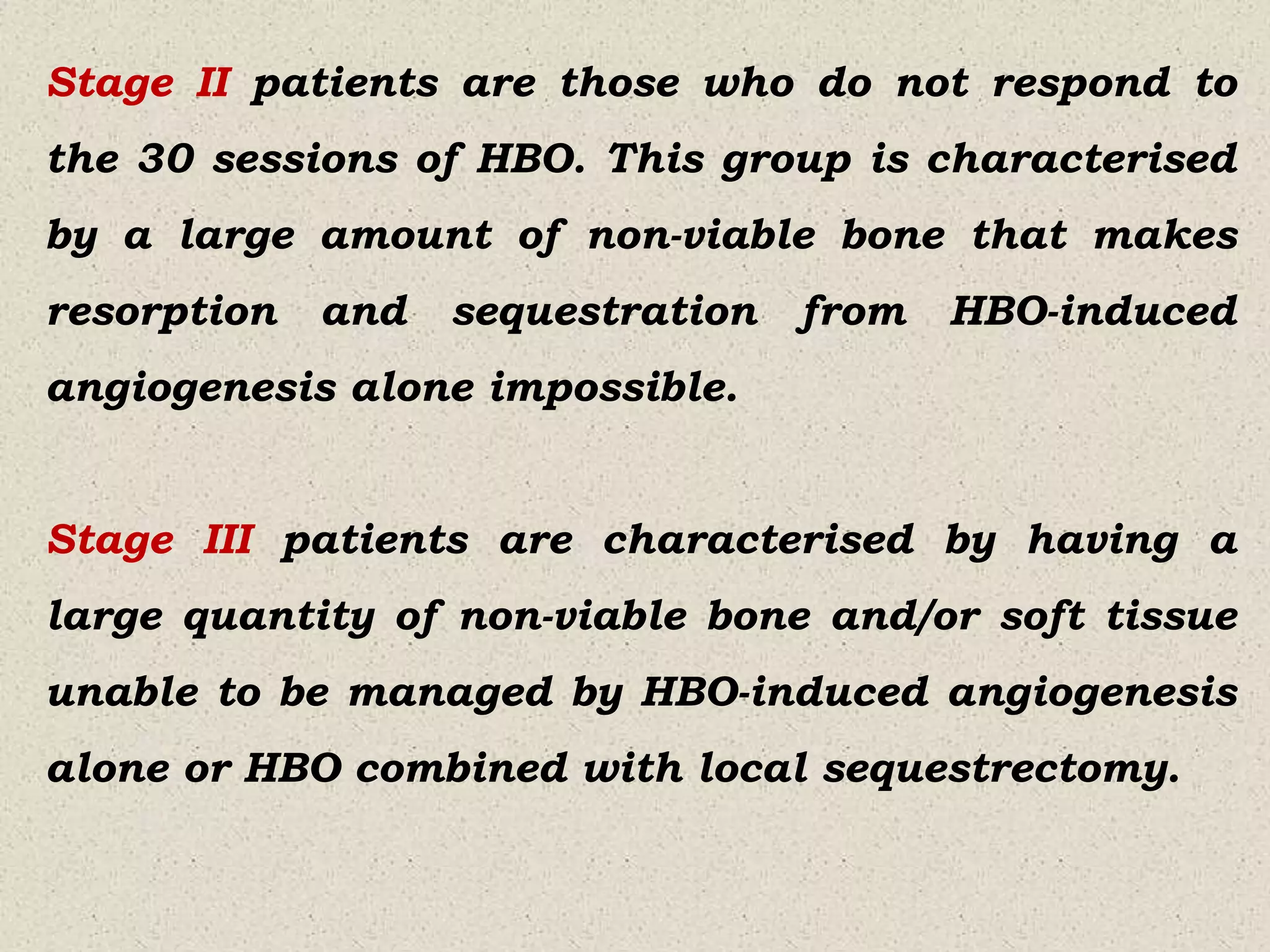
Stage II patients are those who do not respond to
the 30 sessions of HBO. This group is characterised
by a large amount of non-viable bone that makes
resorption and sequestration from HBO-induced
angiogenesis alone impossible.
Stage III patients are characterised by having a
large quantity of non-viable bone and/or soft tissue
unable to be managed by HBO-induced angiogenesis
alone or HBO combined with local sequestrectomy.
Factors that affect the development of ORN
 Primary site of tumor.
 Posterior mandible is more commonly affected
by ORN because of its compact and dense nature.
 Proximity of tumor to bone.
 Extent of mandible included in primary radiation
field.
 State of dentition—odontogenic and periodontal
disease.
 Poor oral hygiene.
 Radiation dose>60 Gy.
 Use of brachytherapy.
 Nutritional status.
 Concomitant chemo-radiation.
 Ill-fitting tissue borne prosthesis resulting in
chronic trauma.
 Acute trauma from surgical procedures to the
jaw.
 Advanced stage tumors.
Pathogenesis of ORN
 ORN affects the small blood vessels of bone,
inducing inflammation (endarteritis), which favors
the generation of small thrombi that obliterate the
vascular lumen and thus interrupt tissue
perfusion.
 Radiation therapy produces an increase in free
radicals and alters collagen synthesis.
The bone loses its normal cellularity and undergoes
fibrosisatrophy with impairment of its repair and
remodeling capacity.
Under such conditions even minimal external
trauma causes ulceration, facilitating
contamination and infection and thus favoring bone
necrosis.
Clinical features of ORN
Mandible more commonly affected than maxilla due
to:
Maxilla rich vascular supply.
Absence of dense cortical plates in maxilla.
Most lesions are perimandibular.
 Posterior mandible affected more readily than
anterior because posterior part of mandible is more
frequently in the direct field of radiation.
 Initially: Trismus, fetid breath, increased
temperature.
 Discomfort or tenderness at the site.
 Bad taste.
 Paresthesia and anesthesia.
 Loss of mucosal covering and exposure of bone.
 Exposed bone is gray to yellow in colour.
 Exposed bone has a rough surface texture that
abrades the adjacent soft tissues and causes
further discomfort.
 Necrosis of exposed bone.
 Tissues surrounding the exposed bone may be
indurated and ulcerated from infections or
recurrent tumors.
 Formation of sequestra.
 Intense pain with intermittent swelling and
drainage extraorally.
Radiological findings
ORN is not usually detectable radiographically in
early stages.
The described radiographic features range from
normal appearance, to localised osteolytic areas,
extensive osteolytic areas, sequestra and fracture.
The most definitive radiographic alterations in early
disease are increased radiodensity, as well as a
mixed radioopaque/radiolucent lesion in which
radiolucent areas represent bone destruction.
In Orthopantomogram (OPT) ORN is depicted as an
undefined radiolucency, without sclerotic
demarcation, which surrounds necrotic zone.
Radiopaque areas can be identified when bone
sequestra are formed. In order to be visible in an
OPT, a substantial alteration in mineral content and
extensive involvement of bone is required and this
only occurs in later stages of ORN.
CT shows osseous abnormalities, such as focal lytic
areas, cortical interruptions and loss of the
spongiosa trabeculation on the symptomatic side,
frequently accompanied by soft-tissue thickening.
In MRI with gadolinium administration, an abnormal
marrow signal, cortical destruction and slight-to-
mild irregular enhancement is demonstrated. MRI
has the advantage of excellent tissue contrast and
high spatial resolution.
Scintigraphy using 99mTc- marked diphosphonates
(99mTc-MDP) allows highly sensitive depiction of
mandibular lesions as a result of their altered
phosphate metabolism.
Dental management of ORN
Pre- irradiation dental care:
 The non-restorable teeth should be extracted a
traumatically under antibiotic coverage.
 Sufficient time to be given for proper healing 7-14
days.
 Judicious alveoloplasty to be done to permit linear
closure of mucoperiostium.
 All sharp bony margins to be contoured because the
irradiated bone does not remodel spontaneously.
 All restorable teeth to be restored.
 Periodontal therapy to be completed within this 2
weeks interval.
 Oral hygiene maintenance instructions.
 Application of fluoride in custom made trays:
1- 0.4% Stannous fluoride gel.
2- 1% sodium fluoride gel.
3- 1% acidulated fluorophosphate gel.
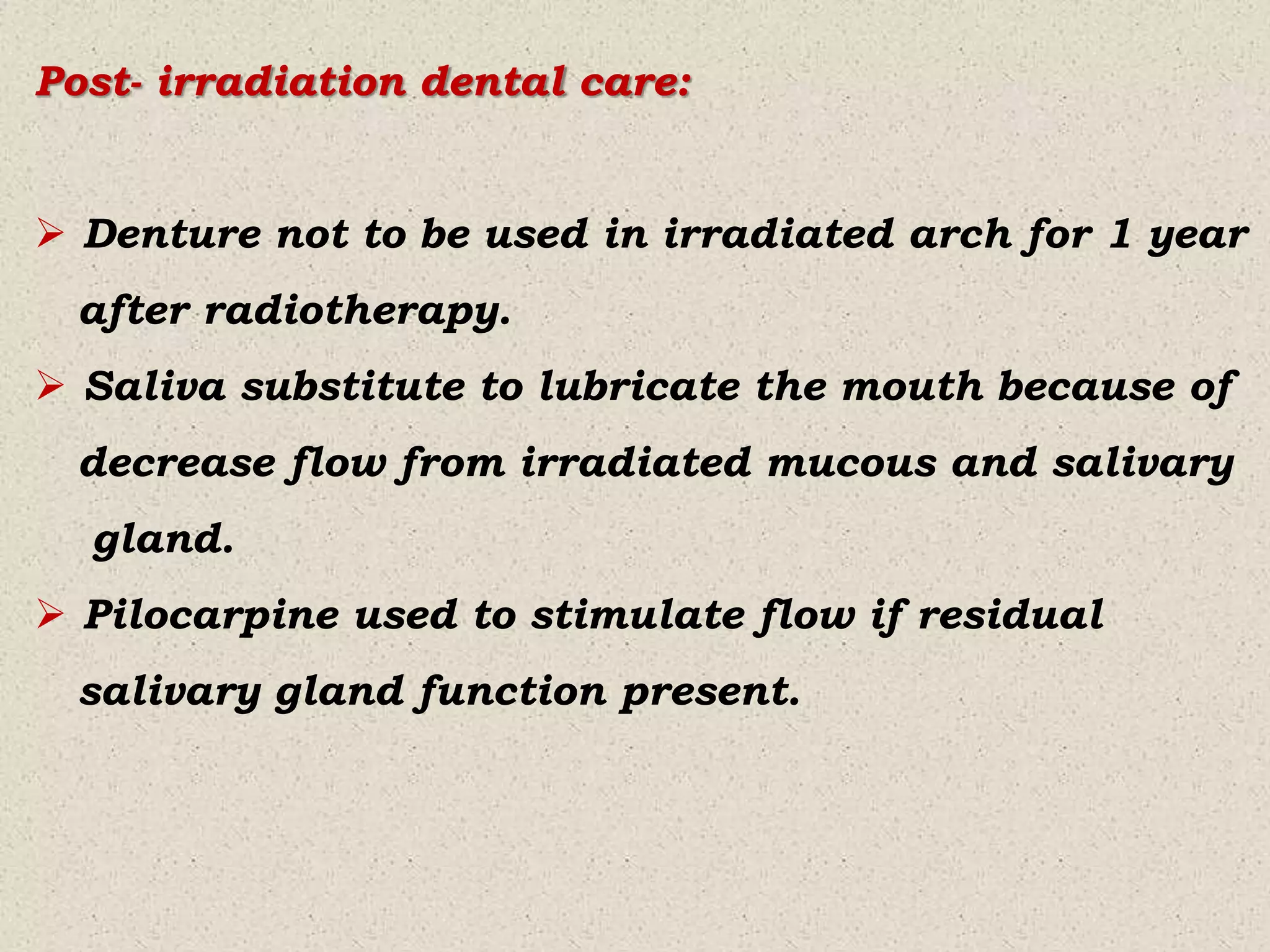
Post- irradiation dental care:
 Denture not to be used in irradiated arch for 1 year
after radiotherapy.
 Saliva substitute to lubricate the mouth because of
decrease flow from irradiated mucous and salivary
gland.
 Pilocarpine used to stimulate flow if residual
salivary gland function present.
 If pulpitis develops, endodontic therapy to be
started, care taken during instrumentation.
 Necessary extraction, limited to 1 or 2 teeth per
appointment.
Management of ORN
When ORN develops:
Avoid mucosal irritants.
Discontinue the use of dental appliances.
Maintain nutritional status.
Stop smoking and alcohol consumption.
Topical antibiotic (tetracycline) and Antiseptic
(chlorhexidine) rinses may reduce the potential local
irritationfrom the microbial flora.
For chronic persisting ORN:
Local wound care:
Penicillin V 500 mg QID X 7 days with
Metronidazole 400 mg QID X 7 days.
or
Clindamycin 300 mg TID X 7days.
Topical tetracycline rinses.
Antiseptic mouthwashes (Chlorhexidine).
Hyperbaric oxygen if needed.
For active progressive ORN:
Appropriate analgesia should be provided.
HBO (hyperbaric oxygen)therapy: 20 to 30dives at
100% oxygen and 2 to 2.5 atmospheres of pressure
for 90 minute sessions, five times a week followed by
additional 10 dives.
Bone resection:
Sequestra managed by resectioning of the segment
of involved bone to prevent occurrence of radiation
compromised skin.
Mandible reconstructed to provide continuity for
esthetic and function.
Ultrasound Therapy:
Non thermal effects used in the stimulation of tissue
regeneration, healing of varicose ulcers, pressure
sores, blood flow in chronically ischemic muscles,
protein synthesis in fibroblasts and tendon repair.
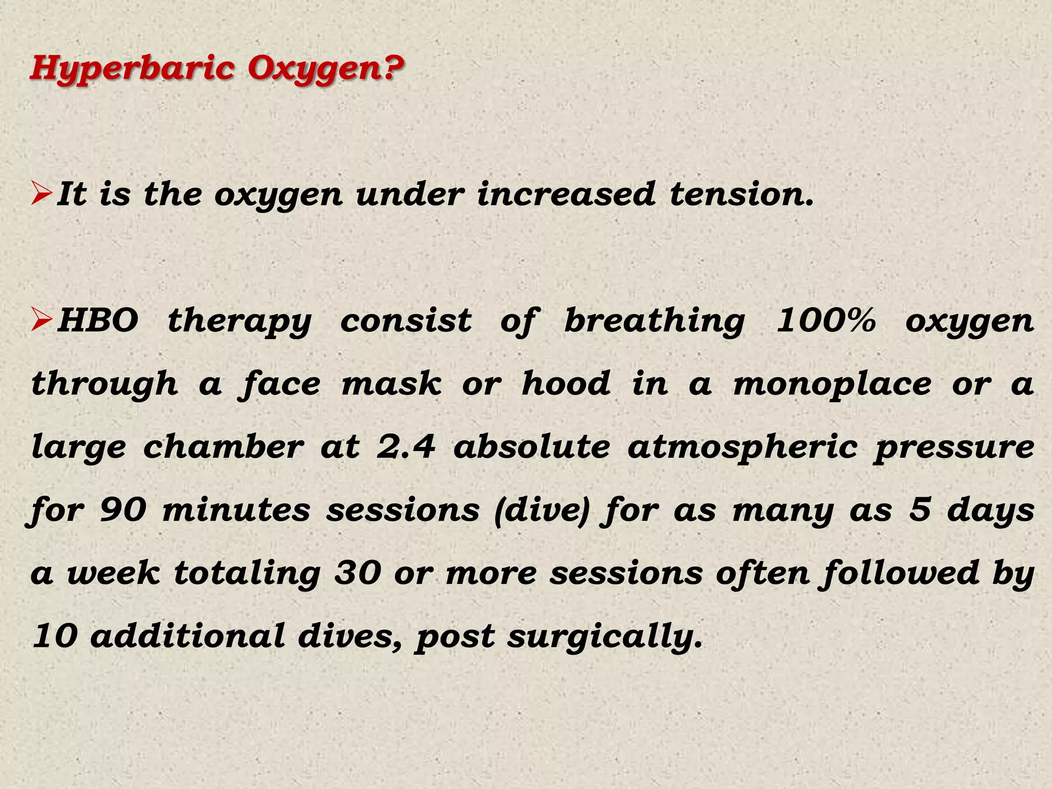
Hyperbaric Oxygen?
It is the oxygen under increased tension.
HBO therapy consist of breathing 100% oxygen
through a face mask or hood in a monoplace or a
large chamber at 2.4 absolute atmospheric pressure
for 90 minutes sessions (dive) for as many as 5 days
a week totaling 30 or more sessions often followed by
10 additional dives, post surgically.
Effects of Hyperbaric Oxygen (HBO)
Increased arterial and venous oxygen tension. The
additional O2 is carried in physical solution in the
plasma.
O2 at high tension enhances the healing by a direct
bacteriostatic effect on the microorganisms that
renders them susceptible to lower antibiotic
concentrations and also enhancing the phagocytic
killing.
Hyperbaric Oxygen (HBO) stimulates:
Neoangiogenesis.
Fibroblastic proliferation
Collagen synthesis.
Proliferation of granulation tissues increases and
advances from increased O2 tension from the non-
diseased periphery into the necrotic bone.
As resorption and replacement of devitalized bone
with healthy tissue progress, formation of sequestra
that may undergo resorption is enhanced.
Indications for prophylactic use of HBO
When surgery is required after radiotherapy.
When patient is at high risk due to high dose
radiation to the bone with a high biologic effect
(Time-Dose Fraction>109).
When extensive surgery is required.
Limitations of HBO therapy
Limited facilities.
Expensive
O2toxicity.
Seizures.
High pressure nervous syndrome.
Trigger Point Injections.
Decompression sickness.
Pneumothorax.
Arterial gas embolism.
Tooth and sinus pain.
Visual change.
Gastric distress.
Contraindications of HBO
Optic neuritis.
Immunosupressive disorders.
COPD.
Claustrophobia.
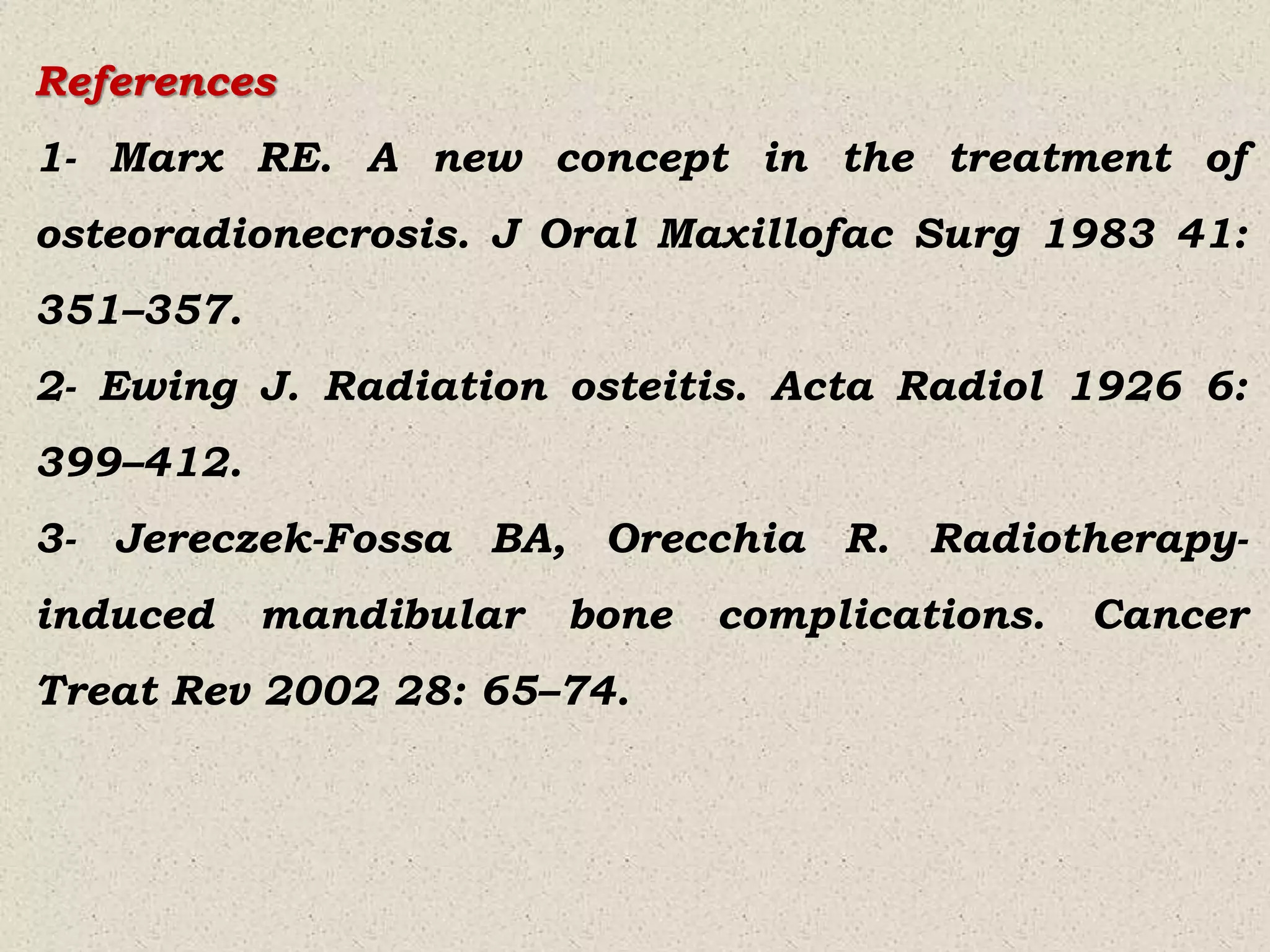
References
1- Marx RE. A new concept in the treatment of
osteoradionecrosis. J Oral Maxillofac Surg 1983 41:
351–357.
2- Ewing J. Radiation osteitis. Acta Radiol 1926 6:
399–412.
3- Jereczek-Fossa BA, Orecchia R. Radiotherapy-
induced mandibular bone complications. Cancer
Treat Rev 2002 28: 65–74.
4- Guttenberg SA. Osteoradionecrosis of the jaw. Am J
Surg 1974:127; 326–332.
5- Epstein JB, Rea G, Wong FL et al. Osteonecrosis-
study of the relationship of dental extractions in
patients receiving radiotherapy. Head Neck Surg
1987 10: 48–54.
6- Hermans R, Fossion E, Ioannides C et al. CT
findings in osteoradionecrosis of the mandible.
Skeletal Radiol 1996 25: 31–36.
7- Ardran G. Bone destruction not demonstrable by
radiography. Br J Radiol 1951 24: 107–109.
8- Khalighi M. Pain Management. Available from:
https://depts.washington. edu/uwmedres/pdf/
hospitalist/Pain_Management.pdf.
9- Carrington C. Oral targeted therapy for cancer
Aust Prescr. 2015;38: 171–176.
10- Nadella KR, Kodali RM, Guttikonda LK,
Jonnalagadda A Osteoradionecrosis of the Jaws:
Clinico-Therapeutic Management: A Literature Review
and Update J. Maxillofac. Oral Surg 2015;14:891–
901.
11- Rathy R, Sunil S, Nivia M Osteoradionecrosis of
mandible: Case report with review of literature
Contemporary Clinical Dentistry.2013;4:251-53.

More Related Content

What's hot

Temporomandibular joint imaging
Temporomandibular joint imagingTemporomandibular joint imaging
Temporomandibular joint imaging
Dr Jinki Singha
 
periapical radiolucencies
 periapical radiolucencies periapical radiolucencies
periapical radiolucencies
vidushiKhanna1
 
Anatomical landmarks in Periapical and Orthopantomogram X-ray
Anatomical landmarks in Periapical and Orthopantomogram X-rayAnatomical landmarks in Periapical and Orthopantomogram X-ray
Anatomical landmarks in Periapical and Orthopantomogram X-ray
Rizgar Saeed
 
Osteoradionecrosis
Osteoradionecrosis Osteoradionecrosis
Osteoradionecrosis
avinash_verma20
 
Cbct
CbctCbct
Osteoradionecrosis
OsteoradionecrosisOsteoradionecrosis
Osteoradionecrosis
lakku_boy
 
Chronic infections of jaws
Chronic infections of jaws  Chronic infections of jaws
Chronic infections of jaws
Mohammed Haneef Farooq
 
mixed radiolucent radiopaque lesions of oral cavity
mixed radiolucent radiopaque lesions of oral cavitymixed radiolucent radiopaque lesions of oral cavity
mixed radiolucent radiopaque lesions of oral cavity
Stephanie Chahrouk
 
General principles of Periodontal surgery
General principles of Periodontal surgeryGeneral principles of Periodontal surgery
General principles of Periodontal surgery
Jignesh Patel
 
Periapical radiolucencies
Periapical radiolucencies Periapical radiolucencies
Periapical radiolucencies
Shabil Mohamed Mustafa
 
Diagnostic aids in endodontics /certified fixed orthodontic courses by India...
Diagnostic aids in endodontics  /certified fixed orthodontic courses by India...Diagnostic aids in endodontics  /certified fixed orthodontic courses by India...
Diagnostic aids in endodontics /certified fixed orthodontic courses by India...
Indian dental academy
 
Oral Submucous Fibrosis - OSMF : Dr Sanjana Ravindra
Oral Submucous Fibrosis - OSMF : Dr Sanjana RavindraOral Submucous Fibrosis - OSMF : Dr Sanjana Ravindra
Oral Submucous Fibrosis - OSMF : Dr Sanjana Ravindra
Dr. Sanjana Ravindra
 
Bsso
BssoBsso
periapical radiopacities
periapical radiopacitiesperiapical radiopacities
periapical radiopacities
Revath Vyas Devulapalli
 
Oral Submucous Fibrosis
Oral Submucous FibrosisOral Submucous Fibrosis
Oral Submucous FibrosisVibhuti Kaul
 
Effects of radiation on oral tissues
Effects of radiation on oral tissuesEffects of radiation on oral tissues
Effects of radiation on oral tissues
Adwiti Vidushi
 
Red and White Lesions
Red and White LesionsRed and White Lesions
Ideal radiograph
Ideal radiographIdeal radiograph
Ideal radiograph
Indian dental academy
 

What's hot (20)

9.(new)osteoradionecrosis
9.(new)osteoradionecrosis9.(new)osteoradionecrosis
9.(new)osteoradionecrosis
 
Temporomandibular joint imaging
Temporomandibular joint imagingTemporomandibular joint imaging
Temporomandibular joint imaging
 
periapical radiolucencies
 periapical radiolucencies periapical radiolucencies
periapical radiolucencies
 
Anatomical landmarks in Periapical and Orthopantomogram X-ray
Anatomical landmarks in Periapical and Orthopantomogram X-rayAnatomical landmarks in Periapical and Orthopantomogram X-ray
Anatomical landmarks in Periapical and Orthopantomogram X-ray
 
Osteoradionecrosis
Osteoradionecrosis Osteoradionecrosis
Osteoradionecrosis
 
Cbct
CbctCbct
Cbct
 
Osteoradionecrosis
OsteoradionecrosisOsteoradionecrosis
Osteoradionecrosis
 
Chronic infections of jaws
Chronic infections of jaws  Chronic infections of jaws
Chronic infections of jaws
 
mixed radiolucent radiopaque lesions of oral cavity
mixed radiolucent radiopaque lesions of oral cavitymixed radiolucent radiopaque lesions of oral cavity
mixed radiolucent radiopaque lesions of oral cavity
 
General principles of Periodontal surgery
General principles of Periodontal surgeryGeneral principles of Periodontal surgery
General principles of Periodontal surgery
 
Periapical radiolucencies
Periapical radiolucencies Periapical radiolucencies
Periapical radiolucencies
 
CBCT
CBCTCBCT
CBCT
 
Diagnostic aids in endodontics /certified fixed orthodontic courses by India...
Diagnostic aids in endodontics  /certified fixed orthodontic courses by India...Diagnostic aids in endodontics  /certified fixed orthodontic courses by India...
Diagnostic aids in endodontics /certified fixed orthodontic courses by India...
 
Oral Submucous Fibrosis - OSMF : Dr Sanjana Ravindra
Oral Submucous Fibrosis - OSMF : Dr Sanjana RavindraOral Submucous Fibrosis - OSMF : Dr Sanjana Ravindra
Oral Submucous Fibrosis - OSMF : Dr Sanjana Ravindra
 
Bsso
BssoBsso
Bsso
 
periapical radiopacities
periapical radiopacitiesperiapical radiopacities
periapical radiopacities
 
Oral Submucous Fibrosis
Oral Submucous FibrosisOral Submucous Fibrosis
Oral Submucous Fibrosis
 
Effects of radiation on oral tissues
Effects of radiation on oral tissuesEffects of radiation on oral tissues
Effects of radiation on oral tissues
 
Red and White Lesions
Red and White LesionsRed and White Lesions
Red and White Lesions
 
Ideal radiograph
Ideal radiographIdeal radiograph
Ideal radiograph
 

Similar to Osteoradionecrosis.pptx

OSTEORADIONECROSIS.pptx
OSTEORADIONECROSIS.pptxOSTEORADIONECROSIS.pptx
OSTEORADIONECROSIS.pptx
Dr. Sumit KUMAR
 
Osteoradionecrosis of the jaws the modern management
Osteoradionecrosis of the jawsthe modern managementOsteoradionecrosis of the jawsthe modern management
Osteoradionecrosis of the jaws the modern management
DR ISHRAT UL EBAD INSTITUTE OF ORAL HEALTH SCIENCES DIKIOHS
 
Infection 3
Infection 3Infection 3
Management of traumatic dental injury of primary teeth
Management of traumatic dental  injury of primary teethManagement of traumatic dental  injury of primary teeth
Management of traumatic dental injury of primary teeth
Dr. Akash Ardeshana
 
Osteoradionecrosis
OsteoradionecrosisOsteoradionecrosis
Osteoradionecrosis
AravindNair71
 
OSTEORADIONECROSIS.pptx
OSTEORADIONECROSIS.pptxOSTEORADIONECROSIS.pptx
OSTEORADIONECROSIS.pptx
Adyasha3
 
osteoradionecrosis-160118051248 new.pptx
osteoradionecrosis-160118051248 new.pptxosteoradionecrosis-160118051248 new.pptx
osteoradionecrosis-160118051248 new.pptx
ShaliniN51
 
Radiation therapy and its prosthodontic implications
Radiation therapy and its prosthodontic implicationsRadiation therapy and its prosthodontic implications
Radiation therapy and its prosthodontic implications
Rani Ranabhatt, KGMC, Lucknow
 
Root fractures and its management
Root fractures and its managementRoot fractures and its management
Root fractures and its management
Vasundhara naik
 
My coronoplasty
My coronoplastyMy coronoplasty
My coronoplasty
hishashwati
 
Treatment and complications of impactions
Treatment and complications of impactionsTreatment and complications of impactions
Treatment and complications of impactions
Suparn Kelkar
 
fenestration.docx
fenestration.docxfenestration.docx
fenestration.docx
Dr.Mohammed Alruby
 
Dental extractions in irradiated patients
Dental extractions in irradiated patientsDental extractions in irradiated patients
Dental extractions in irradiated patientsUjwal Gautam
 
Root Resection – A Dark Horse in Management Offurcation Involved Maxillary Mo...
Root Resection – A Dark Horse in Management Offurcation Involved Maxillary Mo...Root Resection – A Dark Horse in Management Offurcation Involved Maxillary Mo...
Root Resection – A Dark Horse in Management Offurcation Involved Maxillary Mo...
iosrjce
 
endo-perio.ppt
endo-perio.pptendo-perio.ppt
endo-perio.ppt
Pankaj Rama priyadarshi
 
Peripheral Ossifying Fibroma: A Case Report
Peripheral Ossifying Fibroma: A Case ReportPeripheral Ossifying Fibroma: A Case Report
Peripheral Ossifying Fibroma: A Case Report
iosrjce
 
osteomyelitis
osteomyelitisosteomyelitis
Mandibular third moalr impaction
Mandibular third moalr impactionMandibular third moalr impaction
Mandibular third moalr impaction
Ashish Soni
 
Impacted lower 3rd molar
Impacted lower 3rd molar Impacted lower 3rd molar
Impacted lower 3rd molar
OlaMR
 
periodontitis associated with endodontic lesions
periodontitis associated with endodontic lesionsperiodontitis associated with endodontic lesions
periodontitis associated with endodontic lesionsParth Thakkar
 

Similar to Osteoradionecrosis.pptx (20)

OSTEORADIONECROSIS.pptx
OSTEORADIONECROSIS.pptxOSTEORADIONECROSIS.pptx
OSTEORADIONECROSIS.pptx
 
Osteoradionecrosis of the jaws the modern management
Osteoradionecrosis of the jawsthe modern managementOsteoradionecrosis of the jawsthe modern management
Osteoradionecrosis of the jaws the modern management
 
Infection 3
Infection 3Infection 3
Infection 3
 
Management of traumatic dental injury of primary teeth
Management of traumatic dental  injury of primary teethManagement of traumatic dental  injury of primary teeth
Management of traumatic dental injury of primary teeth
 
Osteoradionecrosis
OsteoradionecrosisOsteoradionecrosis
Osteoradionecrosis
 
OSTEORADIONECROSIS.pptx
OSTEORADIONECROSIS.pptxOSTEORADIONECROSIS.pptx
OSTEORADIONECROSIS.pptx
 
osteoradionecrosis-160118051248 new.pptx
osteoradionecrosis-160118051248 new.pptxosteoradionecrosis-160118051248 new.pptx
osteoradionecrosis-160118051248 new.pptx
 
Radiation therapy and its prosthodontic implications
Radiation therapy and its prosthodontic implicationsRadiation therapy and its prosthodontic implications
Radiation therapy and its prosthodontic implications
 
Root fractures and its management
Root fractures and its managementRoot fractures and its management
Root fractures and its management
 
My coronoplasty
My coronoplastyMy coronoplasty
My coronoplasty
 
Treatment and complications of impactions
Treatment and complications of impactionsTreatment and complications of impactions
Treatment and complications of impactions
 
fenestration.docx
fenestration.docxfenestration.docx
fenestration.docx
 
Dental extractions in irradiated patients
Dental extractions in irradiated patientsDental extractions in irradiated patients
Dental extractions in irradiated patients
 
Root Resection – A Dark Horse in Management Offurcation Involved Maxillary Mo...
Root Resection – A Dark Horse in Management Offurcation Involved Maxillary Mo...Root Resection – A Dark Horse in Management Offurcation Involved Maxillary Mo...
Root Resection – A Dark Horse in Management Offurcation Involved Maxillary Mo...
 
endo-perio.ppt
endo-perio.pptendo-perio.ppt
endo-perio.ppt
 
Peripheral Ossifying Fibroma: A Case Report
Peripheral Ossifying Fibroma: A Case ReportPeripheral Ossifying Fibroma: A Case Report
Peripheral Ossifying Fibroma: A Case Report
 
osteomyelitis
osteomyelitisosteomyelitis
osteomyelitis
 
Mandibular third moalr impaction
Mandibular third moalr impactionMandibular third moalr impaction
Mandibular third moalr impaction
 
Impacted lower 3rd molar
Impacted lower 3rd molar Impacted lower 3rd molar
Impacted lower 3rd molar
 
periodontitis associated with endodontic lesions
periodontitis associated with endodontic lesionsperiodontitis associated with endodontic lesions
periodontitis associated with endodontic lesions
 

More from Rama Dental College Hospital and Research Centre

Myofascial Pain Dysfunction Syndrome.ppt
Myofascial Pain Dysfunction Syndrome.pptMyofascial Pain Dysfunction Syndrome.ppt
Myofascial Pain Dysfunction Syndrome.ppt
Rama Dental College Hospital and Research Centre
 
Classical signs in oral and maxillofacial radiology.pptx
Classical signs in oral and maxillofacial radiology.pptxClassical signs in oral and maxillofacial radiology.pptx
Classical signs in oral and maxillofacial radiology.pptx
Rama Dental College Hospital and Research Centre
 
Dentigerous Cyst.ppt
Dentigerous Cyst.pptDentigerous Cyst.ppt
Acute Radiation Syndrome.pptx
Acute Radiation Syndrome.pptxAcute Radiation Syndrome.pptx
Herpes zoster.pptx
Herpes zoster.pptxHerpes zoster.pptx
Recurrent aphthous ulcers (RAS).pptx
Recurrent aphthous ulcers (RAS).pptxRecurrent aphthous ulcers (RAS).pptx
Recurrent aphthous ulcers (RAS).pptx
Rama Dental College Hospital and Research Centre
 
Oral Lichen Planus.pptx
Oral Lichen Planus.pptxOral Lichen Planus.pptx
Trigeminal Neuralgia
Trigeminal NeuralgiaTrigeminal Neuralgia

More from Rama Dental College Hospital and Research Centre (8)

Myofascial Pain Dysfunction Syndrome.ppt
Myofascial Pain Dysfunction Syndrome.pptMyofascial Pain Dysfunction Syndrome.ppt
Myofascial Pain Dysfunction Syndrome.ppt
 
Classical signs in oral and maxillofacial radiology.pptx
Classical signs in oral and maxillofacial radiology.pptxClassical signs in oral and maxillofacial radiology.pptx
Classical signs in oral and maxillofacial radiology.pptx
 
Dentigerous Cyst.ppt
Dentigerous Cyst.pptDentigerous Cyst.ppt
Dentigerous Cyst.ppt
 
Acute Radiation Syndrome.pptx
Acute Radiation Syndrome.pptxAcute Radiation Syndrome.pptx
Acute Radiation Syndrome.pptx
 
Herpes zoster.pptx
Herpes zoster.pptxHerpes zoster.pptx
Herpes zoster.pptx
 
Recurrent aphthous ulcers (RAS).pptx
Recurrent aphthous ulcers (RAS).pptxRecurrent aphthous ulcers (RAS).pptx
Recurrent aphthous ulcers (RAS).pptx
 
Oral Lichen Planus.pptx
Oral Lichen Planus.pptxOral Lichen Planus.pptx
Oral Lichen Planus.pptx
 
Trigeminal Neuralgia
Trigeminal NeuralgiaTrigeminal Neuralgia
Trigeminal Neuralgia
 

Recently uploaded

Ocular injury ppt Upendra pal optometrist upums saifai etawah
Ocular injury  ppt  Upendra pal  optometrist upums saifai etawahOcular injury  ppt  Upendra pal  optometrist upums saifai etawah
Ocular injury ppt Upendra pal optometrist upums saifai etawah
pal078100
 
HOT NEW PRODUCT! BIG SALES FAST SHIPPING NOW FROM CHINA!! EU KU DB BK substit...
HOT NEW PRODUCT! BIG SALES FAST SHIPPING NOW FROM CHINA!! EU KU DB BK substit...HOT NEW PRODUCT! BIG SALES FAST SHIPPING NOW FROM CHINA!! EU KU DB BK substit...
HOT NEW PRODUCT! BIG SALES FAST SHIPPING NOW FROM CHINA!! EU KU DB BK substit...
GL Anaacs
 
Report Back from SGO 2024: What’s the Latest in Cervical Cancer?
Report Back from SGO 2024: What’s the Latest in Cervical Cancer?Report Back from SGO 2024: What’s the Latest in Cervical Cancer?
Report Back from SGO 2024: What’s the Latest in Cervical Cancer?
bkling
 
Charaka Samhita Sutra Sthana 9 Chapter khuddakachatuspadadhyaya
Charaka Samhita Sutra Sthana 9 Chapter khuddakachatuspadadhyayaCharaka Samhita Sutra Sthana 9 Chapter khuddakachatuspadadhyaya
Charaka Samhita Sutra Sthana 9 Chapter khuddakachatuspadadhyaya
Dr KHALID B.M
 
New Directions in Targeted Therapeutic Approaches for Older Adults With Mantl...
New Directions in Targeted Therapeutic Approaches for Older Adults With Mantl...New Directions in Targeted Therapeutic Approaches for Older Adults With Mantl...
New Directions in Targeted Therapeutic Approaches for Older Adults With Mantl...
i3 Health
 
The hemodynamic and autonomic determinants of elevated blood pressure in obes...
The hemodynamic and autonomic determinants of elevated blood pressure in obes...The hemodynamic and autonomic determinants of elevated blood pressure in obes...
The hemodynamic and autonomic determinants of elevated blood pressure in obes...
Catherine Liao
 
Tom Selleck Health: A Comprehensive Look at the Iconic Actor’s Wellness Journey
Tom Selleck Health: A Comprehensive Look at the Iconic Actor’s Wellness JourneyTom Selleck Health: A Comprehensive Look at the Iconic Actor’s Wellness Journey
Tom Selleck Health: A Comprehensive Look at the Iconic Actor’s Wellness Journey
greendigital
 
Maxilla, Mandible & Hyoid Bone & Clinical Correlations by Dr. RIG.pptx
Maxilla, Mandible & Hyoid Bone & Clinical Correlations by Dr. RIG.pptxMaxilla, Mandible & Hyoid Bone & Clinical Correlations by Dr. RIG.pptx
Maxilla, Mandible & Hyoid Bone & Clinical Correlations by Dr. RIG.pptx
Dr. Rabia Inam Gandapore
 
Surat @ℂall @Girls ꧁❤8527049040❤꧂@ℂall @Girls Service Vip Top Model Safe
Surat @ℂall @Girls ꧁❤8527049040❤꧂@ℂall @Girls Service Vip Top Model SafeSurat @ℂall @Girls ꧁❤8527049040❤꧂@ℂall @Girls Service Vip Top Model Safe
Surat @ℂall @Girls ꧁❤8527049040❤꧂@ℂall @Girls Service Vip Top Model Safe
Savita Shen $i11
 
Are There Any Natural Remedies To Treat Syphilis.pdf
Are There Any Natural Remedies To Treat Syphilis.pdfAre There Any Natural Remedies To Treat Syphilis.pdf
Are There Any Natural Remedies To Treat Syphilis.pdf
Little Cross Family Clinic
 
ARTIFICIAL INTELLIGENCE IN HEALTHCARE.pdf
ARTIFICIAL INTELLIGENCE IN  HEALTHCARE.pdfARTIFICIAL INTELLIGENCE IN  HEALTHCARE.pdf
ARTIFICIAL INTELLIGENCE IN HEALTHCARE.pdf
Anujkumaranit
 
Ozempic: Preoperative Management of Patients on GLP-1 Receptor Agonists
Ozempic: Preoperative Management of Patients on GLP-1 Receptor Agonists  Ozempic: Preoperative Management of Patients on GLP-1 Receptor Agonists
Ozempic: Preoperative Management of Patients on GLP-1 Receptor Agonists
Saeid Safari
 
The Normal Electrocardiogram - Part I of II
The Normal Electrocardiogram - Part I of IIThe Normal Electrocardiogram - Part I of II
The Normal Electrocardiogram - Part I of II
MedicoseAcademics
 
Cervical & Brachial Plexus By Dr. RIG.pptx
Cervical & Brachial Plexus By Dr. RIG.pptxCervical & Brachial Plexus By Dr. RIG.pptx
Cervical & Brachial Plexus By Dr. RIG.pptx
Dr. Rabia Inam Gandapore
 
Antiulcer drugs Advance Pharmacology .pptx
Antiulcer drugs Advance Pharmacology .pptxAntiulcer drugs Advance Pharmacology .pptx
Antiulcer drugs Advance Pharmacology .pptx
Rohit chaurpagar
 
KDIGO 2024 guidelines for diabetologists
KDIGO 2024 guidelines for diabetologistsKDIGO 2024 guidelines for diabetologists
KDIGO 2024 guidelines for diabetologists
د.محمود نجيب
 
Hemodialysis: Chapter 3, Dialysis Water Unit - Dr.Gawad
Hemodialysis: Chapter 3, Dialysis Water Unit - Dr.GawadHemodialysis: Chapter 3, Dialysis Water Unit - Dr.Gawad
Hemodialysis: Chapter 3, Dialysis Water Unit - Dr.Gawad
NephroTube - Dr.Gawad
 
BRACHYTHERAPY OVERVIEW AND APPLICATORS
BRACHYTHERAPY OVERVIEW  AND  APPLICATORSBRACHYTHERAPY OVERVIEW  AND  APPLICATORS
BRACHYTHERAPY OVERVIEW AND APPLICATORS
Krishan Murari
 
Superficial & Deep Fascia of the NECK.pptx
Superficial & Deep Fascia of the NECK.pptxSuperficial & Deep Fascia of the NECK.pptx
Superficial & Deep Fascia of the NECK.pptx
Dr. Rabia Inam Gandapore
 
24 Upakrama.pptx class ppt useful in all
24 Upakrama.pptx class ppt useful in all24 Upakrama.pptx class ppt useful in all
24 Upakrama.pptx class ppt useful in all
DrSathishMS1
 

Recently uploaded (20)

Ocular injury ppt Upendra pal optometrist upums saifai etawah
Ocular injury  ppt  Upendra pal  optometrist upums saifai etawahOcular injury  ppt  Upendra pal  optometrist upums saifai etawah
Ocular injury ppt Upendra pal optometrist upums saifai etawah
 
HOT NEW PRODUCT! BIG SALES FAST SHIPPING NOW FROM CHINA!! EU KU DB BK substit...
HOT NEW PRODUCT! BIG SALES FAST SHIPPING NOW FROM CHINA!! EU KU DB BK substit...HOT NEW PRODUCT! BIG SALES FAST SHIPPING NOW FROM CHINA!! EU KU DB BK substit...
HOT NEW PRODUCT! BIG SALES FAST SHIPPING NOW FROM CHINA!! EU KU DB BK substit...
 
Report Back from SGO 2024: What’s the Latest in Cervical Cancer?
Report Back from SGO 2024: What’s the Latest in Cervical Cancer?Report Back from SGO 2024: What’s the Latest in Cervical Cancer?
Report Back from SGO 2024: What’s the Latest in Cervical Cancer?
 
Charaka Samhita Sutra Sthana 9 Chapter khuddakachatuspadadhyaya
Charaka Samhita Sutra Sthana 9 Chapter khuddakachatuspadadhyayaCharaka Samhita Sutra Sthana 9 Chapter khuddakachatuspadadhyaya
Charaka Samhita Sutra Sthana 9 Chapter khuddakachatuspadadhyaya
 
New Directions in Targeted Therapeutic Approaches for Older Adults With Mantl...
New Directions in Targeted Therapeutic Approaches for Older Adults With Mantl...New Directions in Targeted Therapeutic Approaches for Older Adults With Mantl...
New Directions in Targeted Therapeutic Approaches for Older Adults With Mantl...
 
The hemodynamic and autonomic determinants of elevated blood pressure in obes...
The hemodynamic and autonomic determinants of elevated blood pressure in obes...The hemodynamic and autonomic determinants of elevated blood pressure in obes...
The hemodynamic and autonomic determinants of elevated blood pressure in obes...
 
Tom Selleck Health: A Comprehensive Look at the Iconic Actor’s Wellness Journey
Tom Selleck Health: A Comprehensive Look at the Iconic Actor’s Wellness JourneyTom Selleck Health: A Comprehensive Look at the Iconic Actor’s Wellness Journey
Tom Selleck Health: A Comprehensive Look at the Iconic Actor’s Wellness Journey
 
Maxilla, Mandible & Hyoid Bone & Clinical Correlations by Dr. RIG.pptx
Maxilla, Mandible & Hyoid Bone & Clinical Correlations by Dr. RIG.pptxMaxilla, Mandible & Hyoid Bone & Clinical Correlations by Dr. RIG.pptx
Maxilla, Mandible & Hyoid Bone & Clinical Correlations by Dr. RIG.pptx
 
Surat @ℂall @Girls ꧁❤8527049040❤꧂@ℂall @Girls Service Vip Top Model Safe
Surat @ℂall @Girls ꧁❤8527049040❤꧂@ℂall @Girls Service Vip Top Model SafeSurat @ℂall @Girls ꧁❤8527049040❤꧂@ℂall @Girls Service Vip Top Model Safe
Surat @ℂall @Girls ꧁❤8527049040❤꧂@ℂall @Girls Service Vip Top Model Safe
 
Are There Any Natural Remedies To Treat Syphilis.pdf
Are There Any Natural Remedies To Treat Syphilis.pdfAre There Any Natural Remedies To Treat Syphilis.pdf
Are There Any Natural Remedies To Treat Syphilis.pdf
 
ARTIFICIAL INTELLIGENCE IN HEALTHCARE.pdf
ARTIFICIAL INTELLIGENCE IN  HEALTHCARE.pdfARTIFICIAL INTELLIGENCE IN  HEALTHCARE.pdf
ARTIFICIAL INTELLIGENCE IN HEALTHCARE.pdf
 
Ozempic: Preoperative Management of Patients on GLP-1 Receptor Agonists
Ozempic: Preoperative Management of Patients on GLP-1 Receptor Agonists  Ozempic: Preoperative Management of Patients on GLP-1 Receptor Agonists
Ozempic: Preoperative Management of Patients on GLP-1 Receptor Agonists
 
The Normal Electrocardiogram - Part I of II
The Normal Electrocardiogram - Part I of IIThe Normal Electrocardiogram - Part I of II
The Normal Electrocardiogram - Part I of II
 
Cervical & Brachial Plexus By Dr. RIG.pptx
Cervical & Brachial Plexus By Dr. RIG.pptxCervical & Brachial Plexus By Dr. RIG.pptx
Cervical & Brachial Plexus By Dr. RIG.pptx
 
Antiulcer drugs Advance Pharmacology .pptx
Antiulcer drugs Advance Pharmacology .pptxAntiulcer drugs Advance Pharmacology .pptx
Antiulcer drugs Advance Pharmacology .pptx
 
KDIGO 2024 guidelines for diabetologists
KDIGO 2024 guidelines for diabetologistsKDIGO 2024 guidelines for diabetologists
KDIGO 2024 guidelines for diabetologists
 
Hemodialysis: Chapter 3, Dialysis Water Unit - Dr.Gawad
Hemodialysis: Chapter 3, Dialysis Water Unit - Dr.GawadHemodialysis: Chapter 3, Dialysis Water Unit - Dr.Gawad
Hemodialysis: Chapter 3, Dialysis Water Unit - Dr.Gawad
 
BRACHYTHERAPY OVERVIEW AND APPLICATORS
BRACHYTHERAPY OVERVIEW  AND  APPLICATORSBRACHYTHERAPY OVERVIEW  AND  APPLICATORS
BRACHYTHERAPY OVERVIEW AND APPLICATORS
 
Superficial & Deep Fascia of the NECK.pptx
Superficial & Deep Fascia of the NECK.pptxSuperficial & Deep Fascia of the NECK.pptx
Superficial & Deep Fascia of the NECK.pptx
 
24 Upakrama.pptx class ppt useful in all
24 Upakrama.pptx class ppt useful in all24 Upakrama.pptx class ppt useful in all
24 Upakrama.pptx class ppt useful in all
 

Osteoradionecrosis.pptx

  • 1. OSTEORADIONECROSIS Presented by Dr. Rahul Srivastava Professor Rama Dental College Hospital & Research Centre Kanpur
  • 2. OSTEORADIONECROSIS (ORN) Ewing was the first to use the term ‘radiation osteitis’ to describe changes in bone after radiotherapy. In the following years, several terms were used to name these changes in bone, such as radiation osteitis, ORN and avascular bone necrosis.
  • 3. In 1974, Guttenberg proposed the term ‘septic ORN of the mandible’ to describe the stage of necrosis when irradiated bone becomes superficially infected, ending up with a high risk of involvement of deeper structures.
  • 4. In 1983, Marx defined ORN as ‘an area >1 cm of exposed bone in a field of irradiation that failed to show any evidence of healing for at least 6 months’. Marx also reported that superficial contamination and no interstitial infection was present. In 1987, Marx and Johnson suggested the definition of ORN as: ‘The exposure of nonviable bone which fails to heal without intervention’.
  • 5. Epstein et al. defined ORN as ‘an ulceration or necrosis of the mucous membrane, with exposure of necrotic bone for more than 3 months’. Widmark et al. described ORN as ‘a non-healing mucosal or cutaneous ulcer with denuded bone, lasting for more than 3 months’.
  • 6. Wong et al. defined ORN as ‘a slow-healing radiation-induced ischemic necrosis of variable extent occurring in the absence of local primary tumor necrosis, recurrence or metastatic disease’.
  • 7. Classification system of osteoradionecrosis (ORN) The Notani classification, is quickly applicable to all cases of mandibular osteoradionecrosis (ORN) after clinical examination and orthopantogram: Notaniclass Clinical features Stage I ORN confined to dentoalveolar bone Stage II ORN limited to dentoalveolar bone or mandible above the inferior dental canal, or both Stage III ORN involving the mandible below the inferior dental canal, or pathological fracture, or skin fistula
  • 8. Epstein et al. classification of osteoradionecrosis Type I Resolved, healed (A) No pathologic fracture (B) Pathological fracture Type II Chronic persistent (nonprogressive) (A) No pathologic fracture (B) Pathological fracture Type III Active progressive (A) No pathologic fracture (B) Pathological fracture
  • 9. Lyons et al. classification of osteoradionecrosis Stage Description 1 <2.5 cm length of bone affected (damaged or exposed); asymptomatic. Medical treatment only. 2 >2.5 cm length of bone; asymptomatic, including pathological fracture or involvement of inferior dental nerve or both. Medicaltreatment only unless there is dental sepsis or obviously loose, necrotic bone. 3 >2.5 cm length of bone; symptomatic, but with no other features despite medical treatment. Consider debridement of loose or necroticbone, and local pedicled flap. 4 2.5 cm length of bone; pathological fracture, involvement of inferior dental nerve, or orocutaneous fistula, or a combination. Reconstruction with free flap if patient’s overall condition allows
  • 10. Clayman introduced a classification of ORN related to the integrity of the overlying mucosa. According to this classification: Type I includes cases of ORN in which bone lysis occurs under intact gingiva or mucosa. Type II includes more aggressive cases of ORN in which soft tissues break down and the bone is exposed to saliva, causing secondary contamination. This is defined as radiation osteomyelitis.
  • 11. Coffin divided cases of ORN into two groups: Minor. Major. Morton and Simpson subdivided ORN into three groups – Minor. Moderate. Major.
  • 12. In 1983, Marx proposed a three-stage system for ORN: Stage I- If they exhibit exposed bone in a field of radiation that has failed to heal for at least 6 months and do not have a pathological fracture, cutaneous fistula or osteolysis to the inferior border.
  • 13. In Stage I, all patients receive 30 sessions of HBO at 2.4 atmospheres absolute for 90 minutes at depth. Patients who respond to HBO alone demonstrate a softening of the radiated tissues and spontaneous sequestration of exposed bone with formation of granulation tissue. Each Stage I responder undergoes an additional 10 HBO sessions and then the tissues are allowed to heal completely.
  • 14. Stage II patients are those who do not respond to the 30 sessions of HBO. This group is characterised by a large amount of non-viable bone that makes resorption and sequestration from HBO-induced angiogenesis alone impossible. Stage III patients are characterised by having a large quantity of non-viable bone and/or soft tissue unable to be managed by HBO-induced angiogenesis alone or HBO combined with local sequestrectomy.
  • 15. Factors that affect the development of ORN  Primary site of tumor.  Posterior mandible is more commonly affected by ORN because of its compact and dense nature.  Proximity of tumor to bone.  Extent of mandible included in primary radiation field.  State of dentition—odontogenic and periodontal disease.  Poor oral hygiene.  Radiation dose>60 Gy.
  • 16.  Use of brachytherapy.  Nutritional status.  Concomitant chemo-radiation.  Ill-fitting tissue borne prosthesis resulting in chronic trauma.  Acute trauma from surgical procedures to the jaw.  Advanced stage tumors.
  • 17. Pathogenesis of ORN  ORN affects the small blood vessels of bone, inducing inflammation (endarteritis), which favors the generation of small thrombi that obliterate the vascular lumen and thus interrupt tissue perfusion.  Radiation therapy produces an increase in free radicals and alters collagen synthesis.
  • 18. The bone loses its normal cellularity and undergoes fibrosisatrophy with impairment of its repair and remodeling capacity. Under such conditions even minimal external trauma causes ulceration, facilitating contamination and infection and thus favoring bone necrosis.
  • 19. Clinical features of ORN Mandible more commonly affected than maxilla due to: Maxilla rich vascular supply. Absence of dense cortical plates in maxilla. Most lesions are perimandibular.
  • 20.  Posterior mandible affected more readily than anterior because posterior part of mandible is more frequently in the direct field of radiation.  Initially: Trismus, fetid breath, increased temperature.  Discomfort or tenderness at the site.  Bad taste.  Paresthesia and anesthesia.  Loss of mucosal covering and exposure of bone.  Exposed bone is gray to yellow in colour.
  • 21.  Exposed bone has a rough surface texture that abrades the adjacent soft tissues and causes further discomfort.  Necrosis of exposed bone.  Tissues surrounding the exposed bone may be indurated and ulcerated from infections or recurrent tumors.  Formation of sequestra.  Intense pain with intermittent swelling and drainage extraorally.
  • 22. Radiological findings ORN is not usually detectable radiographically in early stages. The described radiographic features range from normal appearance, to localised osteolytic areas, extensive osteolytic areas, sequestra and fracture.
  • 23. The most definitive radiographic alterations in early disease are increased radiodensity, as well as a mixed radioopaque/radiolucent lesion in which radiolucent areas represent bone destruction. In Orthopantomogram (OPT) ORN is depicted as an undefined radiolucency, without sclerotic demarcation, which surrounds necrotic zone.
  • 24. Radiopaque areas can be identified when bone sequestra are formed. In order to be visible in an OPT, a substantial alteration in mineral content and extensive involvement of bone is required and this only occurs in later stages of ORN. CT shows osseous abnormalities, such as focal lytic areas, cortical interruptions and loss of the spongiosa trabeculation on the symptomatic side, frequently accompanied by soft-tissue thickening.
  • 25. In MRI with gadolinium administration, an abnormal marrow signal, cortical destruction and slight-to- mild irregular enhancement is demonstrated. MRI has the advantage of excellent tissue contrast and high spatial resolution. Scintigraphy using 99mTc- marked diphosphonates (99mTc-MDP) allows highly sensitive depiction of mandibular lesions as a result of their altered phosphate metabolism.
  • 26. Dental management of ORN Pre- irradiation dental care:  The non-restorable teeth should be extracted a traumatically under antibiotic coverage.  Sufficient time to be given for proper healing 7-14 days.  Judicious alveoloplasty to be done to permit linear closure of mucoperiostium.  All sharp bony margins to be contoured because the irradiated bone does not remodel spontaneously.
  • 27.  All restorable teeth to be restored.  Periodontal therapy to be completed within this 2 weeks interval.  Oral hygiene maintenance instructions.  Application of fluoride in custom made trays: 1- 0.4% Stannous fluoride gel. 2- 1% sodium fluoride gel. 3- 1% acidulated fluorophosphate gel.
  • 28. Post- irradiation dental care:  Denture not to be used in irradiated arch for 1 year after radiotherapy.  Saliva substitute to lubricate the mouth because of decrease flow from irradiated mucous and salivary gland.  Pilocarpine used to stimulate flow if residual salivary gland function present.
  • 29.  If pulpitis develops, endodontic therapy to be started, care taken during instrumentation.  Necessary extraction, limited to 1 or 2 teeth per appointment.
  • 30. Management of ORN When ORN develops: Avoid mucosal irritants. Discontinue the use of dental appliances. Maintain nutritional status. Stop smoking and alcohol consumption. Topical antibiotic (tetracycline) and Antiseptic (chlorhexidine) rinses may reduce the potential local irritationfrom the microbial flora.
  • 31. For chronic persisting ORN: Local wound care: Penicillin V 500 mg QID X 7 days with Metronidazole 400 mg QID X 7 days. or Clindamycin 300 mg TID X 7days. Topical tetracycline rinses. Antiseptic mouthwashes (Chlorhexidine). Hyperbaric oxygen if needed.
  • 32. For active progressive ORN: Appropriate analgesia should be provided. HBO (hyperbaric oxygen)therapy: 20 to 30dives at 100% oxygen and 2 to 2.5 atmospheres of pressure for 90 minute sessions, five times a week followed by additional 10 dives.
  • 33. Bone resection: Sequestra managed by resectioning of the segment of involved bone to prevent occurrence of radiation compromised skin. Mandible reconstructed to provide continuity for esthetic and function.
  • 34. Ultrasound Therapy: Non thermal effects used in the stimulation of tissue regeneration, healing of varicose ulcers, pressure sores, blood flow in chronically ischemic muscles, protein synthesis in fibroblasts and tendon repair.
  • 35. Hyperbaric Oxygen? It is the oxygen under increased tension. HBO therapy consist of breathing 100% oxygen through a face mask or hood in a monoplace or a large chamber at 2.4 absolute atmospheric pressure for 90 minutes sessions (dive) for as many as 5 days a week totaling 30 or more sessions often followed by 10 additional dives, post surgically.
  • 36. Effects of Hyperbaric Oxygen (HBO) Increased arterial and venous oxygen tension. The additional O2 is carried in physical solution in the plasma. O2 at high tension enhances the healing by a direct bacteriostatic effect on the microorganisms that renders them susceptible to lower antibiotic concentrations and also enhancing the phagocytic killing.
  • 37. Hyperbaric Oxygen (HBO) stimulates: Neoangiogenesis. Fibroblastic proliferation Collagen synthesis. Proliferation of granulation tissues increases and advances from increased O2 tension from the non- diseased periphery into the necrotic bone. As resorption and replacement of devitalized bone with healthy tissue progress, formation of sequestra that may undergo resorption is enhanced.
  • 38. Indications for prophylactic use of HBO When surgery is required after radiotherapy. When patient is at high risk due to high dose radiation to the bone with a high biologic effect (Time-Dose Fraction>109). When extensive surgery is required.
  • 39. Limitations of HBO therapy Limited facilities. Expensive O2toxicity. Seizures. High pressure nervous syndrome. Trigger Point Injections. Decompression sickness. Pneumothorax. Arterial gas embolism. Tooth and sinus pain.
  • 40. Visual change. Gastric distress. Contraindications of HBO Optic neuritis. Immunosupressive disorders. COPD. Claustrophobia.
  • 41. References 1- Marx RE. A new concept in the treatment of osteoradionecrosis. J Oral Maxillofac Surg 1983 41: 351–357. 2- Ewing J. Radiation osteitis. Acta Radiol 1926 6: 399–412. 3- Jereczek-Fossa BA, Orecchia R. Radiotherapy- induced mandibular bone complications. Cancer Treat Rev 2002 28: 65–74.
  • 42. 4- Guttenberg SA. Osteoradionecrosis of the jaw. Am J Surg 1974:127; 326–332. 5- Epstein JB, Rea G, Wong FL et al. Osteonecrosis- study of the relationship of dental extractions in patients receiving radiotherapy. Head Neck Surg 1987 10: 48–54. 6- Hermans R, Fossion E, Ioannides C et al. CT findings in osteoradionecrosis of the mandible. Skeletal Radiol 1996 25: 31–36.
  • 43. 7- Ardran G. Bone destruction not demonstrable by radiography. Br J Radiol 1951 24: 107–109. 8- Khalighi M. Pain Management. Available from: https://depts.washington. edu/uwmedres/pdf/ hospitalist/Pain_Management.pdf. 9- Carrington C. Oral targeted therapy for cancer Aust Prescr. 2015;38: 171–176.
  • 44. 10- Nadella KR, Kodali RM, Guttikonda LK, Jonnalagadda A Osteoradionecrosis of the Jaws: Clinico-Therapeutic Management: A Literature Review and Update J. Maxillofac. Oral Surg 2015;14:891– 901. 11- Rathy R, Sunil S, Nivia M Osteoradionecrosis of mandible: Case report with review of literature Contemporary Clinical Dentistry.2013;4:251-53.