SlideShare a Scribd company logo
1 of 76
Presented by
Rani Ranabhatt
JR II
 Most patients with head
and neck cancer will
receive radiotherapy at
some time during the
course of their disease.
 For some tumors
radiation is applied alone
whereas for others it is
used in combination with
surgery or chemotherapy
 Particularly carcinoma of nasopharynx, base of
tongue, soft palate, tonsillar fossa radiation
therapy is the treatment of choice because of
surgical morbidity, difficult access, and high risk
of regional lymph node involvement.
 Carcinoma of salivary gland and alveolar ridge
should be treated surgically followed by
radiotherapy due to potential for bony infiltration.
 Early carcinoma of glottic larynx and tongue are
equally well controlled by radiation or surgery
but radiation offer a better functional result
 Hard deeply infiltrated carcinoma of tongue are
less likely to be controlled by radiation. (Due to
fixation to the vocal cord)
( superficial / exophytic lesions have higher cure
rate with radiation than deeply infiltrated
lesions)
 Minimize the
postradiation
morbidities
associated with oral
function that affect
the patient’s quality
of life
 Radiation therapy is defined as the therapeutic
use of ionizing radiation.
 Two categories of radiation ;
Electromagnetic
Particulate
John Beumer III, Tohomas A. Curtis, and Russel Nishimura:
Radiation Therapy
Electromagnetic wave of wavelength less
than 1 angstrom are called photons.
-neither mass nor charge.
Measured in electron volt. eg x- rays and
gamma rays.
Particulate radiation have mass and are
charged negatively (electrons), positively (
protons, alpha particles) or are neutral
(neutrons)
Radiation absorption by tissue
Direct
ionizing
Indirect
Ionizing
Direct action results
when secondary
particles interact with
the target molecule ie
DNA
Indirect action results
from interaction with
water molecule which
in turn produces free
radical which reacts
with target molecule
Radiobiological
Principles
Reoxygenation Repopulation
Accelerated
repopulation
Anoxic tissue 3
times resistant to
radiation , due to
formation
nonrestorable
organic peroxides
Cells in the
irradiated tissue
can proliferate
and repopulate,
if given enough
overall treatment
times
Radiation triggers
clonogenic
surviving cells in
a tumor to divide
faster than before
4 basic Radiation
Plans
1.7 to 2.0 Gy,
5 fractions per
week, upto 70
Gy
Reduction of
the no of
fractions with
an increase of
the daily
fraction. Eg. 2.5
Gy, 5 fractions
per week, upto
50 Gy
Increasing
both the no of
fractions and
the total dose.
Eg. 1.1Gy
twice a day, 5
days a week,
to 74 Gy
Hybrid of
hypo and
hyperfraction
ation
Conventional
fractionation
Hypofractionation
Hyperfractionation
Accelerated
fractionation
CONVENTIONAL
RADIOTHERAPY
1. Opposed
parallel fields
2. Oral stent or
shield
3. Facemask
4. Simulation and
inkmarks
INTENSITY MODULATED
RADIATION THERAPY
1. Delivered doses of
radiation are defined for
volumes (planning target
volumes)
2. PTV1 : cancer and
carcinomatous lymph
nodes
3. PTV2 : high risk lymph
nodes
4. PTV3 : subclimical
nodes
BRACHY THERAPY
1. Method of radiation
treatment by
radioactive sources
2. Radioisotopes –
irridium 192, cesium
137, and radium 226
3. Can be interstitial, intra
cavitary, or surface
application (molds)
These are used to optimize the delivery of radiation while
reducing the associated morbidity.
stents
shields
positionerscarriers
splints
 Used to rearrange tissue topography within the
radiation field and displace normal tissues outside the
radiation field.
Useful in;
 tongue and floor of
the mouth lesions.
 inferior positioning of tongue
and mandible enabling to lower the radiation field.
Goel A et al . Use of positioning stents in lingual carcinoma
patients subjected to radiotherapy. Int J Prosthodont 2010. 23;
450-52
 Interocclusal stent
prepared that extends
lingually from both occlusal
tables with a flat plate of
acrylic resin.
 Serves to depress the
tongue
 A hole is made in the
anterior horizontal segment
 Serves as an orientation
hole for reproducible
tongue position.
 Impressions
 Interocclusal record at
half/ two-thirds of
maximum opening
 Mounting
 Base plate wax
attached to mandibular
record base to form the
portion which will
depress the tongue.
 Occlusal index for
comfort and stability
 use to boost radiation over Small
superficial lesions (T1 or T2 in
sizes) in accessible locations in
the oral cavity.
 lesions like floor of mouth,
hard palate, soft palate, or
tongue.
(Spares vital adjacent tissues
such as mandible, teeth and
salivary gland.)
 unilateral dose of radiation.
 Buccal mucosa, skin and alveolar ridge.
 It has been reported that; 1 cm thickness of Cerrobend
alloy will prevent transmission of 95%of an 18 Mev
electron beam radiation exposure to normal structures.
Aggarwal et al. Radiation stents: Minimizing radiation-induced
complications. South Asian Journal
 Lipowitz metal or
cerrobend alloy or
Wood’s Metal is
commonly used to
shield.
 Low fusing alloy
 50% bismuth
 26.7% lead
 13.3% tin
 10% cadmium
 Use of a stent to flatten the lip and corner of the mouth,
thereby placing the entire lip in the same plane to
deliver uniform dosage of radiation.
Useful in;
 treating skin lesions
associated with upper and lower lips.
Radioactive source
(cesium132 or iridium 192).
Preloaded After loaded
 Preloaded (RS position within prosthesis prior to
carrier insertion) medical staffs receives some
exposure.
 After loading technique, isotopes are threaded into
the hollow tubing after the carrier is in
predesigned location reduces the radiation
exposure to medical staff.
Direct implantation of the
radioactive source in the
tumor.
useful in;
Lesion of the tongue and anterior
floor of the mouth, palatal
tumors
 Used to position the source
and also determine the proper
depth of insertion.
 Irregular tissue = uneven radiation dose
 A bolus is a tissue equivalent material placed directly
onto or into irregular tissue contours to produce a more
homogenous dose distribution.
commonly used materials are- saline, wax, acrylic
resins.
Singh BP et al. A simplified technique to fabricate tissue bolus device to
manage dose distribution in maxillectomy patient with orbital exenteration
journal of oral biology and craniofacial research(2013 ) 1 0 2 - 0 4
 Following orbital
exenteration and
maxillectomy
 Irregular contours and
air spaces
 Tissues at greatest risk of
radiation injury: skin
grafts, areas of thin
mucosa over bone and
brain tissue
Singh BP et al. A simplified technique to fabricate tissue bolus device to
manage dose distribution in maxillectomy patient with orbital exenteration
journal of oral biology and craniofacial research(2013 ) 10 2 - 0 4
Singh BP et al. A simplified technique to fabricate tissue bolus device to
manage dose distribution in maxillectomy patient with orbital exenteration
journal of oral biology and craniofacial research(2013 ) 10 2 - 0 4
 Oral mucous membrane
 Taste
 Olfaction
 Edema
 Trismus
 Salivary glands
 Bone
 Periodontium
 Teeth
 Pain and dysphagia resulting in
weight loss .
 Mucositis begins to appear
2-3 weeks after the start of
therapy
and reaches peak toward the
end of therapy.
 After therapy, changes in tissues in
the field of therapy predispose to
tissue breakdown and delayed
healing
 Epithelium thin and less
keratinized
 Submucosa less vascular and
fibrotic
Prevention------frequent daily cleaning of teeth with soft brush , oral
rinses with combination of salt and sodium bicarbonate in water or dilute
solutions of hydrogen peroxide and water
Taste bud shows signs of
degeneration and atrophy at 1000
cGy
 Alteration in taste are discovered
during the second week and continue
throughout the course of treatment.
 Perception of bitter and acid flavors
are more impaired than salt and
sweet.
 Taste gradually return to normal
levels after therapy is completed.
 Xerostomia  decreased recovery of
taste
 Since the olfactory epithelium is high in nasal passage
and not included within the radiation field, the sense of
smell is less affected.
 Edema of tongue, buccal
mucosa, submental and
submandibular area
 Apparent during the early
postradiation period when
scaring and fibrosis are
common
(Impairs patency of both
lymphatic and venous
channel resulting in
obstruction.)
 .
 nasopharyngeal, parotid,
palatal and nasal sinus
tumors in which TMJ and
muscles of mastication are
in radiation field.
 Maximum mouth opening
may be reduced upto 10-
15mm.
 Treatment
 Exercise
 Dynamic bite openers
 Saliva changes in volume, viscosity, pH, inorganic
and organic constituents, predisposing to caries,
periodontal disease, impairment of taste acuity,
poor tolerance of prosthetic restoration, and
difficulty in swallowing.
 Bone is 1.8 times as dense as
soft tissue , thus, it absorbs a
large proportion of radiation
than does a comparable
volume of soft tissue.
 Mandible absorbs more than
maxilla because of increased
density, plus reduced
vascularity accounts for
increase incident of
osteoradionecrosis.
 Periodontal ligament
thickens and fibres
become disoriented.
 Exhibit decreased
cellularity and vascularity
 cementum capacity for
repair and regeneration is
also compromised.
 Pulp shows decrease in
vascular elements, with
accompanying fibrosis and
atrophy.
 Pulpal response to infection,
trauma, and various dental
procedures appears
compromised.
Level as low as 2500 cGy
can have marked effect on
tooth development.
 Radiation field that include substantial portions of
salivary glands leads to significant changes in the
composition of oral flora.
 Increased----streptococcus mutans, lactobacillus and
actinomyces predisposing to dental caries.
 Post therapy candidiasis of corner of mouth and
beneath prosthetic appliance is common.
 Criteria for pre-radiation Extraction-
Dental Disease
Factors
Condition of residual
dentition
Dental compliance of
patient
Radiation Delivery
Factor
•Urgency of treatment
•Mode of therapy
•
Radiation fields
•
Mandible versus maxilla
•
Dose to bone
 Dentition in optimal condition
 Extraction of all teeth with questionable
prognosis before radiation.
 Periodontal status in healthy condition.
Becomes difficult to maintain after treatment;
 reduced salivary output.
 Trismus,
 impaired motor functions,
 and surgical morbidities
(The patient’s oral hygiene at initial examination is
often a reliable indicator of future performance.)
 Urgency of treatment
 Mode of therapy
 Radiation fields
 Mandible versus maxilla
 Dose to bone
Fast proliferating tumors eg; palatal tumors .
 The dentist, radiation therapist and patient must
accept the risk of complications and must attempt
to maintain oral health at optimum level. Control
of tumor obviously is the most important
consideration.
 When external beam therapy is used in
combination with radioactive sources implanted(
brachytherapy) - dose to adjacent tissues is
reduced and more confined.
 When external radiation is the sole mean of
radiation delivery - close scrutiny of the dentition
is mandatory.
 Nasopharynx and posterior soft palate, (includes
both parotid glands) – xerostomia and
postradiation caries.
 Lateral tongue and floor of mouth, (encompass
the entire body of mandible ) - osteoradionecrosis
is high.
 Tonsillar, soft palate , or retromolar trigone
carcinomas, (major salivary glands and a
significant portion of body of mandible.) - caries
and osteoradionecrosis is high in this group.
 Osteoradionecrosis in maxilla is rare -
conservative approach is justified. BETTER
BLOOD SUPPLY
 Almost all osteoradionacrosis occur in mandible -
more aggressive approach is advocated,
Particularly mandibular molars (common site of
osteoradionecrosis). when they are in radiation
beam.
 For tissues treated to the high level of `tolerance,
more aggressive program of extracting teeth prior
to therapy is indicated .
 The type of tumor will also dictate the radiation
levels used in treatment. Eg;- Hodgkin's disease -
4000 to 4500 cGy,
-Squamous cell carcinoma of oral cavity-6500 to
8500cGy.
 Extraction of impacted
mandibular third molars prior to
radiation is not advocated for
most patients. (create large
defects requiring prolonged
periods for healing).
 Patients with partially erupted
mandibular third molars
represent a particularly difficult
and perplexing problem because
of risk of pericoronities.
Operculectomy is useful in
selected cases.
 Radical alveoectomy should
be performed, edges of the
tissue flaps everted, and
primary closure obtained .
 Teeth should be removed in
segments.
 Antibiotic coverage
 7-10 days healing prior to
radiotherapy
 The risk of bone necrosis secondary to dental
extractions in postradiation period has been
debated by many clinicians.

 Following definitive course of radiation therapy -
vascular changes in bone and oral mucosa impair
blood supply and predispose to tissue breakdown
and secondary infections of bone and soft tissue.
Best indicator of potential risk is the
radiation dose to bone in the area of the dentition
being considered for removal.
 If the dose to bone locally is below 5500cGy,
conventional therapies for tooth or teeth in
question can be employed, including root planing
and curettage, crown lengthening and root canal
therapy. However, Periodontal flap surgery is not
recommended.
 When tumor dose exceeds 6500cGy, options are
dependant upon the radiation treatment modality
used.
 If the dental infection involved the molar region
adjacent to implant in absence of exposed bone,
dental extractions are employed only as last resort.
 Endodontic therapy is recommended in order to
maintain mucosal integrity.
 If the infection is periodontal and/ or into the
bifurcation area following the root canal therapy,
the crown can be amputated , thereby providing
access for oral hygiene to this area .
 If the implant increases the dose in these regions
above 5500cGy, hyperbaric oxygen maybe
considered .
Difficulties;
o Rubber dam isolation is complicated by minimal
coronal tooth structure and risk of tissue trauma
and resultant bone exposure.
o Oropharyngeal reflexes compromised , translating
into greater risk for aspiration of files.
o Trismus and small pulp canals make the access for
instrumentation and filling difficult.
 Is not primarily an infectious
process, it is exposure of bone
within radiation treatment
volume of 3 months or longer
in duration.
 It may progress to intractable
pain and pathological fracture
of mandible, often accompanied
by orocutaneous fistula and
requiring resection of major
portion of mandible.
 mandibular bone necrosis developed in 85%
of dentulous patients who received 7500cGy
or more to bone.
 Osteoradionecrosis associated with external beam;
 -
 ,
Dose less than
6500cGy and
localized exposure
Dose to bone above 6500cGy
and exposure extends beyond
the mucogingival junction, or
in association with teeth
If external beam dose to
the bone is below
5500cGy
local irrigation and
packing of idoform
gauze, impregnated
with tincture of
benzoin.
-hyperbaric oxygen combined
with surgical sequestrectomy
should be considered.
conservative therapy are
excellent,
Hyperbaric oxygen ;
2.4 atmospheres with 100%oxygen
Stimulates neovascular proliferation in marginally
necrotic tissues , enhances fibroblastic
proliferation, enhances the bactericidal activity of
white blood cells and increases production of bone
matrix.
Marx protocol for treatment of
osteoradionecrosis;
 Stage I- Osteoradionecrosis but without
pathological fracture, orocutaneous fistula or
radiographic evidence of bone resorption to the
inferior border of mandible.
2.4 atmospheres, 100%oxygen for 90
minutes for 30 treatments.
End of 30 treatments improvement
20 treatments are added.
No clinical improvement non- responder
and advanced to stage II
 Stage II- Surgical sequestrectomy, wound closed
primarily in 3 layers over a base of bleeding bone.
Additional 10 hyperbaric treatments wound
dehisces
non -responder and advanced to stage III.
Stage II Nonresponder with orocutaneous
fistula, pathologic fracture or radiographic
evidence of bone resorption to inferior border of
mandible are considered stage III patients.
 Stage III- Nonvital mandibular bone are resected
transorally with the aid of tetracycline fluorescence
under ultraviolet light. External fixation of
mandibular segment, orocutaneous fistulae closed
and soft tissue deficits restored with local or
distant flaps.
 Another 10 hyperbaric treatments are given and
the patient is advanced to stage IIIR.
 Stage IIIR- Ten weeks after resection, the
mandible is reconstructed with bone grafts , using
transcutaneous exposure. Mandibular fixation is
achieved and maintained for 8 weeks.
 10 hyperbaric treatments are given
postoperatively.
 following treatment with interstitial implants and peroral
cone modalities .
 occurs within 1 year after completion of radiation therapy.
 Intense local discomfort is a clinical symptom that is
sometimes useful in differentiating this lesion from
persistent disease.

 If the radiation fields cover little of denture bearing
surfaces (eg; nasopharyngeal carcinoma ),
dentures can be inserted as soon as mucositis
resolves.
 Most prosthodontists advised the construction of
dentures be deferred for at least 1year after
radiation therapy had been completed.
The status of the residual ridge is an important
clinical factors to be carefully appraised.
 Regular/ irregular mandibular ridge
 Denture base should ensure distribution of
pressure as widely and as equally as possible.
 Occlusal scheme should be to minimize lateral
movement of mandibular denture base.
2 groups of people
1. One already a denture wearer
2. Another new denture wearer
 Information of site of the
tumor, mode of therapy
employed, total dose ,dates
of treatment, radiation
fields, tumor response and
prognosis for disease control
should collected.
 Oral examination,
 Conventional border molding, using
custom tray and modeling plastic
 In xerostomia---- apply thin coating of petrolatum
 Efforts should be to gaining stability and support rather
than retention
 thermoplastic waxes
 Polysulfide
Consideration for reduced vertical dimension of
occlusion.
In patients with clinically significant trismus,
entrance of bolus is more easily accomplished
by increasing the interocclusal space.
Lingualised or monoplane occlusal schemes.
 attain a proper buccal horizontal overlap.
 Some clinicians use only 3 posterior teeth, 1
bicuspid and 2 molars in order to avoid
trauma to the posterior buccal mucosa.
 Pressure indicating paste
 Disclosing wax
 Clinical remount
 24 and 48 hour follow up
 Leave dentures out at
night
 Irradiation predisposes changes
in bone, skin, mucosa which
affect the predictability of
Osseointegrated implants.
 Careful consideration to risk of
osteoradionecrosis
 Osseointegration is impaired in
bone that has received > 5000
cGy
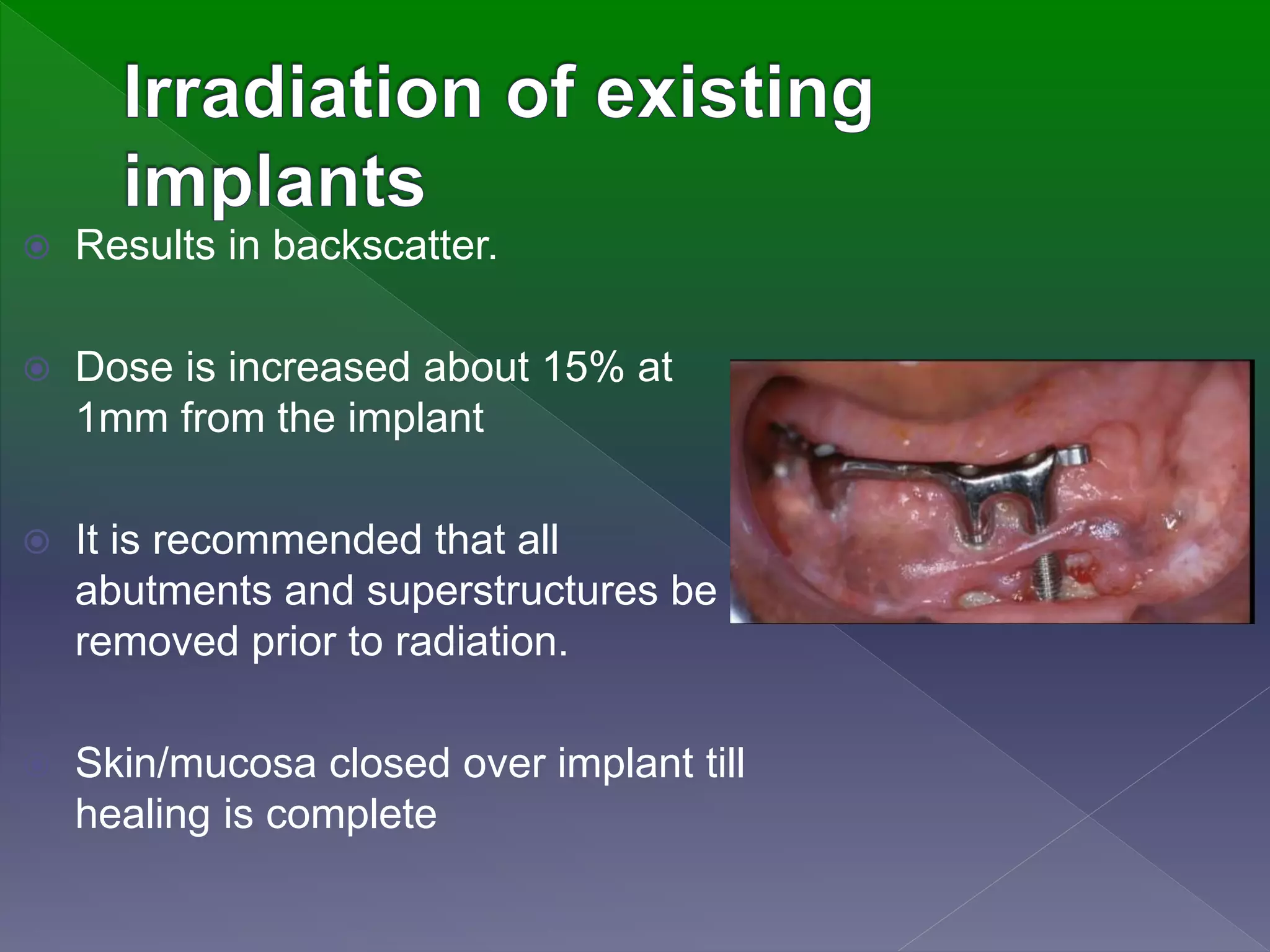
 Results in backscatter.
 Dose is increased about 15% at
1mm from the implant
 It is recommended that all
abutments and superstructures be
removed prior to radiation.
 Skin/mucosa closed over implant till
healing is complete
 Intensity Modulated Radiation Therapy
(IMRT)
 Cyberknife
 Tomotherapy
 Stereotactic radiosurgery
 The cancer patient who is to receive curative
doses of radiation to the head and neck
presents an interesting challenge to the
dentist.
 Dental management of the irradiated patient
is a serious undertaking since the standard
of care has an effect on the patient’s quality
of life.
Radiation therapy and its prosthodontic implications

More Related Content

What's hot

Luting agents used in prosthodontics
Luting agents used in prosthodonticsLuting agents used in prosthodontics
Luting agents used in prosthodonticsaruncs92
 
Resisual Ridge Resorption
Resisual Ridge ResorptionResisual Ridge Resorption
Resisual Ridge ResorptionJehan Dordi
 
An altered cast procedure to improve tissue support
An altered cast procedure to improve tissue supportAn altered cast procedure to improve tissue support
An altered cast procedure to improve tissue supportCPGIDSH
 
Diagnosis & Treatment Planning in FPD
Diagnosis & Treatment Planning in FPDDiagnosis & Treatment Planning in FPD
Diagnosis & Treatment Planning in FPDDr. Anshul Sahu
 
Selective grinding
Selective grindingSelective grinding
Selective grindingshari kurup
 
Osseointegration in Dental Implants
Osseointegration in Dental ImplantsOsseointegration in Dental Implants
Osseointegration in Dental ImplantsNaveed AnJum
 
posterior palatal seal
 posterior palatal seal posterior palatal seal
posterior palatal sealParth Thakkar
 
Functionally Generated Pathway
Functionally Generated Pathway Functionally Generated Pathway
Functionally Generated Pathway Sabnoor Aujla
 
MANAGEMENT OF SEVERELY RESORBED RIDGES
MANAGEMENT OF SEVERELY RESORBED RIDGES MANAGEMENT OF SEVERELY RESORBED RIDGES
MANAGEMENT OF SEVERELY RESORBED RIDGES Kate Maundu
 
impression making-theories and techniques in complete denture
impression making-theories and techniques in complete dentureimpression making-theories and techniques in complete denture
impression making-theories and techniques in complete denturePriyanka Makkar
 
Prosthetic options in implant dentistry
Prosthetic options in implant dentistryProsthetic options in implant dentistry
Prosthetic options in implant dentistryNAMITHA ANAND
 
special/ unconventional dentures
special/ unconventional denturesspecial/ unconventional dentures
special/ unconventional denturesDr. PRAGATI AGRAWAL
 
Resin Bonded Bridges
Resin Bonded BridgesResin Bonded Bridges
Resin Bonded BridgesDr. Almas A
 
OCCLUSION IN COMPLETE DENTURES
OCCLUSION IN COMPLETE DENTURESOCCLUSION IN COMPLETE DENTURES
OCCLUSION IN COMPLETE DENTURESpranav verma
 
Implant supported overdentures
Implant supported overdenturesImplant supported overdentures
Implant supported overdenturesMurtaza Kaderi
 

What's hot (20)

Obturator ppt
Obturator pptObturator ppt
Obturator ppt
 
Luting agents used in prosthodontics
Luting agents used in prosthodonticsLuting agents used in prosthodontics
Luting agents used in prosthodontics
 
Resisual Ridge Resorption
Resisual Ridge ResorptionResisual Ridge Resorption
Resisual Ridge Resorption
 
An altered cast procedure to improve tissue support
An altered cast procedure to improve tissue supportAn altered cast procedure to improve tissue support
An altered cast procedure to improve tissue support
 
Diagnosis & Treatment Planning in FPD
Diagnosis & Treatment Planning in FPDDiagnosis & Treatment Planning in FPD
Diagnosis & Treatment Planning in FPD
 
Selective grinding
Selective grindingSelective grinding
Selective grinding
 
Osseointegration in Dental Implants
Osseointegration in Dental ImplantsOsseointegration in Dental Implants
Osseointegration in Dental Implants
 
posterior palatal seal
 posterior palatal seal posterior palatal seal
posterior palatal seal
 
Functionally Generated Pathway
Functionally Generated Pathway Functionally Generated Pathway
Functionally Generated Pathway
 
MANAGEMENT OF SEVERELY RESORBED RIDGES
MANAGEMENT OF SEVERELY RESORBED RIDGES MANAGEMENT OF SEVERELY RESORBED RIDGES
MANAGEMENT OF SEVERELY RESORBED RIDGES
 
impression making-theories and techniques in complete denture
impression making-theories and techniques in complete dentureimpression making-theories and techniques in complete denture
impression making-theories and techniques in complete denture
 
Surgical and Interim Obturation
Surgical and Interim ObturationSurgical and Interim Obturation
Surgical and Interim Obturation
 
Prosthetic options in implant dentistry
Prosthetic options in implant dentistryProsthetic options in implant dentistry
Prosthetic options in implant dentistry
 
special/ unconventional dentures
special/ unconventional denturesspecial/ unconventional dentures
special/ unconventional dentures
 
Precision attachments
Precision attachmentsPrecision attachments
Precision attachments
 
Failures in FPD
Failures in FPDFailures in FPD
Failures in FPD
 
Resin Bonded Bridges
Resin Bonded BridgesResin Bonded Bridges
Resin Bonded Bridges
 
OCCLUSION IN COMPLETE DENTURES
OCCLUSION IN COMPLETE DENTURESOCCLUSION IN COMPLETE DENTURES
OCCLUSION IN COMPLETE DENTURES
 
Precision attachments
Precision attachmentsPrecision attachments
Precision attachments
 
Implant supported overdentures
Implant supported overdenturesImplant supported overdentures
Implant supported overdentures
 

Viewers also liked

Viewers also liked (6)

Maxillofacial prosthesis of soft cleft palate
Maxillofacial prosthesis of soft cleft palateMaxillofacial prosthesis of soft cleft palate
Maxillofacial prosthesis of soft cleft palate
 
NSAIDs IN DENTISTRY
NSAIDs IN DENTISTRYNSAIDs IN DENTISTRY
NSAIDs IN DENTISTRY
 
2.use of splints and stents during radiation therapy
2.use of splints and stents during radiation therapy2.use of splints and stents during radiation therapy
2.use of splints and stents during radiation therapy
 
Guided Tissue Regeneration
Guided Tissue RegenerationGuided Tissue Regeneration
Guided Tissue Regeneration
 
Denture base materials
Denture base materials Denture base materials
Denture base materials
 
Atlas prothése partielle
Atlas prothése partielleAtlas prothése partielle
Atlas prothése partielle
 

Similar to Radiation therapy and its prosthodontic implications

Protocol of Dental Treatment in Radiotherapy Indicated Patients.pptx
Protocol of Dental Treatment in Radiotherapy Indicated Patients.pptxProtocol of Dental Treatment in Radiotherapy Indicated Patients.pptx
Protocol of Dental Treatment in Radiotherapy Indicated Patients.pptxHoor-E-Jannath Prity
 
Radiation for head and neck cancer video
Radiation for head and neck cancer videoRadiation for head and neck cancer video
Radiation for head and neck cancer videoRobert J Miller MD
 
Symposium Vocal Nodules And Polyp
Symposium Vocal Nodules And PolypSymposium Vocal Nodules And Polyp
Symposium Vocal Nodules And PolypRohit Sinha
 
Managementof Head & Neck Radiotherapy Patients
Managementof Head & Neck Radiotherapy PatientsManagementof Head & Neck Radiotherapy Patients
Managementof Head & Neck Radiotherapy Patientsfondas vakalis
 
Effects of radiation on oral tissues
Effects of radiation on oral tissuesEffects of radiation on oral tissues
Effects of radiation on oral tissuesAdwiti Vidushi
 
5. radiotherapy and chemotherapy-doaa elkady.pptx
5. radiotherapy and chemotherapy-doaa elkady.pptx5. radiotherapy and chemotherapy-doaa elkady.pptx
5. radiotherapy and chemotherapy-doaa elkady.pptxMaiMohamedMohamedAbd
 
OSTEORADIONECROSIS.pptx
OSTEORADIONECROSIS.pptxOSTEORADIONECROSIS.pptx
OSTEORADIONECROSIS.pptxAdyasha3
 
Implants in irradiated jaw
Implants in irradiated jawImplants in irradiated jaw
Implants in irradiated jawZubair_ahmd
 
Carcinomatongue 150622043025-lva1-app6891
Carcinomatongue 150622043025-lva1-app6891Carcinomatongue 150622043025-lva1-app6891
Carcinomatongue 150622043025-lva1-app6891Liju Rajan
 
patients undergoing RT for pdf (2).mmpdf
patients undergoing RT for pdf (2).mmpdfpatients undergoing RT for pdf (2).mmpdf
patients undergoing RT for pdf (2).mmpdfmk5415610
 
CA Tongue and its management.pptx
CA Tongue and its management.pptxCA Tongue and its management.pptx
CA Tongue and its management.pptxMuradOthman5
 
Oral cavity & neck
Oral cavity & neckOral cavity & neck
Oral cavity & neckAnkita Singh
 
Preventive Prosthodontics for Head and Neck Radiotherapy.pptx
Preventive Prosthodontics for Head and Neck Radiotherapy.pptxPreventive Prosthodontics for Head and Neck Radiotherapy.pptx
Preventive Prosthodontics for Head and Neck Radiotherapy.pptxDrApoorwaAwasthi
 
2. Periimplantitis 2017.pptx
2. Periimplantitis 2017.pptx2. Periimplantitis 2017.pptx
2. Periimplantitis 2017.pptxPriyaD36
 
Carcinoma tongue
Carcinoma tongueCarcinoma tongue
Carcinoma tongueViswa Kumar
 

Similar to Radiation therapy and its prosthodontic implications (20)

OSTEORADIONECROSIS.pptx
OSTEORADIONECROSIS.pptxOSTEORADIONECROSIS.pptx
OSTEORADIONECROSIS.pptx
 
Protocol of Dental Treatment in Radiotherapy Indicated Patients.pptx
Protocol of Dental Treatment in Radiotherapy Indicated Patients.pptxProtocol of Dental Treatment in Radiotherapy Indicated Patients.pptx
Protocol of Dental Treatment in Radiotherapy Indicated Patients.pptx
 
Radiation for head and neck cancer video
Radiation for head and neck cancer videoRadiation for head and neck cancer video
Radiation for head and neck cancer video
 
Osteoradionecrosis.pptx
Osteoradionecrosis.pptxOsteoradionecrosis.pptx
Osteoradionecrosis.pptx
 
Symposium Vocal Nodules And Polyp
Symposium Vocal Nodules And PolypSymposium Vocal Nodules And Polyp
Symposium Vocal Nodules And Polyp
 
Managementof Head & Neck Radiotherapy Patients
Managementof Head & Neck Radiotherapy PatientsManagementof Head & Neck Radiotherapy Patients
Managementof Head & Neck Radiotherapy Patients
 
Effects of radiation on oral tissues
Effects of radiation on oral tissuesEffects of radiation on oral tissues
Effects of radiation on oral tissues
 
5. radiotherapy and chemotherapy-doaa elkady.pptx
5. radiotherapy and chemotherapy-doaa elkady.pptx5. radiotherapy and chemotherapy-doaa elkady.pptx
5. radiotherapy and chemotherapy-doaa elkady.pptx
 
OSTEORADIONECROSIS.pptx
OSTEORADIONECROSIS.pptxOSTEORADIONECROSIS.pptx
OSTEORADIONECROSIS.pptx
 
Implants in irradiated jaw
Implants in irradiated jawImplants in irradiated jaw
Implants in irradiated jaw
 
Carcinomatongue 150622043025-lva1-app6891
Carcinomatongue 150622043025-lva1-app6891Carcinomatongue 150622043025-lva1-app6891
Carcinomatongue 150622043025-lva1-app6891
 
patients undergoing RT for pdf (2).mmpdf
patients undergoing RT for pdf (2).mmpdfpatients undergoing RT for pdf (2).mmpdf
patients undergoing RT for pdf (2).mmpdf
 
CA Tongue and its management.pptx
CA Tongue and its management.pptxCA Tongue and its management.pptx
CA Tongue and its management.pptx
 
Oral cavity & neck
Oral cavity & neckOral cavity & neck
Oral cavity & neck
 
Preventive Prosthodontics for Head and Neck Radiotherapy.pptx
Preventive Prosthodontics for Head and Neck Radiotherapy.pptxPreventive Prosthodontics for Head and Neck Radiotherapy.pptx
Preventive Prosthodontics for Head and Neck Radiotherapy.pptx
 
Oral caner
Oral canerOral caner
Oral caner
 
RADIOTHERAPY MANAGEMENT OF ORAL CANCER
RADIOTHERAPY MANAGEMENT OF ORAL CANCERRADIOTHERAPY MANAGEMENT OF ORAL CANCER
RADIOTHERAPY MANAGEMENT OF ORAL CANCER
 
2. Periimplantitis 2017.pptx
2. Periimplantitis 2017.pptx2. Periimplantitis 2017.pptx
2. Periimplantitis 2017.pptx
 
Oral Cancer
Oral CancerOral Cancer
Oral Cancer
 
Carcinoma tongue
Carcinoma tongueCarcinoma tongue
Carcinoma tongue
 

Recently uploaded

Introduction to ArtificiaI Intelligence in Higher Education
Introduction to ArtificiaI Intelligence in Higher EducationIntroduction to ArtificiaI Intelligence in Higher Education
Introduction to ArtificiaI Intelligence in Higher Educationpboyjonauth
 
ECONOMIC CONTEXT - LONG FORM TV DRAMA - PPT
ECONOMIC CONTEXT - LONG FORM TV DRAMA - PPTECONOMIC CONTEXT - LONG FORM TV DRAMA - PPT
ECONOMIC CONTEXT - LONG FORM TV DRAMA - PPTiammrhaywood
 
Concept of Vouching. B.Com(Hons) /B.Compdf
Concept of Vouching. B.Com(Hons) /B.CompdfConcept of Vouching. B.Com(Hons) /B.Compdf
Concept of Vouching. B.Com(Hons) /B.CompdfUmakantAnnand
 
Class 11 Legal Studies Ch-1 Concept of State .pdf
Class 11 Legal Studies Ch-1 Concept of State .pdfClass 11 Legal Studies Ch-1 Concept of State .pdf
Class 11 Legal Studies Ch-1 Concept of State .pdfakmcokerachita
 
A Critique of the Proposed National Education Policy Reform
A Critique of the Proposed National Education Policy ReformA Critique of the Proposed National Education Policy Reform
A Critique of the Proposed National Education Policy ReformChameera Dedduwage
 
Mastering the Unannounced Regulatory Inspection
Mastering the Unannounced Regulatory InspectionMastering the Unannounced Regulatory Inspection
Mastering the Unannounced Regulatory InspectionSafetyChain Software
 
Separation of Lanthanides/ Lanthanides and Actinides
Separation of Lanthanides/ Lanthanides and ActinidesSeparation of Lanthanides/ Lanthanides and Actinides
Separation of Lanthanides/ Lanthanides and ActinidesFatimaKhan178732
 
Hybridoma Technology ( Production , Purification , and Application )
Hybridoma Technology  ( Production , Purification , and Application  ) Hybridoma Technology  ( Production , Purification , and Application  )
Hybridoma Technology ( Production , Purification , and Application ) Sakshi Ghasle
 
_Math 4-Q4 Week 5.pptx Steps in Collecting Data
_Math 4-Q4 Week 5.pptx Steps in Collecting Data_Math 4-Q4 Week 5.pptx Steps in Collecting Data
_Math 4-Q4 Week 5.pptx Steps in Collecting DataJhengPantaleon
 
Incoming and Outgoing Shipments in 1 STEP Using Odoo 17
Incoming and Outgoing Shipments in 1 STEP Using Odoo 17Incoming and Outgoing Shipments in 1 STEP Using Odoo 17
Incoming and Outgoing Shipments in 1 STEP Using Odoo 17Celine George
 
Accessible design: Minimum effort, maximum impact
Accessible design: Minimum effort, maximum impactAccessible design: Minimum effort, maximum impact
Accessible design: Minimum effort, maximum impactdawncurless
 
Alper Gobel In Media Res Media Component
Alper Gobel In Media Res Media ComponentAlper Gobel In Media Res Media Component
Alper Gobel In Media Res Media ComponentInMediaRes1
 
Organic Name Reactions for the students and aspirants of Chemistry12th.pptx
Organic Name Reactions  for the students and aspirants of Chemistry12th.pptxOrganic Name Reactions  for the students and aspirants of Chemistry12th.pptx
Organic Name Reactions for the students and aspirants of Chemistry12th.pptxVS Mahajan Coaching Centre
 
Presiding Officer Training module 2024 lok sabha elections
Presiding Officer Training module 2024 lok sabha electionsPresiding Officer Training module 2024 lok sabha elections
Presiding Officer Training module 2024 lok sabha electionsanshu789521
 
Crayon Activity Handout For the Crayon A
Crayon Activity Handout For the Crayon ACrayon Activity Handout For the Crayon A
Crayon Activity Handout For the Crayon AUnboundStockton
 
18-04-UA_REPORT_MEDIALITERAСY_INDEX-DM_23-1-final-eng.pdf
18-04-UA_REPORT_MEDIALITERAСY_INDEX-DM_23-1-final-eng.pdf18-04-UA_REPORT_MEDIALITERAСY_INDEX-DM_23-1-final-eng.pdf
18-04-UA_REPORT_MEDIALITERAСY_INDEX-DM_23-1-final-eng.pdfssuser54595a
 
URLs and Routing in the Odoo 17 Website App
URLs and Routing in the Odoo 17 Website AppURLs and Routing in the Odoo 17 Website App
URLs and Routing in the Odoo 17 Website AppCeline George
 
Sanyam Choudhary Chemistry practical.pdf
Sanyam Choudhary Chemistry practical.pdfSanyam Choudhary Chemistry practical.pdf
Sanyam Choudhary Chemistry practical.pdfsanyamsingh5019
 

Recently uploaded (20)

Model Call Girl in Bikash Puri Delhi reach out to us at 🔝9953056974🔝
Model Call Girl in Bikash Puri  Delhi reach out to us at 🔝9953056974🔝Model Call Girl in Bikash Puri  Delhi reach out to us at 🔝9953056974🔝
Model Call Girl in Bikash Puri Delhi reach out to us at 🔝9953056974🔝
 
Introduction to ArtificiaI Intelligence in Higher Education
Introduction to ArtificiaI Intelligence in Higher EducationIntroduction to ArtificiaI Intelligence in Higher Education
Introduction to ArtificiaI Intelligence in Higher Education
 
ECONOMIC CONTEXT - LONG FORM TV DRAMA - PPT
ECONOMIC CONTEXT - LONG FORM TV DRAMA - PPTECONOMIC CONTEXT - LONG FORM TV DRAMA - PPT
ECONOMIC CONTEXT - LONG FORM TV DRAMA - PPT
 
Concept of Vouching. B.Com(Hons) /B.Compdf
Concept of Vouching. B.Com(Hons) /B.CompdfConcept of Vouching. B.Com(Hons) /B.Compdf
Concept of Vouching. B.Com(Hons) /B.Compdf
 
Model Call Girl in Tilak Nagar Delhi reach out to us at 🔝9953056974🔝
Model Call Girl in Tilak Nagar Delhi reach out to us at 🔝9953056974🔝Model Call Girl in Tilak Nagar Delhi reach out to us at 🔝9953056974🔝
Model Call Girl in Tilak Nagar Delhi reach out to us at 🔝9953056974🔝
 
Class 11 Legal Studies Ch-1 Concept of State .pdf
Class 11 Legal Studies Ch-1 Concept of State .pdfClass 11 Legal Studies Ch-1 Concept of State .pdf
Class 11 Legal Studies Ch-1 Concept of State .pdf
 
A Critique of the Proposed National Education Policy Reform
A Critique of the Proposed National Education Policy ReformA Critique of the Proposed National Education Policy Reform
A Critique of the Proposed National Education Policy Reform
 
Mastering the Unannounced Regulatory Inspection
Mastering the Unannounced Regulatory InspectionMastering the Unannounced Regulatory Inspection
Mastering the Unannounced Regulatory Inspection
 
Separation of Lanthanides/ Lanthanides and Actinides
Separation of Lanthanides/ Lanthanides and ActinidesSeparation of Lanthanides/ Lanthanides and Actinides
Separation of Lanthanides/ Lanthanides and Actinides
 
Hybridoma Technology ( Production , Purification , and Application )
Hybridoma Technology  ( Production , Purification , and Application  ) Hybridoma Technology  ( Production , Purification , and Application  )
Hybridoma Technology ( Production , Purification , and Application )
 
_Math 4-Q4 Week 5.pptx Steps in Collecting Data
_Math 4-Q4 Week 5.pptx Steps in Collecting Data_Math 4-Q4 Week 5.pptx Steps in Collecting Data
_Math 4-Q4 Week 5.pptx Steps in Collecting Data
 
Incoming and Outgoing Shipments in 1 STEP Using Odoo 17
Incoming and Outgoing Shipments in 1 STEP Using Odoo 17Incoming and Outgoing Shipments in 1 STEP Using Odoo 17
Incoming and Outgoing Shipments in 1 STEP Using Odoo 17
 
Accessible design: Minimum effort, maximum impact
Accessible design: Minimum effort, maximum impactAccessible design: Minimum effort, maximum impact
Accessible design: Minimum effort, maximum impact
 
Alper Gobel In Media Res Media Component
Alper Gobel In Media Res Media ComponentAlper Gobel In Media Res Media Component
Alper Gobel In Media Res Media Component
 
Organic Name Reactions for the students and aspirants of Chemistry12th.pptx
Organic Name Reactions  for the students and aspirants of Chemistry12th.pptxOrganic Name Reactions  for the students and aspirants of Chemistry12th.pptx
Organic Name Reactions for the students and aspirants of Chemistry12th.pptx
 
Presiding Officer Training module 2024 lok sabha elections
Presiding Officer Training module 2024 lok sabha electionsPresiding Officer Training module 2024 lok sabha elections
Presiding Officer Training module 2024 lok sabha elections
 
Crayon Activity Handout For the Crayon A
Crayon Activity Handout For the Crayon ACrayon Activity Handout For the Crayon A
Crayon Activity Handout For the Crayon A
 
18-04-UA_REPORT_MEDIALITERAСY_INDEX-DM_23-1-final-eng.pdf
18-04-UA_REPORT_MEDIALITERAСY_INDEX-DM_23-1-final-eng.pdf18-04-UA_REPORT_MEDIALITERAСY_INDEX-DM_23-1-final-eng.pdf
18-04-UA_REPORT_MEDIALITERAСY_INDEX-DM_23-1-final-eng.pdf
 
URLs and Routing in the Odoo 17 Website App
URLs and Routing in the Odoo 17 Website AppURLs and Routing in the Odoo 17 Website App
URLs and Routing in the Odoo 17 Website App
 
Sanyam Choudhary Chemistry practical.pdf
Sanyam Choudhary Chemistry practical.pdfSanyam Choudhary Chemistry practical.pdf
Sanyam Choudhary Chemistry practical.pdf
 

Radiation therapy and its prosthodontic implications

  • 2.  Most patients with head and neck cancer will receive radiotherapy at some time during the course of their disease.  For some tumors radiation is applied alone whereas for others it is used in combination with surgery or chemotherapy
  • 3.  Particularly carcinoma of nasopharynx, base of tongue, soft palate, tonsillar fossa radiation therapy is the treatment of choice because of surgical morbidity, difficult access, and high risk of regional lymph node involvement.  Carcinoma of salivary gland and alveolar ridge should be treated surgically followed by radiotherapy due to potential for bony infiltration.
  • 4.  Early carcinoma of glottic larynx and tongue are equally well controlled by radiation or surgery but radiation offer a better functional result  Hard deeply infiltrated carcinoma of tongue are less likely to be controlled by radiation. (Due to fixation to the vocal cord) ( superficial / exophytic lesions have higher cure rate with radiation than deeply infiltrated lesions)
  • 5.  Minimize the postradiation morbidities associated with oral function that affect the patient’s quality of life
  • 6.  Radiation therapy is defined as the therapeutic use of ionizing radiation.  Two categories of radiation ; Electromagnetic Particulate John Beumer III, Tohomas A. Curtis, and Russel Nishimura: Radiation Therapy
  • 7. Electromagnetic wave of wavelength less than 1 angstrom are called photons. -neither mass nor charge. Measured in electron volt. eg x- rays and gamma rays. Particulate radiation have mass and are charged negatively (electrons), positively ( protons, alpha particles) or are neutral (neutrons)
  • 8. Radiation absorption by tissue Direct ionizing Indirect Ionizing Direct action results when secondary particles interact with the target molecule ie DNA Indirect action results from interaction with water molecule which in turn produces free radical which reacts with target molecule
  • 9. Radiobiological Principles Reoxygenation Repopulation Accelerated repopulation Anoxic tissue 3 times resistant to radiation , due to formation nonrestorable organic peroxides Cells in the irradiated tissue can proliferate and repopulate, if given enough overall treatment times Radiation triggers clonogenic surviving cells in a tumor to divide faster than before
  • 10. 4 basic Radiation Plans 1.7 to 2.0 Gy, 5 fractions per week, upto 70 Gy Reduction of the no of fractions with an increase of the daily fraction. Eg. 2.5 Gy, 5 fractions per week, upto 50 Gy Increasing both the no of fractions and the total dose. Eg. 1.1Gy twice a day, 5 days a week, to 74 Gy Hybrid of hypo and hyperfraction ation Conventional fractionation Hypofractionation Hyperfractionation Accelerated fractionation
  • 11. CONVENTIONAL RADIOTHERAPY 1. Opposed parallel fields 2. Oral stent or shield 3. Facemask 4. Simulation and inkmarks INTENSITY MODULATED RADIATION THERAPY 1. Delivered doses of radiation are defined for volumes (planning target volumes) 2. PTV1 : cancer and carcinomatous lymph nodes 3. PTV2 : high risk lymph nodes 4. PTV3 : subclimical nodes BRACHY THERAPY 1. Method of radiation treatment by radioactive sources 2. Radioisotopes – irridium 192, cesium 137, and radium 226 3. Can be interstitial, intra cavitary, or surface application (molds)
  • 12.
  • 13. These are used to optimize the delivery of radiation while reducing the associated morbidity. stents shields positionerscarriers splints
  • 14.  Used to rearrange tissue topography within the radiation field and displace normal tissues outside the radiation field. Useful in;  tongue and floor of the mouth lesions.  inferior positioning of tongue and mandible enabling to lower the radiation field. Goel A et al . Use of positioning stents in lingual carcinoma patients subjected to radiotherapy. Int J Prosthodont 2010. 23; 450-52
  • 15.  Interocclusal stent prepared that extends lingually from both occlusal tables with a flat plate of acrylic resin.  Serves to depress the tongue  A hole is made in the anterior horizontal segment  Serves as an orientation hole for reproducible tongue position.
  • 16.  Impressions  Interocclusal record at half/ two-thirds of maximum opening  Mounting  Base plate wax attached to mandibular record base to form the portion which will depress the tongue.  Occlusal index for comfort and stability
  • 17.  use to boost radiation over Small superficial lesions (T1 or T2 in sizes) in accessible locations in the oral cavity.  lesions like floor of mouth, hard palate, soft palate, or tongue. (Spares vital adjacent tissues such as mandible, teeth and salivary gland.)
  • 18.  unilateral dose of radiation.  Buccal mucosa, skin and alveolar ridge.  It has been reported that; 1 cm thickness of Cerrobend alloy will prevent transmission of 95%of an 18 Mev electron beam radiation exposure to normal structures. Aggarwal et al. Radiation stents: Minimizing radiation-induced complications. South Asian Journal
  • 19.  Lipowitz metal or cerrobend alloy or Wood’s Metal is commonly used to shield.  Low fusing alloy  50% bismuth  26.7% lead  13.3% tin  10% cadmium
  • 20.  Use of a stent to flatten the lip and corner of the mouth, thereby placing the entire lip in the same plane to deliver uniform dosage of radiation. Useful in;  treating skin lesions associated with upper and lower lips.
  • 21. Radioactive source (cesium132 or iridium 192). Preloaded After loaded  Preloaded (RS position within prosthesis prior to carrier insertion) medical staffs receives some exposure.  After loading technique, isotopes are threaded into the hollow tubing after the carrier is in predesigned location reduces the radiation exposure to medical staff.
  • 22. Direct implantation of the radioactive source in the tumor. useful in; Lesion of the tongue and anterior floor of the mouth, palatal tumors  Used to position the source and also determine the proper depth of insertion.
  • 23.
  • 24.  Irregular tissue = uneven radiation dose  A bolus is a tissue equivalent material placed directly onto or into irregular tissue contours to produce a more homogenous dose distribution. commonly used materials are- saline, wax, acrylic resins. Singh BP et al. A simplified technique to fabricate tissue bolus device to manage dose distribution in maxillectomy patient with orbital exenteration journal of oral biology and craniofacial research(2013 ) 1 0 2 - 0 4
  • 25.  Following orbital exenteration and maxillectomy  Irregular contours and air spaces  Tissues at greatest risk of radiation injury: skin grafts, areas of thin mucosa over bone and brain tissue Singh BP et al. A simplified technique to fabricate tissue bolus device to manage dose distribution in maxillectomy patient with orbital exenteration journal of oral biology and craniofacial research(2013 ) 10 2 - 0 4
  • 26. Singh BP et al. A simplified technique to fabricate tissue bolus device to manage dose distribution in maxillectomy patient with orbital exenteration journal of oral biology and craniofacial research(2013 ) 10 2 - 0 4
  • 27.  Oral mucous membrane  Taste  Olfaction  Edema  Trismus  Salivary glands  Bone  Periodontium  Teeth
  • 28.  Pain and dysphagia resulting in weight loss .  Mucositis begins to appear 2-3 weeks after the start of therapy and reaches peak toward the end of therapy.
  • 29.  After therapy, changes in tissues in the field of therapy predispose to tissue breakdown and delayed healing  Epithelium thin and less keratinized  Submucosa less vascular and fibrotic Prevention------frequent daily cleaning of teeth with soft brush , oral rinses with combination of salt and sodium bicarbonate in water or dilute solutions of hydrogen peroxide and water
  • 30. Taste bud shows signs of degeneration and atrophy at 1000 cGy  Alteration in taste are discovered during the second week and continue throughout the course of treatment.  Perception of bitter and acid flavors are more impaired than salt and sweet.  Taste gradually return to normal levels after therapy is completed.  Xerostomia  decreased recovery of taste
  • 31.  Since the olfactory epithelium is high in nasal passage and not included within the radiation field, the sense of smell is less affected.
  • 32.  Edema of tongue, buccal mucosa, submental and submandibular area  Apparent during the early postradiation period when scaring and fibrosis are common (Impairs patency of both lymphatic and venous channel resulting in obstruction.)  .
  • 33.  nasopharyngeal, parotid, palatal and nasal sinus tumors in which TMJ and muscles of mastication are in radiation field.  Maximum mouth opening may be reduced upto 10- 15mm.  Treatment  Exercise  Dynamic bite openers
  • 34.  Saliva changes in volume, viscosity, pH, inorganic and organic constituents, predisposing to caries, periodontal disease, impairment of taste acuity, poor tolerance of prosthetic restoration, and difficulty in swallowing.
  • 35.  Bone is 1.8 times as dense as soft tissue , thus, it absorbs a large proportion of radiation than does a comparable volume of soft tissue.  Mandible absorbs more than maxilla because of increased density, plus reduced vascularity accounts for increase incident of osteoradionecrosis.
  • 36.  Periodontal ligament thickens and fibres become disoriented.  Exhibit decreased cellularity and vascularity  cementum capacity for repair and regeneration is also compromised.
  • 37.  Pulp shows decrease in vascular elements, with accompanying fibrosis and atrophy.  Pulpal response to infection, trauma, and various dental procedures appears compromised. Level as low as 2500 cGy can have marked effect on tooth development.
  • 38.  Radiation field that include substantial portions of salivary glands leads to significant changes in the composition of oral flora.  Increased----streptococcus mutans, lactobacillus and actinomyces predisposing to dental caries.  Post therapy candidiasis of corner of mouth and beneath prosthetic appliance is common.
  • 39.  Criteria for pre-radiation Extraction- Dental Disease Factors Condition of residual dentition Dental compliance of patient Radiation Delivery Factor •Urgency of treatment •Mode of therapy • Radiation fields • Mandible versus maxilla • Dose to bone
  • 40.  Dentition in optimal condition  Extraction of all teeth with questionable prognosis before radiation.  Periodontal status in healthy condition.
  • 41. Becomes difficult to maintain after treatment;  reduced salivary output.  Trismus,  impaired motor functions,  and surgical morbidities (The patient’s oral hygiene at initial examination is often a reliable indicator of future performance.)
  • 42.  Urgency of treatment  Mode of therapy  Radiation fields  Mandible versus maxilla  Dose to bone
  • 43. Fast proliferating tumors eg; palatal tumors .  The dentist, radiation therapist and patient must accept the risk of complications and must attempt to maintain oral health at optimum level. Control of tumor obviously is the most important consideration.
  • 44.  When external beam therapy is used in combination with radioactive sources implanted( brachytherapy) - dose to adjacent tissues is reduced and more confined.  When external radiation is the sole mean of radiation delivery - close scrutiny of the dentition is mandatory.
  • 45.  Nasopharynx and posterior soft palate, (includes both parotid glands) – xerostomia and postradiation caries.  Lateral tongue and floor of mouth, (encompass the entire body of mandible ) - osteoradionecrosis is high.  Tonsillar, soft palate , or retromolar trigone carcinomas, (major salivary glands and a significant portion of body of mandible.) - caries and osteoradionecrosis is high in this group.
  • 46.  Osteoradionecrosis in maxilla is rare - conservative approach is justified. BETTER BLOOD SUPPLY  Almost all osteoradionacrosis occur in mandible - more aggressive approach is advocated, Particularly mandibular molars (common site of osteoradionecrosis). when they are in radiation beam.
  • 47.  For tissues treated to the high level of `tolerance, more aggressive program of extracting teeth prior to therapy is indicated .  The type of tumor will also dictate the radiation levels used in treatment. Eg;- Hodgkin's disease - 4000 to 4500 cGy, -Squamous cell carcinoma of oral cavity-6500 to 8500cGy.
  • 48.  Extraction of impacted mandibular third molars prior to radiation is not advocated for most patients. (create large defects requiring prolonged periods for healing).  Patients with partially erupted mandibular third molars represent a particularly difficult and perplexing problem because of risk of pericoronities. Operculectomy is useful in selected cases.
  • 49.  Radical alveoectomy should be performed, edges of the tissue flaps everted, and primary closure obtained .  Teeth should be removed in segments.  Antibiotic coverage  7-10 days healing prior to radiotherapy
  • 50.  The risk of bone necrosis secondary to dental extractions in postradiation period has been debated by many clinicians.   Following definitive course of radiation therapy - vascular changes in bone and oral mucosa impair blood supply and predispose to tissue breakdown and secondary infections of bone and soft tissue. Best indicator of potential risk is the radiation dose to bone in the area of the dentition being considered for removal.
  • 51.  If the dose to bone locally is below 5500cGy, conventional therapies for tooth or teeth in question can be employed, including root planing and curettage, crown lengthening and root canal therapy. However, Periodontal flap surgery is not recommended.  When tumor dose exceeds 6500cGy, options are dependant upon the radiation treatment modality used.
  • 52.  If the dental infection involved the molar region adjacent to implant in absence of exposed bone, dental extractions are employed only as last resort.  Endodontic therapy is recommended in order to maintain mucosal integrity.  If the infection is periodontal and/ or into the bifurcation area following the root canal therapy, the crown can be amputated , thereby providing access for oral hygiene to this area .  If the implant increases the dose in these regions above 5500cGy, hyperbaric oxygen maybe considered .
  • 53. Difficulties; o Rubber dam isolation is complicated by minimal coronal tooth structure and risk of tissue trauma and resultant bone exposure. o Oropharyngeal reflexes compromised , translating into greater risk for aspiration of files. o Trismus and small pulp canals make the access for instrumentation and filling difficult.
  • 54.  Is not primarily an infectious process, it is exposure of bone within radiation treatment volume of 3 months or longer in duration.  It may progress to intractable pain and pathological fracture of mandible, often accompanied by orocutaneous fistula and requiring resection of major portion of mandible.
  • 55.  mandibular bone necrosis developed in 85% of dentulous patients who received 7500cGy or more to bone.
  • 56.  Osteoradionecrosis associated with external beam;  -  , Dose less than 6500cGy and localized exposure Dose to bone above 6500cGy and exposure extends beyond the mucogingival junction, or in association with teeth If external beam dose to the bone is below 5500cGy local irrigation and packing of idoform gauze, impregnated with tincture of benzoin. -hyperbaric oxygen combined with surgical sequestrectomy should be considered. conservative therapy are excellent,
  • 57. Hyperbaric oxygen ; 2.4 atmospheres with 100%oxygen Stimulates neovascular proliferation in marginally necrotic tissues , enhances fibroblastic proliferation, enhances the bactericidal activity of white blood cells and increases production of bone matrix.
  • 58. Marx protocol for treatment of osteoradionecrosis;  Stage I- Osteoradionecrosis but without pathological fracture, orocutaneous fistula or radiographic evidence of bone resorption to the inferior border of mandible. 2.4 atmospheres, 100%oxygen for 90 minutes for 30 treatments. End of 30 treatments improvement 20 treatments are added. No clinical improvement non- responder and advanced to stage II
  • 59.  Stage II- Surgical sequestrectomy, wound closed primarily in 3 layers over a base of bleeding bone. Additional 10 hyperbaric treatments wound dehisces non -responder and advanced to stage III. Stage II Nonresponder with orocutaneous fistula, pathologic fracture or radiographic evidence of bone resorption to inferior border of mandible are considered stage III patients.
  • 60.  Stage III- Nonvital mandibular bone are resected transorally with the aid of tetracycline fluorescence under ultraviolet light. External fixation of mandibular segment, orocutaneous fistulae closed and soft tissue deficits restored with local or distant flaps.  Another 10 hyperbaric treatments are given and the patient is advanced to stage IIIR.  Stage IIIR- Ten weeks after resection, the mandible is reconstructed with bone grafts , using transcutaneous exposure. Mandibular fixation is achieved and maintained for 8 weeks.  10 hyperbaric treatments are given postoperatively.
  • 61.  following treatment with interstitial implants and peroral cone modalities .  occurs within 1 year after completion of radiation therapy.  Intense local discomfort is a clinical symptom that is sometimes useful in differentiating this lesion from persistent disease. 
  • 62.  If the radiation fields cover little of denture bearing surfaces (eg; nasopharyngeal carcinoma ), dentures can be inserted as soon as mucositis resolves.  Most prosthodontists advised the construction of dentures be deferred for at least 1year after radiation therapy had been completed.
  • 63. The status of the residual ridge is an important clinical factors to be carefully appraised.  Regular/ irregular mandibular ridge  Denture base should ensure distribution of pressure as widely and as equally as possible.  Occlusal scheme should be to minimize lateral movement of mandibular denture base. 2 groups of people 1. One already a denture wearer 2. Another new denture wearer
  • 64.  Information of site of the tumor, mode of therapy employed, total dose ,dates of treatment, radiation fields, tumor response and prognosis for disease control should collected.  Oral examination,
  • 65.  Conventional border molding, using custom tray and modeling plastic  In xerostomia---- apply thin coating of petrolatum  Efforts should be to gaining stability and support rather than retention  thermoplastic waxes  Polysulfide
  • 66. Consideration for reduced vertical dimension of occlusion. In patients with clinically significant trismus, entrance of bolus is more easily accomplished by increasing the interocclusal space.
  • 67.
  • 68.
  • 69. Lingualised or monoplane occlusal schemes.  attain a proper buccal horizontal overlap.  Some clinicians use only 3 posterior teeth, 1 bicuspid and 2 molars in order to avoid trauma to the posterior buccal mucosa.
  • 70.  Pressure indicating paste  Disclosing wax  Clinical remount  24 and 48 hour follow up  Leave dentures out at night
  • 71.  Irradiation predisposes changes in bone, skin, mucosa which affect the predictability of Osseointegrated implants.  Careful consideration to risk of osteoradionecrosis  Osseointegration is impaired in bone that has received > 5000 cGy
  • 72.  Results in backscatter.  Dose is increased about 15% at 1mm from the implant  It is recommended that all abutments and superstructures be removed prior to radiation.  Skin/mucosa closed over implant till healing is complete
  • 73.  Intensity Modulated Radiation Therapy (IMRT)  Cyberknife  Tomotherapy  Stereotactic radiosurgery
  • 74.  The cancer patient who is to receive curative doses of radiation to the head and neck presents an interesting challenge to the dentist.  Dental management of the irradiated patient is a serious undertaking since the standard of care has an effect on the patient’s quality of life.