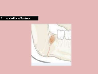
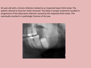
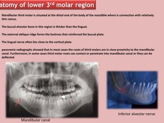
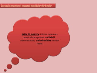
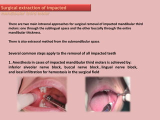
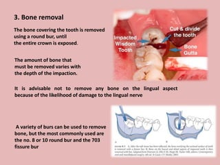
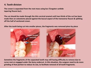
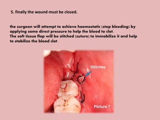
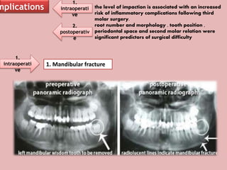
A 65-year-old patient presented with a chronic infection related to an impacted lower third molar. Despite recommendations for removal, the patient refused treatment, resulting in progression of the deep bone infection and a pathologic fracture of the jaw. Factors such as age, medical comorbidities, proximity to adjacent structures, and risk of damage during surgery are considered when determining if an impacted third molar should be removed. Surgical extraction involves raising flaps, removing bone, dividing and extracting the tooth, and closing the wound. Postoperative care may include antibiotics, analgesics, and steroids. Complications can be intraoperative such as nerve injury, fracture, or bleeding, or postoperative like pain, swelling, and infection.
Detailed description on management of impacted maxillary and mandibular third molars. Surgical approaches and complications are also discussed in details.
Classification of Impaction and Methods & Techniques of Third molar/Manidibular impaction removal,Flap designs of impaction removal techniques and more
Dr. Ahmed M. Adawy, Professor Emeritus, Dep. Oral & Maxillofacial Surgery. Former Dean, Faculty of Dental Medicine
Al-Azhar University. Impacted teeth can be defined as those prevented from eruption at the expected time due to physical barrier. The etiology, frequency of impactions are given. Classification systems are based on the depth, angulation, and available space. Complications associated with lower third molar impaction are discussed and methods of treatment are explained. Comparison between maxillary third molar and mandibular one is given. Upper canine is the second most commonly impacted tooth after third molars. It form the foundation of an esthetic smile. The management of impacted canine is interdisciplinary management comprises of a team of an orthodontist, oral surgeon, and periodontist.
Wisdom teeth are the third and last molars on each side of the upper and lower jaws. They are also the final teeth to erupt; they usually appear when a person is in their late teens or early twenties
Terminology in Orthodontics
Copyright by Department of Orthodontics
University of Dental Medicine, Yangon
Feel free to request to take it down this slide if you are copyright owner.
presentation about impacted canine incidence, prevalence,classification,diagnosis, localization and treatment options including surgical and non surgical modalities
Fenestration
Dehiscence: lack of cortical bone at the level of dental root, at least 4mm apical to the margin of interproximal bone, the measurement was per found by graduated probe.
Or: lowering the crestal bone margin to expose root surface.
Fenestration: localized defect in the alveolar bone that exposed the root surface usually the apical or the medium third but did not involve the alveolar margin.
Or: is isolated areas in which the root is denuded of bone and the root surface is covered only by periosteum and overlying gingiva.
Fenestration is a term derived from Latin ward fenestra, meaning window, describe area of alveolar process devoid of bone, creating a window exposing the root surface.
Gingival recession: is the apical movement of the marginal soft tissue surrounding the tooth exposing root surface.
According to some studies that irreversible recession can be caused by fixed appliances for 1.3% to 10.0% of treated cases.
We can use CBCT to measure the labial and lingual thickness of the bone related to roots.
ABT: apical buccal thickness. MBT: middle buccal thickness. CBT: cervical buccal thickness.
ALT: apical lingual thickness. MLT; middle lingual thickness. CLT: cervical lingual thickness.
== naturally occurring alveolar bone dehiscence and fenestration are common finding in different type of malocclusion especially in anterior region of class III.
== fenestration and dehiscence may lead to gingival recession and additional bone loss during orthodontic treatment in addition to large amount of labial inclination such as decompensation in class III malocclusion may pose a greater risk of periodontal complication such as:
Alveolar dehiscence, fenestration, gingival recession.
Clinically gingival recession, is always accompanied by alveolar bone dehiscence whether underlying bone dehiscence is developed before or parallel to gingival recession.
N:B- gingival recession; is described as exposure of root surface by an apical shift in the position of gingiva.
Factors affect recession increase:
1- Difficulty of plaque control due to fixed appliances
2- Proclination of teeth.
3- Coronally attached Frenum.
4- Muscle attachment.
5- Abnormal tooth position.
6- Overhanging restoration or crowns.
7- Fenestration and dehiscence.
** Classification of fenestration according to their apicocoronal location in relation to root length to:
1- At the level of the apical third of the dental root 48% all in maxilla.
2- At the level of the middle third of dental root 28% in maxilla and mandible.
3- At the level of the coronal third of the dental root 19% all in mandible.
4- Extending from the apical to the middle third of the dental root 4.3% of them located in maxilla.
** Etiology of dehiscence:
1- Ectopically positioned teeth which are outside of the bony limits of the alveolus are often lacking the normal amount of bone on the overlying facial surface.
2- Roots of the toot
Treatment of Extremely Displaced and Impacted Second Premolar in the MandibleAbu-Hussein Muhamad
The mandibular second premolar is one of the most frequently impacted teeth. The recommended treatment is to extract the second primary molar with or without removing the bone along the eruption path, to uncover the tooth surgically and move it into the arch by orthodontic treatment. The purpose of this article is to review the principles of case management of soft tissue impacted second premolars mandibular and to illustrate their potential to respond well to the treatment. Although the scope of treatment may depend on a varying range of factors, this case report demonstrates the inherent potential for good treatment outcome in cases of soft tissue impactions.
The Indian Dental Academy is the Leader in continuing dental education , training dentists in all aspects of dentistry and
offering a wide range of dental certified courses in different formats.
Impaction is the cessation of eruption of a tooth caused by a physical barrier or ectopic positioning of a tooth.
unerupted tooth is a tooth lying within the jaw bone, entirely covered by soft tissue, and partially or completely covered by bone.
A partially erupted tooth is a tooth that has failed to erupt fully into a normal position
These lecture slides, by Dr Sidra Arshad, offer a quick overview of physiological basis of a normal electrocardiogram.
Learning objectives:
1. Define an electrocardiogram (ECG) and electrocardiography
2. Describe how dipoles generated by the heart produce the waveforms of the ECG
3. Describe the components of a normal electrocardiogram of a typical bipolar leads (limb II)
4. Differentiate between intervals and segments
5. Enlist some common indications for obtaining an ECG
Study Resources:
1. Chapter 11, Guyton and Hall Textbook of Medical Physiology, 14th edition
2. Chapter 9, Human Physiology - From Cells to Systems, Lauralee Sherwood, 9th edition
3. Chapter 29, Ganong’s Review of Medical Physiology, 26th edition
4. Electrocardiogram, StatPearls - https://www.ncbi.nlm.nih.gov/books/NBK549803/
5. ECG in Medical Practice by ABM Abdullah, 4th edition
6. ECG Basics, http://www.nataliescasebook.com/tag/e-c-g-basics
- Video recording of this lecture in English language: https://youtu.be/lK81BzxMqdo
- Video recording of this lecture in Arabic language: https://youtu.be/Ve4P0COk9OI
- Link to download the book free: https://nephrotube.blogspot.com/p/nephrotube-nephrology-books.html
- Link to NephroTube website: www.NephroTube.com
- Link to NephroTube social media accounts: https://nephrotube.blogspot.com/p/join-nephrotube-on-social-media.html
Flu Vaccine Alert in Bangalore Karnatakaaddon Scans
As flu season approaches, health officials in Bangalore, Karnataka, are urging residents to get their flu vaccinations. The seasonal flu, while common, can lead to severe health complications, particularly for vulnerable populations such as young children, the elderly, and those with underlying health conditions.
Dr. Vidisha Kumari, a leading epidemiologist in Bangalore, emphasizes the importance of getting vaccinated. "The flu vaccine is our best defense against the influenza virus. It not only protects individuals but also helps prevent the spread of the virus in our communities," he says.
This year, the flu season is expected to coincide with a potential increase in other respiratory illnesses. The Karnataka Health Department has launched an awareness campaign highlighting the significance of flu vaccinations. They have set up multiple vaccination centers across Bangalore, making it convenient for residents to receive their shots.
To encourage widespread vaccination, the government is also collaborating with local schools, workplaces, and community centers to facilitate vaccination drives. Special attention is being given to ensuring that the vaccine is accessible to all, including marginalized communities who may have limited access to healthcare.
Residents are reminded that the flu vaccine is safe and effective. Common side effects are mild and may include soreness at the injection site, mild fever, or muscle aches. These side effects are generally short-lived and far less severe than the flu itself.
Healthcare providers are also stressing the importance of continuing COVID-19 precautions. Wearing masks, practicing good hand hygiene, and maintaining social distancing are still crucial, especially in crowded places.
Protect yourself and your loved ones by getting vaccinated. Together, we can help keep Bangalore healthy and safe this flu season. For more information on vaccination centers and schedules, residents can visit the Karnataka Health Department’s official website or follow their social media pages.
Stay informed, stay safe, and get your flu shot today!
ARTIFICIAL INTELLIGENCE IN HEALTHCARE.pdfAnujkumaranit
Artificial intelligence (AI) refers to the simulation of human intelligence processes by machines, especially computer systems. It encompasses tasks such as learning, reasoning, problem-solving, perception, and language understanding. AI technologies are revolutionizing various fields, from healthcare to finance, by enabling machines to perform tasks that typically require human intelligence.
Lung Cancer: Artificial Intelligence, Synergetics, Complex System Analysis, S...Oleg Kshivets
RESULTS: Overall life span (LS) was 2252.1±1742.5 days and cumulative 5-year survival (5YS) reached 73.2%, 10 years – 64.8%, 20 years – 42.5%. 513 LCP lived more than 5 years (LS=3124.6±1525.6 days), 148 LCP – more than 10 years (LS=5054.4±1504.1 days).199 LCP died because of LC (LS=562.7±374.5 days). 5YS of LCP after bi/lobectomies was significantly superior in comparison with LCP after pneumonectomies (78.1% vs.63.7%, P=0.00001 by log-rank test). AT significantly improved 5YS (66.3% vs. 34.8%) (P=0.00000 by log-rank test) only for LCP with N1-2. Cox modeling displayed that 5YS of LCP significantly depended on: phase transition (PT) early-invasive LC in terms of synergetics, PT N0—N12, cell ratio factors (ratio between cancer cells- CC and blood cells subpopulations), G1-3, histology, glucose, AT, blood cell circuit, prothrombin index, heparin tolerance, recalcification time (P=0.000-0.038). Neural networks, genetic algorithm selection and bootstrap simulation revealed relationships between 5YS and PT early-invasive LC (rank=1), PT N0—N12 (rank=2), thrombocytes/CC (3), erythrocytes/CC (4), eosinophils/CC (5), healthy cells/CC (6), lymphocytes/CC (7), segmented neutrophils/CC (8), stick neutrophils/CC (9), monocytes/CC (10); leucocytes/CC (11). Correct prediction of 5YS was 100% by neural networks computing (area under ROC curve=1.0; error=0.0).
CONCLUSIONS: 5YS of LCP after radical procedures significantly depended on: 1) PT early-invasive cancer; 2) PT N0--N12; 3) cell ratio factors; 4) blood cell circuit; 5) biochemical factors; 6) hemostasis system; 7) AT; 8) LC characteristics; 9) LC cell dynamics; 10) surgery type: lobectomy/pneumonectomy; 11) anthropometric data. Optimal diagnosis and treatment strategies for LC are: 1) screening and early detection of LC; 2) availability of experienced thoracic surgeons because of complexity of radical procedures; 3) aggressive en block surgery and adequate lymph node dissection for completeness; 4) precise prediction; 5) adjuvant chemoimmunoradiotherapy for LCP with unfavorable prognosis.
Title: Sense of Smell
Presenter: Dr. Faiza, Assistant Professor of Physiology
Qualifications:
MBBS (Best Graduate, AIMC Lahore)
FCPS Physiology
ICMT, CHPE, DHPE (STMU)
MPH (GC University, Faisalabad)
MBA (Virtual University of Pakistan)
Learning Objectives:
Describe the primary categories of smells and the concept of odor blindness.
Explain the structure and location of the olfactory membrane and mucosa, including the types and roles of cells involved in olfaction.
Describe the pathway and mechanisms of olfactory signal transmission from the olfactory receptors to the brain.
Illustrate the biochemical cascade triggered by odorant binding to olfactory receptors, including the role of G-proteins and second messengers in generating an action potential.
Identify different types of olfactory disorders such as anosmia, hyposmia, hyperosmia, and dysosmia, including their potential causes.
Key Topics:
Olfactory Genes:
3% of the human genome accounts for olfactory genes.
400 genes for odorant receptors.
Olfactory Membrane:
Located in the superior part of the nasal cavity.
Medially: Folds downward along the superior septum.
Laterally: Folds over the superior turbinate and upper surface of the middle turbinate.
Total surface area: 5-10 square centimeters.
Olfactory Mucosa:
Olfactory Cells: Bipolar nerve cells derived from the CNS (100 million), with 4-25 olfactory cilia per cell.
Sustentacular Cells: Produce mucus and maintain ionic and molecular environment.
Basal Cells: Replace worn-out olfactory cells with an average lifespan of 1-2 months.
Bowman’s Gland: Secretes mucus.
Stimulation of Olfactory Cells:
Odorant dissolves in mucus and attaches to receptors on olfactory cilia.
Involves a cascade effect through G-proteins and second messengers, leading to depolarization and action potential generation in the olfactory nerve.
Quality of a Good Odorant:
Small (3-20 Carbon atoms), volatile, water-soluble, and lipid-soluble.
Facilitated by odorant-binding proteins in mucus.
Membrane Potential and Action Potential:
Resting membrane potential: -55mV.
Action potential frequency in the olfactory nerve increases with odorant strength.
Adaptation Towards the Sense of Smell:
Rapid adaptation within the first second, with further slow adaptation.
Psychological adaptation greater than receptor adaptation, involving feedback inhibition from the central nervous system.
Primary Sensations of Smell:
Camphoraceous, Musky, Floral, Pepperminty, Ethereal, Pungent, Putrid.
Odor Detection Threshold:
Examples: Hydrogen sulfide (0.0005 ppm), Methyl-mercaptan (0.002 ppm).
Some toxic substances are odorless at lethal concentrations.
Characteristics of Smell:
Odor blindness for single substances due to lack of appropriate receptor protein.
Behavioral and emotional influences of smell.
Transmission of Olfactory Signals:
From olfactory cells to glomeruli in the olfactory bulb, involving lateral inhibition.
Primitive, less old, and new olfactory systems with different path
Anti ulcer drugs and their Advance pharmacology ||
Anti-ulcer drugs are medications used to prevent and treat ulcers in the stomach and upper part of the small intestine (duodenal ulcers). These ulcers are often caused by an imbalance between stomach acid and the mucosal lining, which protects the stomach lining.
||Scope: Overview of various classes of anti-ulcer drugs, their mechanisms of action, indications, side effects, and clinical considerations.
2. introduction
An impacted tooth defined as a tooth that is prevented from erupting into
position
Causesof
impaction
lack of space
angulated roots were
more common in
impacted mandibular
third molars
hereditary
factors
lack of sufficient
eruption force for
third molars
regression of the jaw
size
3. lassifications
Classification describes wisdom tooth relation to the adjacent anatomical structures: mandibular
ramus, second molar, alveolar crest, mandibular canal.
Impacted wisdom teeth are classified by the direction and depth of impaction, the amount of
available space for tooth eruption, and the amount of soft tissue or bone (or both) that covers them.
The classification structure helps clinicians estimate the risks for impaction, infections and
complications associated with wisdom teeth removal.
Depth of impaction
Relationship with the mandibular ramus
Angulation of impaction
4. Depth of impaction
According to the Pell and Gregory classification, the relation of the
cementoenamel junction (CEJ) of the third molar with the bone level is
categorized as follows:
Level A:
• Not buried in bone
Level B
• Partially buried in bone if any part of
CEJ was lower than bone level
Level C
• Completely buried in bone
5. Relationship with the mandibular ramus
According to the Pell and Gregory classification, the position of the distal surface of the third molar
crown in relation to the anterior border of the ascending ramus is categorized as follows:
Class I: Anterior to the
anterior border;
Class II: Half of the crown
is covered by the anterior
border
Class III: The crown is
fully covered by the
anterior border
6. Angulation of impaction
Based on Winter's classification, the angle between the
longitudinal axis of the second and third molars
Mesioangular
impaction
Horizontal impaction
Vertical
impaction
Distoangular
impaction
11. MANAGEMENT OF UNERUPTED AND IMPACTED THIRD MOLAR TEETH
CLINICAL ASSESSMENT
Clinical assessment should be carried out with the aim of assessing the status
of the third molars and excluding other causes of the symptoms. A complete
examination should include assessment of:
• the eruption status of the third molar
• the presence of local infection
• caries in, or resorption of, the third molar and the adjacent tooth
• periodontal status
• orientation and relationship of the tooth to the inferior dental canal (IDC)
• occlusal relationship
• temporomandibular joint function
• regional lymph nodes.
Any associated pathology should also be noted.
12. RADIOLOGICAL EVALUATION
The purpose of a careful radiological evaluation is to complement the clinical examination by providing
additional information about the third molar, the related teeth and anatomical features, and the surrounding
bone. This is necessary in order to make a sound decision about the proposed surgical procedure.
the type and orientation of impaction and the access to the tooth.
the crown size and condition
the root number and morphology
The alveolar bone level
the periodontal status
the relationship or proximity of lower third molars to the inferior dental canal.
ing information should be noted:
13. To remove OR
not
1. Impacted teeth with pericoronitis should be extracted electively because
of their known potential for repetitive infection and morbidity.
ation for extraction
18. 6. preceding dental work with fixed or
removable appliances
7. for chronic facial pain.
wding of the dental arch
ation of the opposing soft tissue
19. 65 year-old with a chronic infection related to an impacted lower third molar. The
patient refused to have her tooth removed. The delay in proper treatment resulted in
progression of the deep bone infection caused by the impacted third molar. This
eventually resulted in a pathologic fracture of the jaw.
22. Mandibular third molar is situated at the distal end of the body of the mandible where is connection with relatively
thin ramus.
The buccal alveolar bone in this region is thicker than the lingual.
The external oblique ridge forms the buttress that reinforced the buccal plate.
The lingual nerve often lies close to the cortical plate.
panoramic radiographs showed that in most cases the roots of third molars are in close proximity to the mandibular
canal. Furthermore, in some cases third molar roots can contact or penetrate into mandibular canal or they can be
deflected.
atomy of lower 3rd molar region
Inferior alveolar nerve
Mandibular canal
23. prior to surgery, interim measures
may include systemic antibiotic
administration, chlorhexidine mouth
rinses
24. Surgical extraction of impacted
mandibular third molar
Several common steps apply to the removal of all impacted teeth
1. Anesthesia in cases of impacted mandibular third molars is achieved by:
inferior alveolar nerve block, buccal nerve block , lingual nerve block,
and local infiltration for hemostasis in the surgical field
There are two main intraoral approaches for surgical removal of impacted mandibular third
molars: one through the sublingual space and the other buccally through the entire
mandibular thickness.
There is also extraoral method from the submandibular space.
25. 2. Adequate flaps must be reflected for accessibility, Accessibility is a key issue in
removal of impacted teeth.
A full-thickness mucoperiosteal flap must be elevated to allow for visualization and
placement of retractors, drilling equipment, elevators, and forceps.
26. However there are many modifications of flap
techniques including :
1.Triangular flap (L-shaped)
27. 2. Variation of the triangular flap (bayonet)
3. Envelope flap
28. 3. Bone removal
The bone covering the tooth is removed
using a round bur, until
the entire crown is exposed.
The amount of bone that
must be removed varies with
the depth of the impaction.
It is advisable not to remove any bone on the lingual aspect
because of the likelihood of damage to the lingual nerve
A variety of burs can be used to remove
bone, but the most commonly used are
the no. 8 or 10 round bur and the 703
fissure bur
29. 4. Tooth division
After the tooth being cut into smaller pieces, the fragments was removed piece by piece
Sometime the fragments of the separated tooth may still having difficulty to remove due to
curve root or engaged under the bony undercut. In this situation, the surgeon need to do more
grinding of that portion to reduce its size, to facilitate removal of all tooth fragments.
The crown is separated from the root mass using bur (Tungsten carbide
tapering fissure bur).
The cut should be made through the thin cervical enamel until two thirds of the cut has been
made then an osteotome placed against the buccal aspect of the transverse fissure & splitting
off the half of sectioned crown.
30. 5. finally the wound must be closed.
the surgeon will attempt to achieve haemostatic (stop bleeding) by
applying some direct pressure to help the blood to clot.
The soft tissue flap will be stitched (suture) to immobilize it and help
to stabilize the blood clot.
31.
32. POSTOPERATIVE DRUG THERAPY
Antibiotic
Analgesia
steroid
•Where there is significant
bone removal, prolonged
operation time, or the
patient is at increased risk
of infection
•Normal practice is to
prescribe or advise oral
analgesics such as
paracetamol or ibuprofen.
•Where there is a risk of
significant postoperative
swelling
33. mplications
1.
intraoperati
ve
2.
postoperativ
e
the level of impaction is associated with an increased
risk of inflammatory complications following third
molar surgery.
root number and morphology , tooth position ,
periodontal space and second molar relation were
significant predictors of surgical difficulty
1.
intraoperati
ve
1. Mandibular fracture
34. 2. damage of adjacent teeth,
(In cases if the excessive intraoral force was
applied or/and part of bone was removed, risk
of mandibular fracture or damage of adjacent
teeth is increased)
3. tooth or tooth fragments displacement into soft tissues
can occur in case of wrong operation technique
4. bleeding.
35. The most serious and unpleasant iatrogenic complication that arise from third molar surgery is
inferior alveolar and/or lingual nerve injury and neurosensory function disturbance.
Inferior alveolar nerve injury can cause paresthesia to complete numbness and/or pain in the
region of the skin of the mental area, the lower lip, mucous membranes, and the gingiva as far
posteriorly as the second premolar . Furthermore this commonly interferes with speech, eating,
and drinking.
The injury of the lingual nerve leads to numbness of the ipsilateral anterior two thirds of the
tongue and taste disturbance .
Eruption status of the lower third molar is important risk factor for inferior alveolar
nerve injury. The risk of nerve injury is increasing with the depth of the impacted
mandibular wisdom teeth.
In general the proximity of the mandibular third molar to the mandibular canal is
considered a risk factor for damage to the inferior alveolar nerve.
Studies demonstrated that the most important parameters for inferior alveolar nerve
injury prediction are third molar root apices inside or in contact with the mandibular
canal.
Iatrogenic injury to the lingual nerve may happen during third molar surgery due to the
anatomical proximity of the cortex region of the molar to the nerve, being separated from
it by the periosteum alone.
5. inferior alveolar and/or lingual nerve injury and neurosensory function disturbance
36. 2.
postoperativ
e
are pain, swelling, bruising, trismus , osteitis and
surgical site infection .
Furthermore, the prevalence of post-extraction
complications correlated with the absence of
cortication around the mandibular canal
swelling after wisdom teeth removal
37.
38. Other possible complications will be prolonged pain & discomfort, usually due to dense
socket bone around the wisdom tooth that restrain the blood clot formation ( Dry
Socket )