Downloaded 146 times
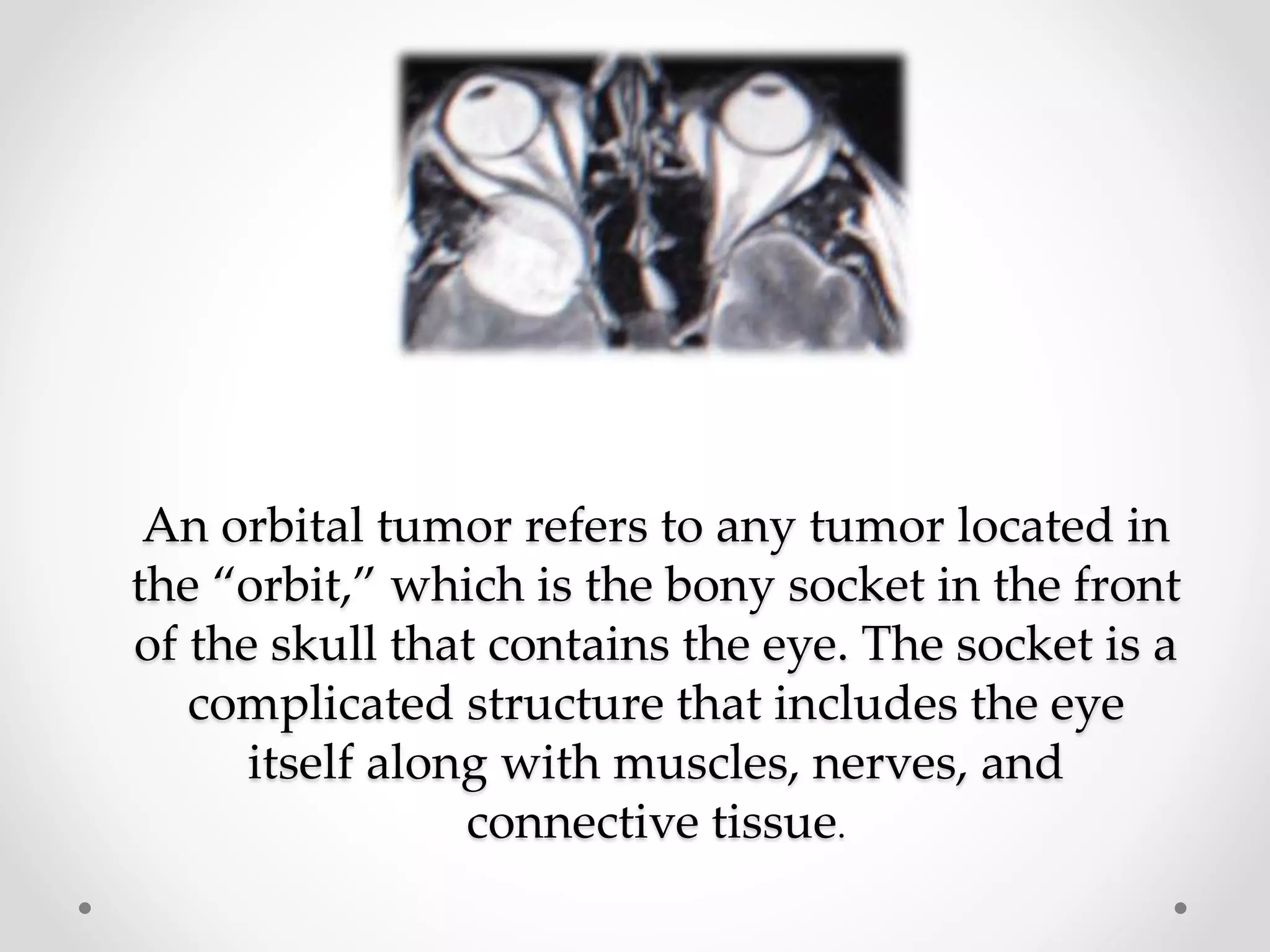


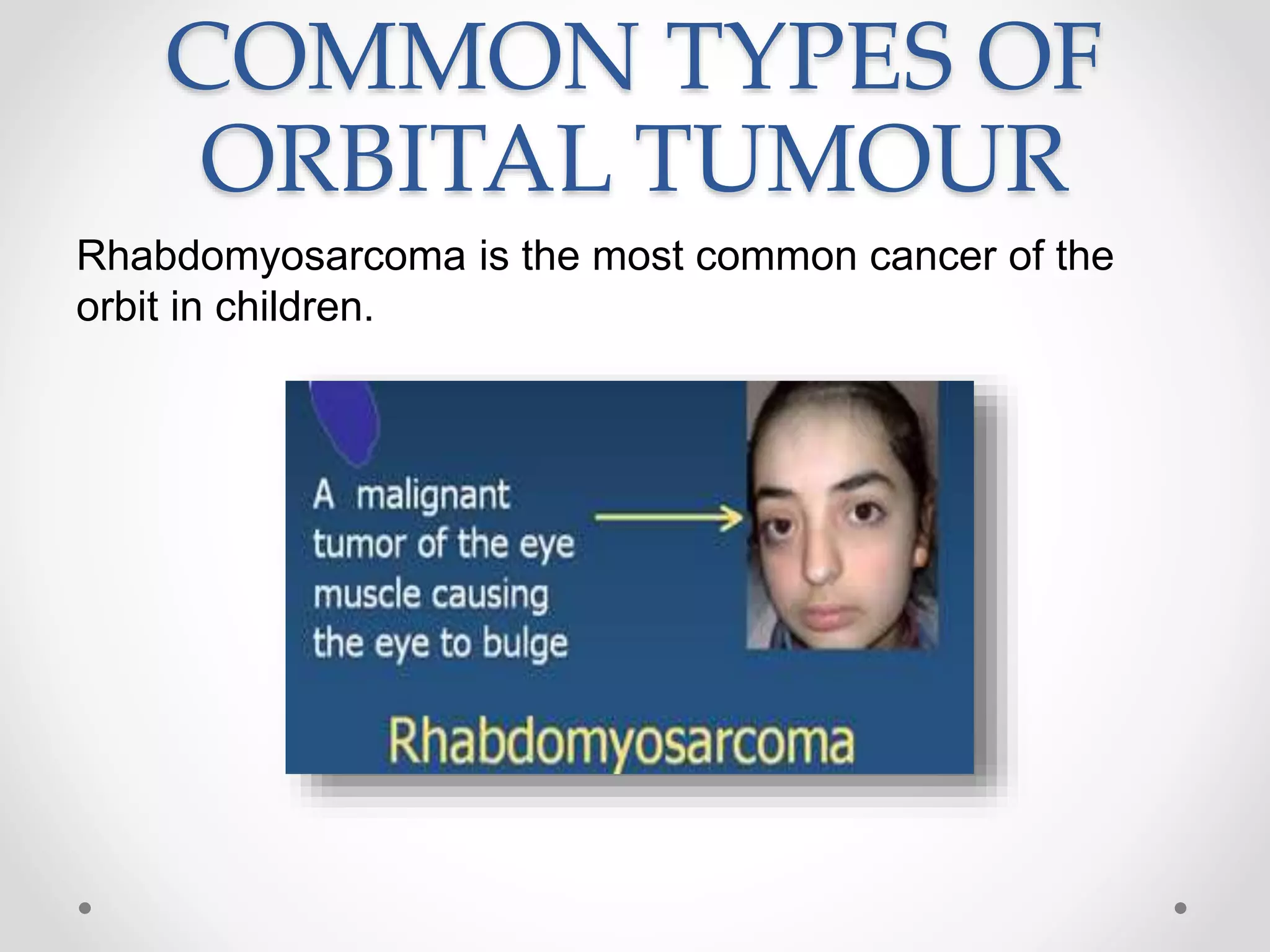
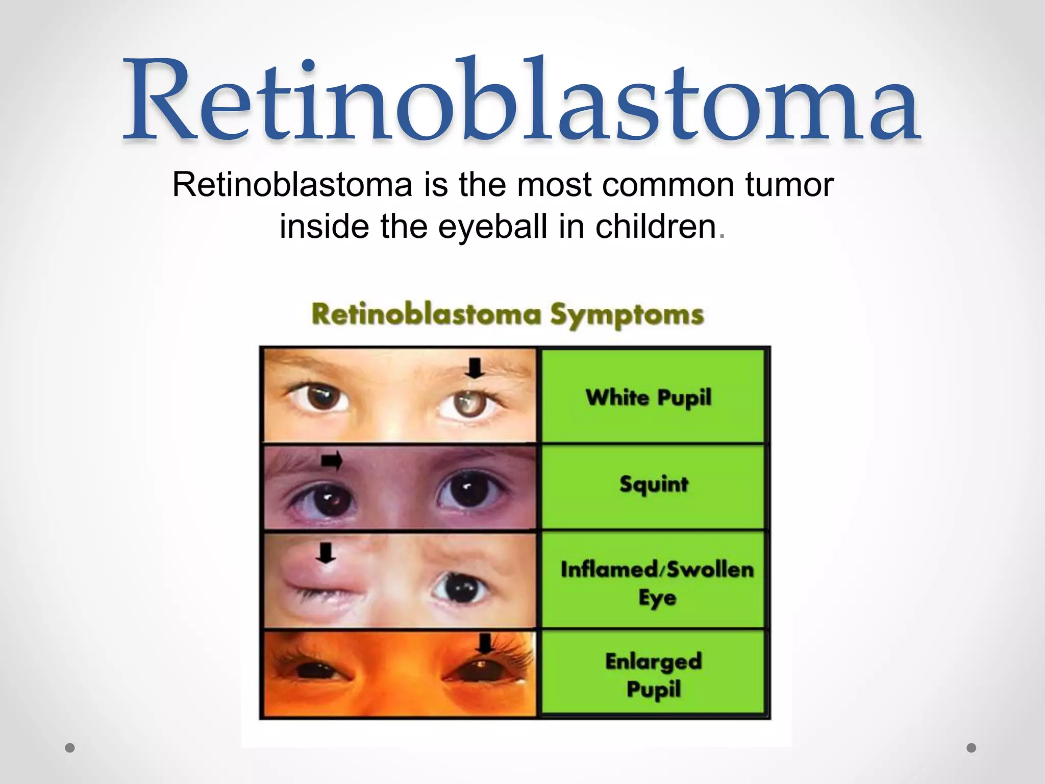

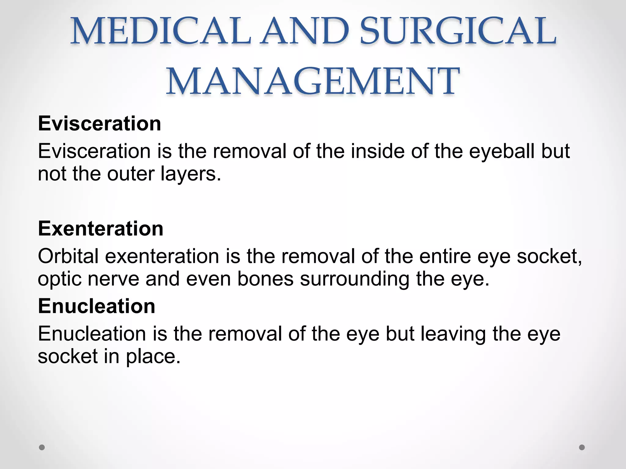


The document discusses orbital tumors, which are tumors located in the bony socket of the skull that contains the eye, outlining their causes, symptoms, diagnostic tests, and treatment options. It highlights common types of orbital tumors in children and adults, details the TNM staging system for cancer, and reviews various surgical and medical management options available. The prognosis varies depending on the tumor type and may require therapies like surgery, radiotherapy, and chemotherapy.


![Apporach to lung biopsy [Auto-saved].pptx latest](https://cdn.slidesharecdn.com/ss_thumbnails/apporachtolungbiopsyauto-saved-251211225655-93258539-thumbnail.jpg?width=640&height=640&fit=bounds)


