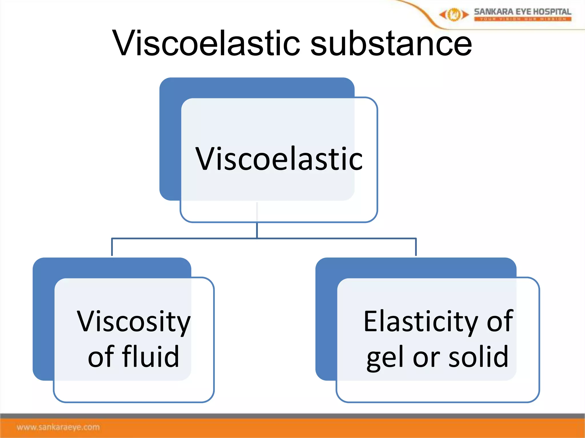
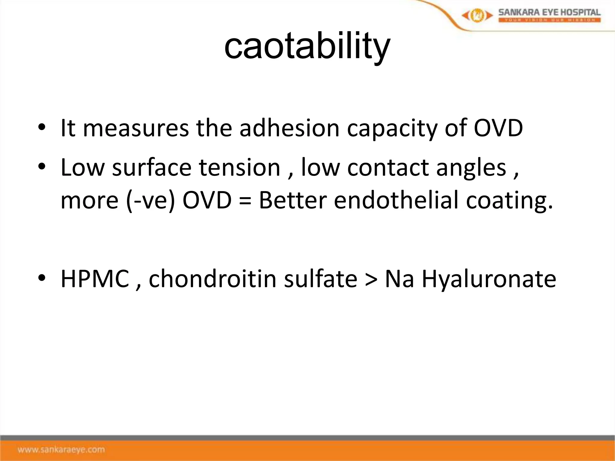
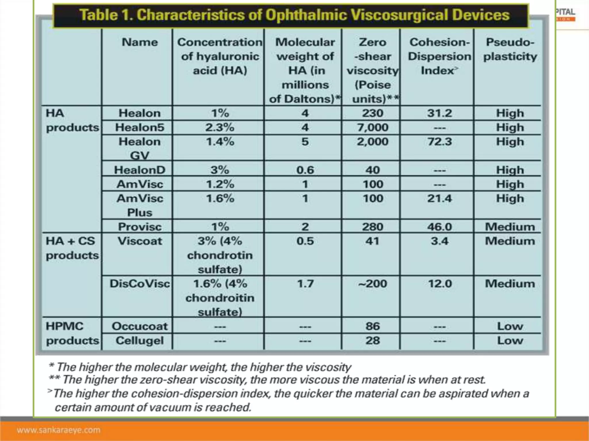
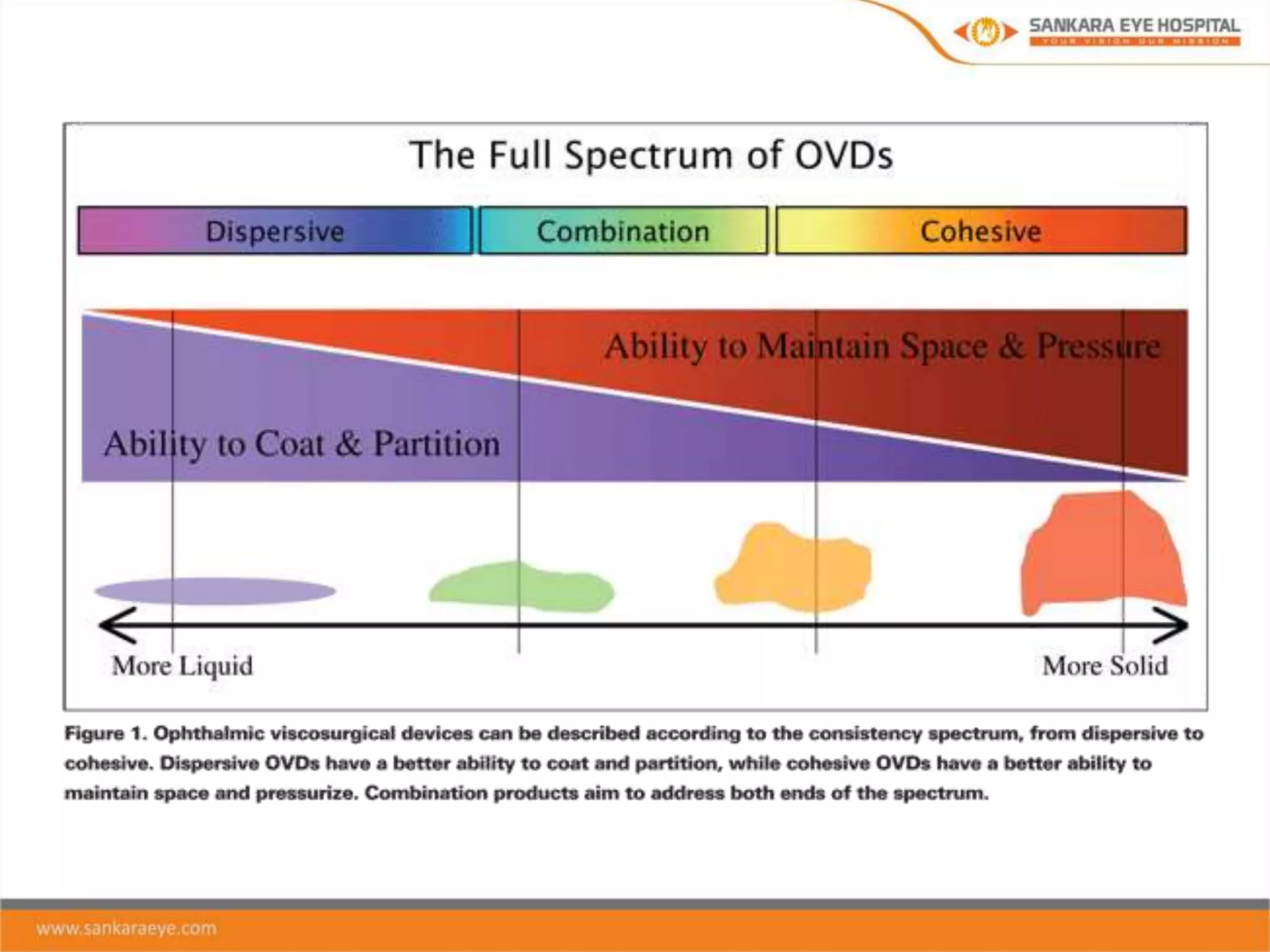
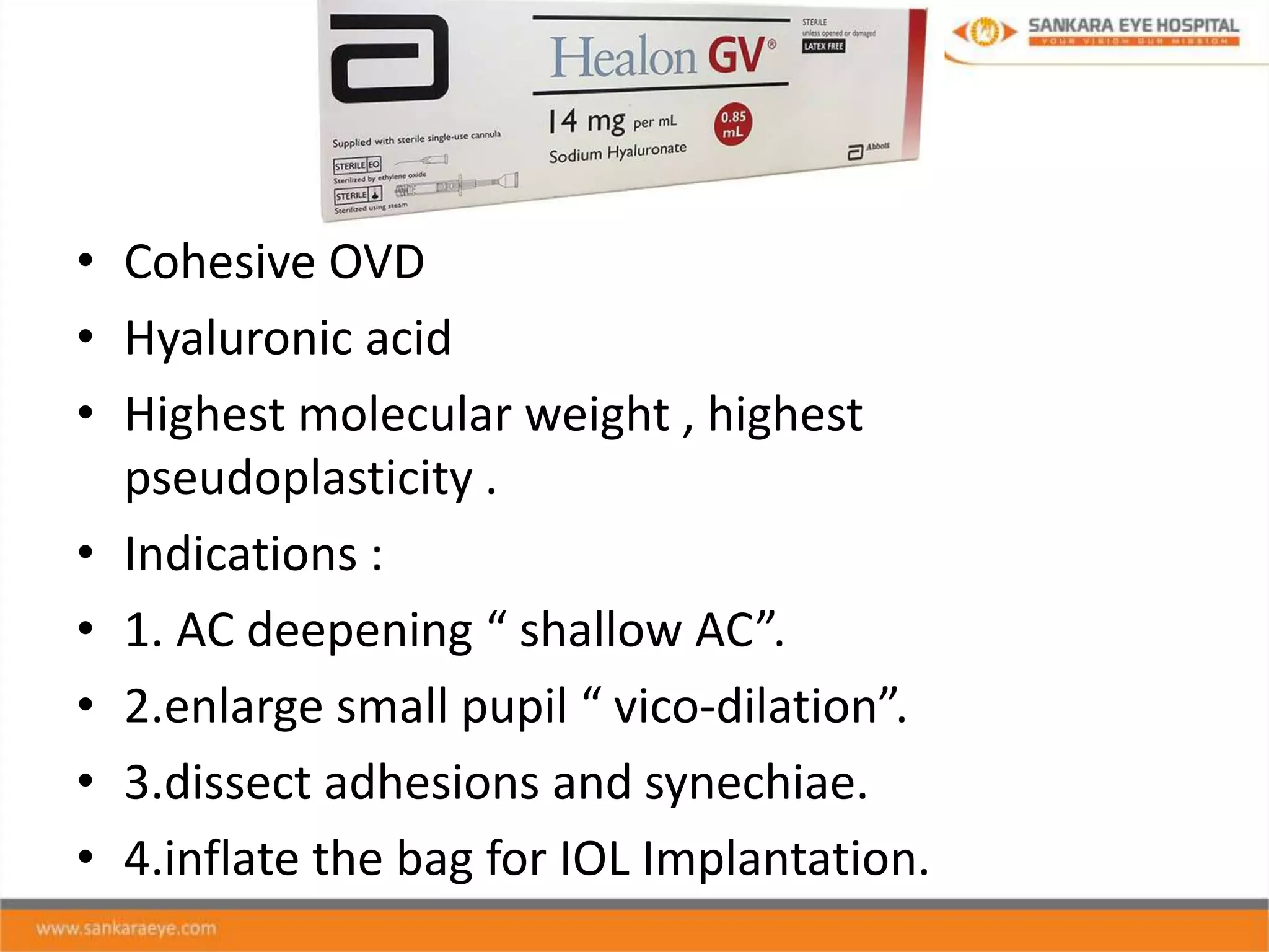
OVD
Ophthalmic Viscosurgical Devices
Presented by : Dr. Amr Mohamed
Moderator : Dr. Vidhya C
Outline
• Historical overview.
• Characteristics of ideal viscoelastic.
• Rheological properties.
• Classification of OVD’s.
• Commonly used OVD’s.
• Clinical uses .
• Complications associated with OVD use.
Historical overview
Viscoelastic substance
Viscoelastic
Viscosity
of fluid
Elasticity of
gel or solid
Characteristics of ideal viscoelastic
• Inert , iso-osmotic , non-toxic .
• Optically clear
• Ease of infusion and removal
• Retention under positive pressure in eye and
during phaco.
• Endothelium protection.
• Cost effective .
Rheological properties
Viscosity
Pseudoplasticity
Elasticity
Coatability
Cohesiveness
Dispersiveness
Viscosity
pseudoplasticity
• It refers to changes in viscosity with different shear rates.
• It is the ability of the solution to transform from gel like
state in to liquid like state (viscocity reduces) under
pressure ( shear force).
Low shear High shear
Substance at rest Substance under force
Viscosity increases Viscosity decreases
Gel form Liquid form
Elasticity
caotability
• It measures the adhesion capacity of OVD
• Low surface tension , low contact angles ,
more (-ve) OVD = Better endothelial coating.
• HPMC , chondroitin sulfate > Na Hyaluronate
Cohesiveness
Dispersiveness
• It is the tendency of a material to disperse
when injected in to AC.
OVD
viscosity
pseudoplasticity
Elastisicity
cohesiveness
coatability
dispersiveness
Composition of OVD
• Sodium hyalouronate
• Chondroitin sulfate
• Hydroxypropyl methyl cellulose ( HPMC)
Sodium hyalouronate
• It is a glycosaminoglycan and long
chain polymer of disaccharide
units of Na-glucuronate-n-
acetylglucosamine.
• Naturally in many connective
tissues throughout the body .
• Mainly present in visco-cohesives.
• High molecular weight
Chondroitin sulfate
• Sulfated glycosaminoglycan “ GAG”
• A chain of alternating sugars ( n-
acetylgalactosamine and glucouronic
acid).
• Obtained from shark fish cartilage .
• Coats tissues but poor space
maintainer.
• Low molecular weight .
Hydroxypropyl methylcellulose “
HPMC”
• Widely disrtibuted in plant fibers.
• Easy availability.
• Ease of preparation.
• Storage at room temperature.
• Ability to with stand autoclaving.
• Main components in dispersives.
Classification of OVD
Cohesive Viscoadaptive Dispersive
Cohesive OVD
• High viscosity .
• Able to pressurize the eye .
• Creating and maintaining spaces.
• They act like a gel .
• Eg : cohesive Na-Ha.
Dispersive OVD
• Low viscosity
• Ability to coat intraocular structures
• They tend to stay in fluidic movements of
phaco sx.
• Eg : HPMC , chondroitin sulfate.
• Cohesive OVD
• Hyaluronic acid
• Highest molecular weight , highest
pseudoplasticity .
• Indications :
• 1. AC deepening “ shallow AC”.
• 2.enlarge small pupil “ vico-dilation”.
• 3.dissect adhesions and synechiae.
• 4.inflate the bag for IOL Implantation.
Viscoat
• Dispersive OVD
• Chondroitin sulfate + Na-Hya
• Low molecular weight
• Used mainly in patients with
compromised corneas , dense
cataracts.
Advantages Advantages
Maintain spaces at low shear rates Excellent coating and give superior
endothelial protection
Easily removed Disadvantages
low risk of IOP rise Difficult removal
Disadvantages Do not maintain or stabilise spaces
Minimal coating , less endothelial
protection.
Can form microbubbles and obscure the
view.
High risk of post op IOP rise.
Viscoadaptive OVD
• Changes it’s behaviour at different flow rates.
• Eg: Healon 5
• In low flow rate = ↑ viscosity and cohesive.
• In high flow rate = becomes dispersive and
protects endothelium.
Clinical uses of OVD
• Cataract surgery
• Glaucoma surgery
• Keratoplasty
• Posterior segment surgery
• Ocular trauma surgery
Cataract surgery
• maintain a stable anterior chamber depth and
protect the corneal endothelial cells from
being damaged during the entire surgical
procedure.
Soft shell technique
This technique is effective in reducing corneal
endothelial cell loss after phacoemulsification
surgery, especially in eyes with dense nuclear
opacity
Soft shell technique
• Soft-shell Viscoelastic Technique for Cataract
Surgery.mp4
Ultimate soft shell technique
Glaucoma surgery
• trabeculectomy and goniotomy
• non-penetrating glaucoma surgery
(viscocanalostomy).
Complications associated with OVD use
• Post-op increase in IOP
- Occurs in 1st 24 hrs and resolves within 72 hrs
- Due to mechanical resistance at TM
Capsular block syndrome
• Characterized by accumulation of liquified
substance within a closed chamber inside the
capsular bag , formed because the lens nucleus or
PCIOL optic occludes the anterior capsule opening
“ capsulorrhexis”.
• Intraoperative CBS
• Early post operative CBS
• Late post operative CBS
Conclusion
• OVD use in various ocular surgeries is extremely
important in maintaining space during surgery
and protecting the endothelium , in addition to
making the surgery easier and more controlled.
• Knowing different types of OVD , Their chemical ,
rheological properties is important for choosing
which OVD is suitable for your procedure.
• Importance of Knowing complications associated
with OVD use and ways to avoid and treat safely .
ophthalmic viscosurgical devices

ophthalmic viscosurgical devices

  • 1.
    OVD Ophthalmic Viscosurgical Devices Presentedby : Dr. Amr Mohamed Moderator : Dr. Vidhya C
  • 2.
    Outline • Historical overview. •Characteristics of ideal viscoelastic. • Rheological properties. • Classification of OVD’s. • Commonly used OVD’s. • Clinical uses . • Complications associated with OVD use.
  • 3.
  • 4.
  • 5.
    Characteristics of idealviscoelastic • Inert , iso-osmotic , non-toxic . • Optically clear • Ease of infusion and removal • Retention under positive pressure in eye and during phaco. • Endothelium protection. • Cost effective .
  • 6.
  • 7.
  • 8.
    pseudoplasticity • It refersto changes in viscosity with different shear rates. • It is the ability of the solution to transform from gel like state in to liquid like state (viscocity reduces) under pressure ( shear force). Low shear High shear Substance at rest Substance under force Viscosity increases Viscosity decreases Gel form Liquid form
  • 9.
  • 10.
    caotability • It measuresthe adhesion capacity of OVD • Low surface tension , low contact angles , more (-ve) OVD = Better endothelial coating. • HPMC , chondroitin sulfate > Na Hyaluronate
  • 11.
  • 12.
    Dispersiveness • It isthe tendency of a material to disperse when injected in to AC.
  • 13.
  • 14.
    Composition of OVD •Sodium hyalouronate • Chondroitin sulfate • Hydroxypropyl methyl cellulose ( HPMC)
  • 15.
    Sodium hyalouronate • Itis a glycosaminoglycan and long chain polymer of disaccharide units of Na-glucuronate-n- acetylglucosamine. • Naturally in many connective tissues throughout the body . • Mainly present in visco-cohesives. • High molecular weight
  • 18.
    Chondroitin sulfate • Sulfatedglycosaminoglycan “ GAG” • A chain of alternating sugars ( n- acetylgalactosamine and glucouronic acid). • Obtained from shark fish cartilage . • Coats tissues but poor space maintainer. • Low molecular weight .
  • 19.
    Hydroxypropyl methylcellulose “ HPMC” •Widely disrtibuted in plant fibers. • Easy availability. • Ease of preparation. • Storage at room temperature. • Ability to with stand autoclaving. • Main components in dispersives.
  • 21.
    Classification of OVD CohesiveViscoadaptive Dispersive
  • 23.
    Cohesive OVD • Highviscosity . • Able to pressurize the eye . • Creating and maintaining spaces. • They act like a gel . • Eg : cohesive Na-Ha.
  • 24.
    Dispersive OVD • Lowviscosity • Ability to coat intraocular structures • They tend to stay in fluidic movements of phaco sx. • Eg : HPMC , chondroitin sulfate.
  • 26.
    • Cohesive OVD •Hyaluronic acid • Highest molecular weight , highest pseudoplasticity . • Indications : • 1. AC deepening “ shallow AC”. • 2.enlarge small pupil “ vico-dilation”. • 3.dissect adhesions and synechiae. • 4.inflate the bag for IOL Implantation.
  • 27.
    Viscoat • Dispersive OVD •Chondroitin sulfate + Na-Hya • Low molecular weight • Used mainly in patients with compromised corneas , dense cataracts.
  • 28.
    Advantages Advantages Maintain spacesat low shear rates Excellent coating and give superior endothelial protection Easily removed Disadvantages low risk of IOP rise Difficult removal Disadvantages Do not maintain or stabilise spaces Minimal coating , less endothelial protection. Can form microbubbles and obscure the view. High risk of post op IOP rise.
  • 29.
    Viscoadaptive OVD • Changesit’s behaviour at different flow rates. • Eg: Healon 5 • In low flow rate = ↑ viscosity and cohesive. • In high flow rate = becomes dispersive and protects endothelium.
  • 30.
    Clinical uses ofOVD • Cataract surgery • Glaucoma surgery • Keratoplasty • Posterior segment surgery • Ocular trauma surgery
  • 31.
    Cataract surgery • maintaina stable anterior chamber depth and protect the corneal endothelial cells from being damaged during the entire surgical procedure.
  • 32.
    Soft shell technique Thistechnique is effective in reducing corneal endothelial cell loss after phacoemulsification surgery, especially in eyes with dense nuclear opacity
  • 33.
    Soft shell technique •Soft-shell Viscoelastic Technique for Cataract Surgery.mp4
  • 34.
  • 35.
    Glaucoma surgery • trabeculectomyand goniotomy • non-penetrating glaucoma surgery (viscocanalostomy).
  • 36.
    Complications associated withOVD use • Post-op increase in IOP - Occurs in 1st 24 hrs and resolves within 72 hrs - Due to mechanical resistance at TM
  • 37.
    Capsular block syndrome •Characterized by accumulation of liquified substance within a closed chamber inside the capsular bag , formed because the lens nucleus or PCIOL optic occludes the anterior capsule opening “ capsulorrhexis”. • Intraoperative CBS • Early post operative CBS • Late post operative CBS
  • 39.
    Conclusion • OVD usein various ocular surgeries is extremely important in maintaining space during surgery and protecting the endothelium , in addition to making the surgery easier and more controlled. • Knowing different types of OVD , Their chemical , rheological properties is important for choosing which OVD is suitable for your procedure. • Importance of Knowing complications associated with OVD use and ways to avoid and treat safely .

Editor's Notes

  • #4 In the shift from ICCE to ECCE and with the introduction of IOL with the high rates of endothelial decompensations and other complications . Dr. endre balazs introduced OVD , He worked on Na hyalouronate .
  • #5 As the name denotes = visco” viscosity” + elastic”elasticity” But OVD have more properties we will know about in the next few slides
  • #7 Rheology is the branch of physics in which we study the way in which materials deform or flow in response to applied forces or stresses. The material properties that govern the specific way in which these deformation or flow behaviors occur are called rheological properties
  • #8 Viscosity reflects a solution’s resistance to flow , a function of the molecular weight of the substance. The higher the solution’s molecular weight , the more it resists to flow .
  • #9 This property enables easy injection and removal of an agent
  • #10 Elastic substances are good for maintaining space High molecular weight highly elastic
  • #12 Cohesion the degree to which the material adheres to itself the more cohesive an OVD , the lower the flow rate and hence they are good for maintaining and easily removed
  • #35 The dispersive OVD is injected through the phaco incision to form a central mound on the surface of the anterior capsule, stopping once the anterior chamber (AC) is about 25% full A second viscoadaptive layer is then injected beneath the dispersive onto the surface of the anterior capsule This displaces the dispersive shell upward against the corneal endothelium, creating a smooth, protective shell Injection of viscoadaptive should continue until the pupil stops dilating but before the eye becomes firm. 3. Balanced salt solution, or the xylocaine-phenylephrine mixture, is then injected slowly underneath the viscoadaptive layer, with the cannula directed downward toward the lens surface This creates a pool of low viscosity fluid directly on the lenticular surface.
  • #38 Intraoperative CBS is felt to result from high irrigation pressures during hydrodissection which hyperdistend the posterior capsule, increasing the risk for capsule rupture. Early postoperative CBS is associated with incomplete viscoelastic removal posterior to the IOL with resulting anterior IOL displacement leading to a myopic shift, anterior chamber shallowing, and IOP elevation. Late CBS classically does not exhibit these symptoms and is only noticed due to the decreased visual acuity recognized by the patient.