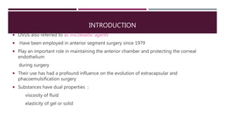
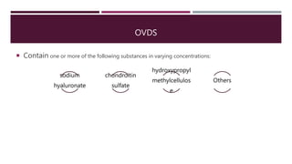
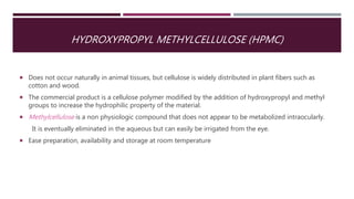
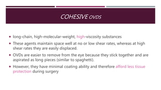
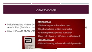
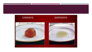
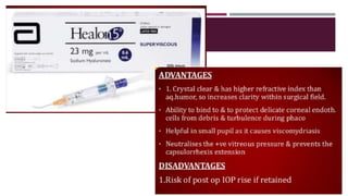
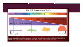
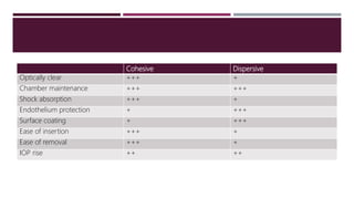
OCULAR VISCOELASTIC DEVICES (OVDS)
WAQAR QABBA’A
MD
LAYOUT
 Introduction
 Physical properties
 Contents
 Classification
 Indication
INTRODUCTION
 OVDs also referred to as viscoelastic agents
 Have been employed in anterior segment surgery since 1979
 Play an important role in maintaining the anterior chamber and protecting the corneal
endothelium
during surgery
 Their use has had a profound influence on the evolution of extracapsular and
phacoemulsification surgery
 Substances have dual properties :
viscosity of fluid
elasticity of gel or solid
IDEAL VISCOLELASTIC SUBSTANCE
 Ease of infusion
 Retention under positive pressure in eye
 Retention during phaco
 Easy removal/no removal needed
 Doesn’t interfere with instrument insertion
 Protect endothelium
 Nontoxic
 Doesn’t obstruct aqueous flow
PHYSICAL PROPERTIES
Elasticity
pseudo plasticity
Viscosity
Surface tension
• They are the result of chain
length and molecular
interactions both within chains
and between chains and
ocular tissue, not due
composition
• 4 general physical properties
PHYSICAL PROPERTIES
 Viscosity describes resistance to flow and can be thought of as the
“thickness” or “thinness” of a fluid.
 It is determined primarily by molecular weight and concentration, so that
substances with high molecular weight and high concentration have the
highest viscosity.
 The higher the viscosity, the better the OVD is at displacing tissue and
staying in place.
 Measured in centipoise (cPs) or centistokes(cSt)
PHYSICAL PROPERTIES
 Elasticity refers to the ability of a material to return to its original
shape after being stressed.
 It describes the OVD’s ability to re-form after an external force is
applied to the anterior chamber and then removed.
 A highly elastic substance is excellent for maintaining space
PHYSICAL PROPERTIES
 Pseudo plasticity refers to the ease with which a material can change
from being highly viscous at rest to being watery at increasing rates of
shear stress.
 This property is found in certain everyday substances such as
toothpaste; when squeezed out of a tube, toothpaste flows easily, but
it retains its shape when it is at rest on the toothbrush.
 In clinical terms, at zero shear force an OVD is a lubricant and coats
tissues well, but when forced through a small-gauge cannula it
functions like a liquid.
PHYSICAL PROPERTIES
 Surface tension describes how the surface of a fluid tends to stick to another
surface. This property is also referred to as coatability, which is inversely
proportional to surface tension.
 Thus, an OVD with low surface tension is better at coating tissue but is
harder to remove from the eye.
OVDS
 Contain one or more of the following substances in varying concentrations:
sodium
hyaluronate
chondroitin
sulfate
hydroxypropyl
methylcellulos
e
Others
SODIUM HYALURONATE
 Biopolymer, disaccharides
 occurs in many connective tissues throughout the body, such as synovial
(joint) fluid and vitreous humor
 It was originally isolated from human umbilical cord and rooster combs
 Has a half-life of approximately 1 day in aqueous and 3 days in vitreous
CHONDROITIN SULFATE
 Sulfated glycosaminoglycan, which is an important component of cartilage.
 Obtained from shark fin cartilage
 Eleminated from AC 24 to 30 hours
HYDROXYPROPYL METHYLCELLULOSE (HPMC)
 Does not occur naturally in animal tissues, but cellulose is widely distributed in plant fibers such as
cotton and wood.
 The commercial product is a cellulose polymer modified by the addition of hydroxypropyl and methyl
groups to increase the hydrophilic property of the material.
 Methylcellulose is a non physiologic compound that does not appear to be metabolized intraocularly.
It is eventually eliminated in the aqueous but can easily be irrigated from the eye.
 Ease preparation, availability and storage at room temperature
CLASSIFICATION
OVDS
Cohesive
Dispersive
COHESIVE OVDS
 long-chain, high-molecular-weight, high-viscosity substances
 These agents maintain space well at no or low shear rates, whereas at high
shear rates they are easily displaced.
 OVDs are easier to remove from the eye because they stick together and are
aspirated as long pieces (similar to spaghetti).
 However, they have minimal coating ability and therefore afford less tissue
protection during surgery
COHESIVE OVDS
 include Healon, Healon GV (Abbott Medical Optics, Santa Ana, CA); Amvisc,
Amvisc Plus (Bausch + Lomb, Rochester, NY); and Provisc (Alcon, Ft Worth, TX)
 HYALURONATE PRODUCTS
DISPERSIVE OVDS
 short-chain, low-molecular-weight, low-viscosity substances with low surface
tension.
 These agents provide excellent coating and protection at high shear rates;
however, they are more difficult to remove from the eye because they do not
stick together and are aspirated in short fragments (similar to macaroni).
 They are more likely to be retained in the eye after cataract surgery, increasing
the likelihood of angle obstruction with reduced outflow and subsequent IOP
elevation
 include OcuCoat (Bausch + Lomb), Viscoat (Alcon), and Healon Endocoat
(Abbott Medical Optics). Discovisc (Alcon) combines qualities of dispersive
and cohesive agents
 MOST OF THEM HPMC
DISPERSIVE OVDS
OTHERS
 Some additional OVDs, such as the viscoadaptive agent Healon5 (Abbott
Medical Optics), may need separate classiffcation.
 Healon5 is a long, fragile chain with high molecular weight that changes its
behavior at diferent flow rates. The lower the flow rate, the more viscous and
cohesive the OVD is, and the higher the flow rate, the more the chains
fracture.
 As a result, this OVD acts as a pseudodispersive agent. However, it must be
carefully removed at the end of surgery because it can cause extremely
elevated IOP if left in the eye
Cohesive Dispersive
Optically clear +++ +
Chamber maintenance +++ +++
Shock absorption +++ +
Endothelium protection + +++
Surface coating + +++
Ease of insertion +++ +
Ease of removal +++ +
IOP rise ++ ++
SOFT SHELL TECHNIQUE
 High myopia
 Floppy iris syndrome
 Cases of broken zonules
 https://www.youtube.com/watch?v=
dX_YT-
uqzAw&feature=player_embedded_
uturn
USES OF OPHTHALMIC VISCOSURGICAL DEVICES
 The space maintenance ability of OVDs keeps the anterior chamber formed despite the presence of
one or more corneal incisions.
 With expansion of the chamber, manipulations can be made away from the corneal endothelium and
posterior lens capsule
 A cohesive OVD can be used to enlarge a marginally dilated pupil (viscomydriasis)
 It can also be used to keep the plane of the anterior capsule flat to assist a controlled continuous
curvilinear capsulorrhexis
 Lens implantation is less traumatic to the zonular fibers and the posterior capsule when the capsular
bag is inflated with an OVD
CONT..
 The coatability of OVDs can be used to protect the corneal endothelium from phaco energy,
particularly in dense cataracts or during long operations. The surgeon must take care to
remove dispersive OVDs completely to reduce the risk of an ocular hypertensive period
caused by angle outflow obstruction.
 In the presence of an open posterior lens capsule, a dispersive OVD can be injected over the
tear to provide a vitreous tamponade and prevent prolapse of vitreous anteriorly.
 The optical clarity of OVDs has prompted some surgeons to use a layer of OVD on the surface
of the cornea. When slightly moistened with balanced salt solution, the agent coats the
epithelium. This maneuver prevents drying and eliminates the need to irrigate the corneal
surface. It also provides a slightly magnified view of anterior segment structures.
CONT…
 The choice of OVD varies depending on the clinical scenario. A survey showed
that 97% of surgeons vary their choice of OVD in complicated cases. For
example, in pediatric cataracts or cases with a low endothelial cell count,
shallow anterior chamber, or intraoperative floppy iris syndrome, the choice of
OVD can play a critical role in management.
INDICATIONS
 Cataract surgery
 Penetrating keratoplasty
 Glaucoma surgery
 Corneal laceration
 Retinal detachment surgery
 Thank you 

Ovds

  • 1.
    OCULAR VISCOELASTIC DEVICES(OVDS) WAQAR QABBA’A MD
  • 2.
    LAYOUT  Introduction  Physicalproperties  Contents  Classification  Indication
  • 3.
    INTRODUCTION  OVDs alsoreferred to as viscoelastic agents  Have been employed in anterior segment surgery since 1979  Play an important role in maintaining the anterior chamber and protecting the corneal endothelium during surgery  Their use has had a profound influence on the evolution of extracapsular and phacoemulsification surgery  Substances have dual properties : viscosity of fluid elasticity of gel or solid
  • 4.
    IDEAL VISCOLELASTIC SUBSTANCE Ease of infusion  Retention under positive pressure in eye  Retention during phaco  Easy removal/no removal needed  Doesn’t interfere with instrument insertion  Protect endothelium  Nontoxic  Doesn’t obstruct aqueous flow
  • 5.
    PHYSICAL PROPERTIES Elasticity pseudo plasticity Viscosity Surfacetension • They are the result of chain length and molecular interactions both within chains and between chains and ocular tissue, not due composition • 4 general physical properties
  • 6.
    PHYSICAL PROPERTIES  Viscositydescribes resistance to flow and can be thought of as the “thickness” or “thinness” of a fluid.  It is determined primarily by molecular weight and concentration, so that substances with high molecular weight and high concentration have the highest viscosity.  The higher the viscosity, the better the OVD is at displacing tissue and staying in place.  Measured in centipoise (cPs) or centistokes(cSt)
  • 7.
    PHYSICAL PROPERTIES  Elasticityrefers to the ability of a material to return to its original shape after being stressed.  It describes the OVD’s ability to re-form after an external force is applied to the anterior chamber and then removed.  A highly elastic substance is excellent for maintaining space
  • 8.
    PHYSICAL PROPERTIES  Pseudoplasticity refers to the ease with which a material can change from being highly viscous at rest to being watery at increasing rates of shear stress.  This property is found in certain everyday substances such as toothpaste; when squeezed out of a tube, toothpaste flows easily, but it retains its shape when it is at rest on the toothbrush.  In clinical terms, at zero shear force an OVD is a lubricant and coats tissues well, but when forced through a small-gauge cannula it functions like a liquid.
  • 9.
    PHYSICAL PROPERTIES  Surfacetension describes how the surface of a fluid tends to stick to another surface. This property is also referred to as coatability, which is inversely proportional to surface tension.  Thus, an OVD with low surface tension is better at coating tissue but is harder to remove from the eye.
  • 10.
    OVDS  Contain oneor more of the following substances in varying concentrations: sodium hyaluronate chondroitin sulfate hydroxypropyl methylcellulos e Others
  • 11.
    SODIUM HYALURONATE  Biopolymer,disaccharides  occurs in many connective tissues throughout the body, such as synovial (joint) fluid and vitreous humor  It was originally isolated from human umbilical cord and rooster combs  Has a half-life of approximately 1 day in aqueous and 3 days in vitreous
  • 12.
    CHONDROITIN SULFATE  Sulfatedglycosaminoglycan, which is an important component of cartilage.  Obtained from shark fin cartilage  Eleminated from AC 24 to 30 hours
  • 13.
    HYDROXYPROPYL METHYLCELLULOSE (HPMC) Does not occur naturally in animal tissues, but cellulose is widely distributed in plant fibers such as cotton and wood.  The commercial product is a cellulose polymer modified by the addition of hydroxypropyl and methyl groups to increase the hydrophilic property of the material.  Methylcellulose is a non physiologic compound that does not appear to be metabolized intraocularly. It is eventually eliminated in the aqueous but can easily be irrigated from the eye.  Ease preparation, availability and storage at room temperature
  • 14.
  • 15.
    COHESIVE OVDS  long-chain,high-molecular-weight, high-viscosity substances  These agents maintain space well at no or low shear rates, whereas at high shear rates they are easily displaced.  OVDs are easier to remove from the eye because they stick together and are aspirated as long pieces (similar to spaghetti).  However, they have minimal coating ability and therefore afford less tissue protection during surgery
  • 16.
    COHESIVE OVDS  includeHealon, Healon GV (Abbott Medical Optics, Santa Ana, CA); Amvisc, Amvisc Plus (Bausch + Lomb, Rochester, NY); and Provisc (Alcon, Ft Worth, TX)  HYALURONATE PRODUCTS
  • 17.
    DISPERSIVE OVDS  short-chain,low-molecular-weight, low-viscosity substances with low surface tension.  These agents provide excellent coating and protection at high shear rates; however, they are more difficult to remove from the eye because they do not stick together and are aspirated in short fragments (similar to macaroni).  They are more likely to be retained in the eye after cataract surgery, increasing the likelihood of angle obstruction with reduced outflow and subsequent IOP elevation  include OcuCoat (Bausch + Lomb), Viscoat (Alcon), and Healon Endocoat (Abbott Medical Optics). Discovisc (Alcon) combines qualities of dispersive and cohesive agents  MOST OF THEM HPMC
  • 18.
  • 20.
    OTHERS  Some additionalOVDs, such as the viscoadaptive agent Healon5 (Abbott Medical Optics), may need separate classiffcation.  Healon5 is a long, fragile chain with high molecular weight that changes its behavior at diferent flow rates. The lower the flow rate, the more viscous and cohesive the OVD is, and the higher the flow rate, the more the chains fracture.  As a result, this OVD acts as a pseudodispersive agent. However, it must be carefully removed at the end of surgery because it can cause extremely elevated IOP if left in the eye
  • 23.
    Cohesive Dispersive Optically clear+++ + Chamber maintenance +++ +++ Shock absorption +++ + Endothelium protection + +++ Surface coating + +++ Ease of insertion +++ + Ease of removal +++ + IOP rise ++ ++
  • 24.
    SOFT SHELL TECHNIQUE High myopia  Floppy iris syndrome  Cases of broken zonules  https://www.youtube.com/watch?v= dX_YT- uqzAw&feature=player_embedded_ uturn
  • 25.
    USES OF OPHTHALMICVISCOSURGICAL DEVICES  The space maintenance ability of OVDs keeps the anterior chamber formed despite the presence of one or more corneal incisions.  With expansion of the chamber, manipulations can be made away from the corneal endothelium and posterior lens capsule  A cohesive OVD can be used to enlarge a marginally dilated pupil (viscomydriasis)  It can also be used to keep the plane of the anterior capsule flat to assist a controlled continuous curvilinear capsulorrhexis  Lens implantation is less traumatic to the zonular fibers and the posterior capsule when the capsular bag is inflated with an OVD
  • 26.
    CONT..  The coatabilityof OVDs can be used to protect the corneal endothelium from phaco energy, particularly in dense cataracts or during long operations. The surgeon must take care to remove dispersive OVDs completely to reduce the risk of an ocular hypertensive period caused by angle outflow obstruction.  In the presence of an open posterior lens capsule, a dispersive OVD can be injected over the tear to provide a vitreous tamponade and prevent prolapse of vitreous anteriorly.  The optical clarity of OVDs has prompted some surgeons to use a layer of OVD on the surface of the cornea. When slightly moistened with balanced salt solution, the agent coats the epithelium. This maneuver prevents drying and eliminates the need to irrigate the corneal surface. It also provides a slightly magnified view of anterior segment structures.
  • 27.
    CONT…  The choiceof OVD varies depending on the clinical scenario. A survey showed that 97% of surgeons vary their choice of OVD in complicated cases. For example, in pediatric cataracts or cases with a low endothelial cell count, shallow anterior chamber, or intraoperative floppy iris syndrome, the choice of OVD can play a critical role in management.
  • 28.
    INDICATIONS  Cataract surgery Penetrating keratoplasty  Glaucoma surgery  Corneal laceration  Retinal detachment surgery
  • 29.