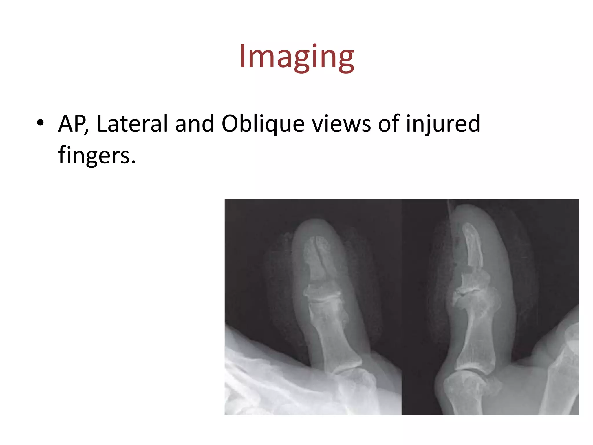
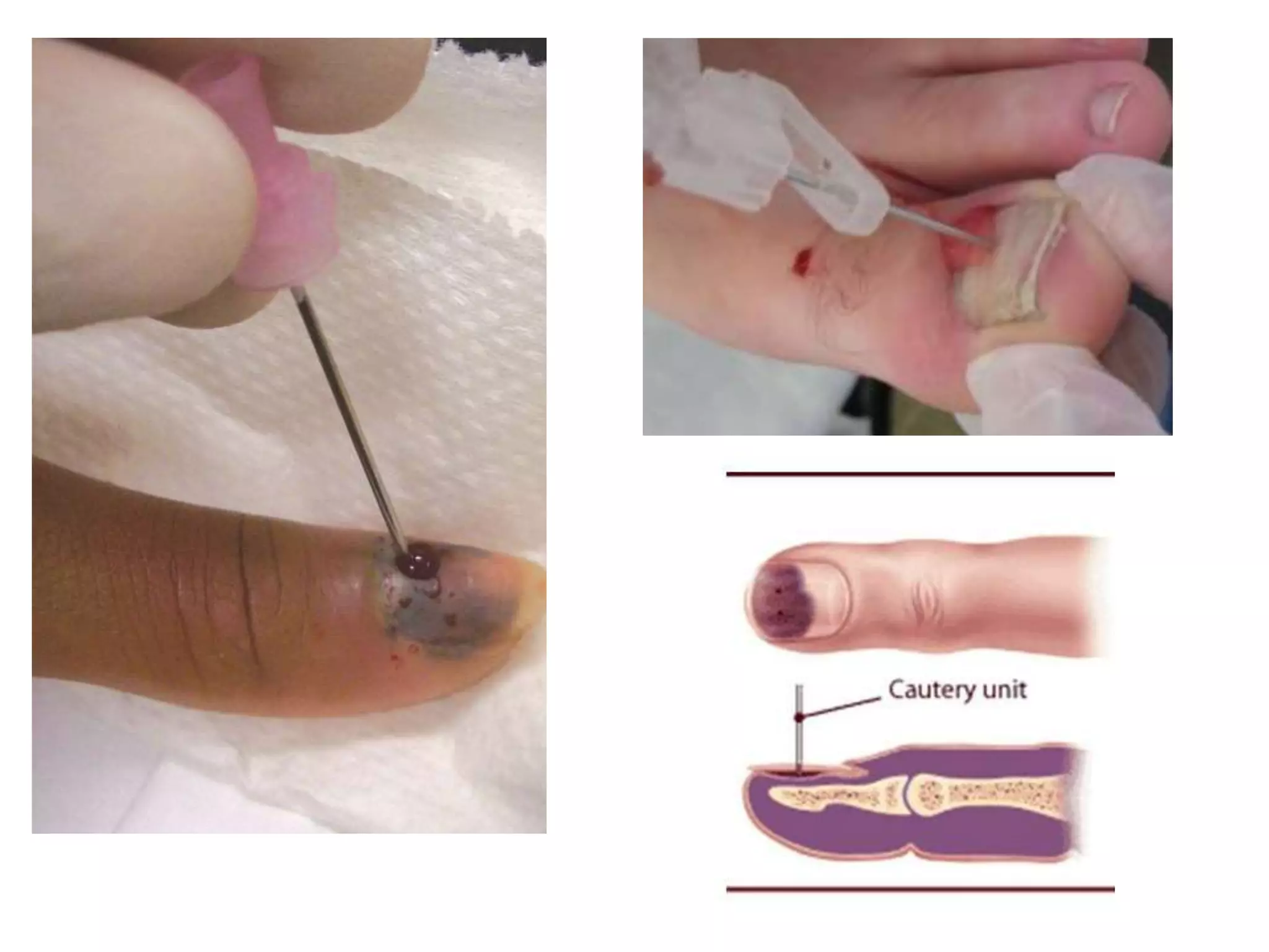
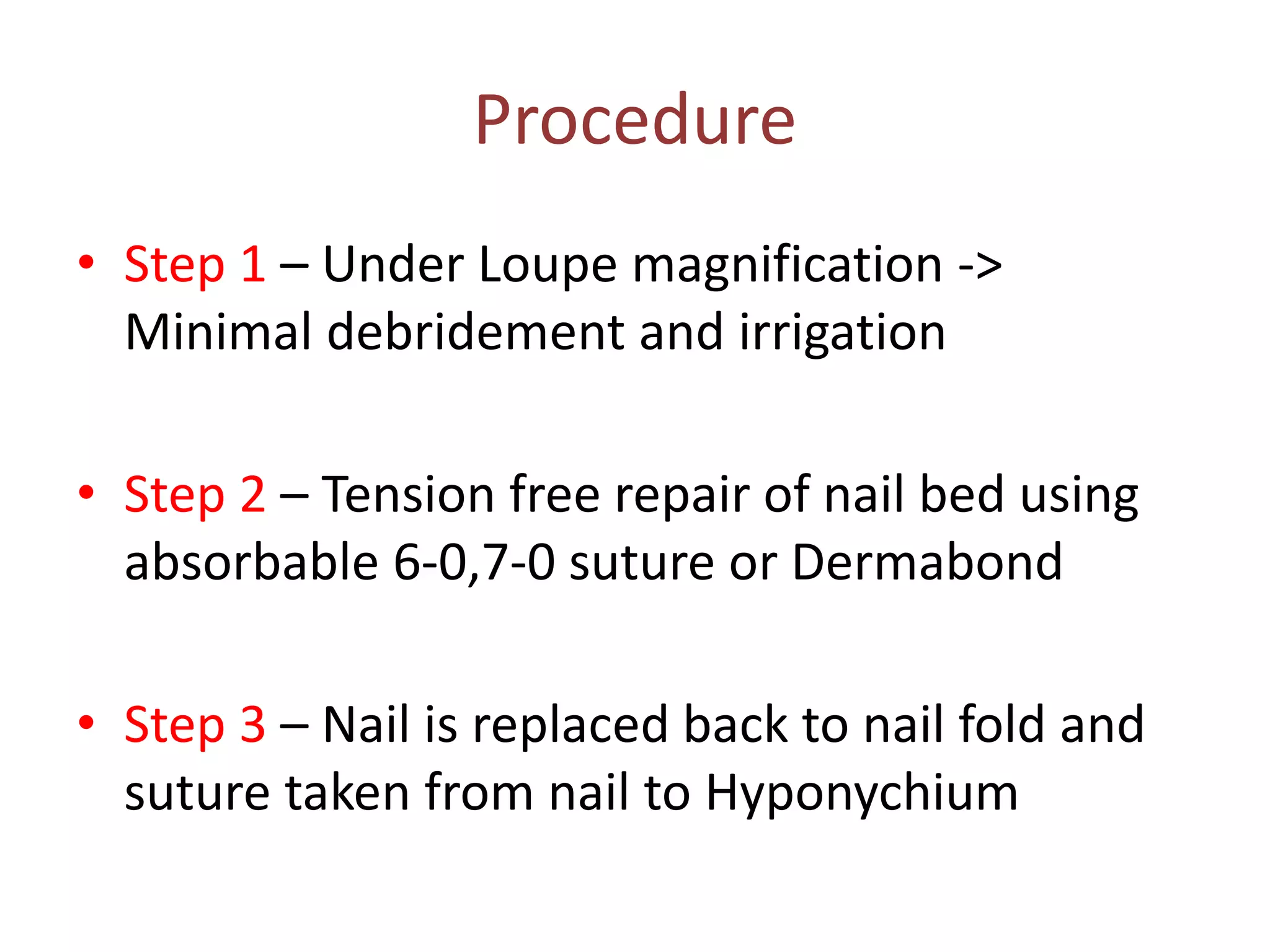
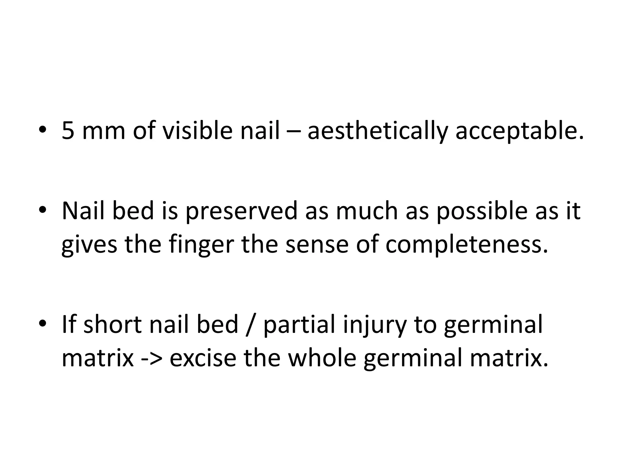
NAIL BED INJURY
SEMINAR PRESENTATION
Dr Prajwal K Rao
Anatomy
• Perinochyium – Nail, Nail bed, Surrounding
skin
• Paronychium – Lateral nail folds
• Hyponychium – Skin distal and palmar to nail
• Eponychium – Dorsal nail fold
• Lunula
• Matrix - Sterile and Germinal
• Germinal Matrix – Production of Nail Plate
• Sterile Matrix – Adds Squamous cells to Nail
• Dorsal Roof Matrix – Luster of Nail Plate
• Nail growth is 0.1mm/day
Nail bed Injury
• 75% males
• Long fingers commonly involved
• Common age : 4-30 years
• Cause – Deforming Force
Clinical examination
• Sensory examination
• Pattern of nail bed laceration
• Involvement of germinal and dorsal roof
matrix
• Associated Subtotal Pulp amputation
Imaging
• AP, Lateral and Oblique views of injured
fingers.
Classification
• Subungual hematoma
• Simple lacerations
• Stellate lacerations
• Severe Crush
• Avulsion
Trephination
• Helps to reduce pain, do not fasten healing
process
• Drainage of hematoma by perforation if < 50
% of nail involved.
• Nail removal, Debridement and Nail bed
repair if >50 % of nail involved
Nail bed Lacerations
Indications for Nail bed repair
• Open nail bed laceration
• Closed nail bed laceration with Subungual
hematoma involving > 50 percent of nail
• Closed nail bed laceration with displaced distal
phalangeal fracture
Positioning and Exposure
• Under tourniquet control and Local anesthesia
• Freer elevator inserted b/w nail plate and nail
bed from distal to proximal
• Elevation of dorsal roof matrix to expose
laceration under nail fold or germinal matrix
Procedure
• Step 1 – Under Loupe magnification ->
Minimal debridement and irrigation
• Step 2 – Tension free repair of nail bed using
absorbable 6-0,7-0 suture or Dermabond
• Step 3 – Nail is replaced back to nail fold and
suture taken from nail to Hyponychium
• If nail is not available!
OPTIONS:
• Artificial Silicone nail
• Suture Package
Remember to make couple of holes in the nail
• 5 mm of visible nail – aesthetically acceptable.
• Nail bed is preserved as much as possible as it
gives the finger the sense of completeness.
• If short nail bed / partial injury to germinal
matrix -> excise the whole germinal matrix.
Nail bed graft
Loss of nail bed or gap more than 4 to 5mm.
Loss of sterile matrix
• Split Thickness Sterile Matrix Graft, from
adjacent nail bed, amputed fingers or great
toe.
Loss of Germinal Matrix
• Full thickness graft
• Free toe pulp including nail complex
Avulsion laceration
• Cause: High energy injuries
• Associated condition: Distal phalanx fracture
• Treatment: Nail removal, Nail bed repair with
+/- fixation.
If there is significant loss of nail matrix
then split thickness graft or nail matrix
transfer is done.
Germinal Matrix Avulsion
• Step 1 –> Nail bed flap reduced back and Splint in
nail fold
• Step 2 –> Elevate dorsal roof matrix, Pass 5-0
nonabsorbable suture from dorsum, horizontal
bite on nail bed and brought back to dorsum and
held in mosquito forceps.
• Step 3 –> 2 or 3sutures passed, then these sutures
are tied on the dorsum, reducing the nailbed.
Tuft fracture
• Nail injury in combination with finger tip
fracture.
Options:
• Repair of nail bed and placing nail back on the
bed.
• K wire through the medullary cavity.
Postoperative Care
• Non adherent dressing
• Finger splint immobilizing DIP joint
• Complete first nail regeneration takes 4 to 6
months with lack of sheen.
• Quality of nail depends on initial injury to matrix
and age of patient.
Complications
• Hook nail – due to advancement of nail matrix
to obtain coverage without adequate bony
support.
• Nail Ridge
• Split nail – caused by scarring of matrix.
• Non adherence (onycholysis)
• Nail absence
• Cornified nail bed
• Nail spikes/cysts
Reference
Thank you

Nail bed injury

  • 1.
    NAIL BED INJURY SEMINARPRESENTATION Dr Prajwal K Rao
  • 3.
    Anatomy • Perinochyium –Nail, Nail bed, Surrounding skin • Paronychium – Lateral nail folds • Hyponychium – Skin distal and palmar to nail • Eponychium – Dorsal nail fold • Lunula • Matrix - Sterile and Germinal
  • 4.
    • Germinal Matrix– Production of Nail Plate • Sterile Matrix – Adds Squamous cells to Nail • Dorsal Roof Matrix – Luster of Nail Plate • Nail growth is 0.1mm/day
  • 5.
    Nail bed Injury •75% males • Long fingers commonly involved • Common age : 4-30 years • Cause – Deforming Force
  • 6.
    Clinical examination • Sensoryexamination • Pattern of nail bed laceration • Involvement of germinal and dorsal roof matrix • Associated Subtotal Pulp amputation
  • 7.
    Imaging • AP, Lateraland Oblique views of injured fingers.
  • 8.
    Classification • Subungual hematoma •Simple lacerations • Stellate lacerations • Severe Crush • Avulsion
  • 10.
    Trephination • Helps toreduce pain, do not fasten healing process • Drainage of hematoma by perforation if < 50 % of nail involved. • Nail removal, Debridement and Nail bed repair if >50 % of nail involved
  • 12.
  • 13.
    Indications for Nailbed repair • Open nail bed laceration • Closed nail bed laceration with Subungual hematoma involving > 50 percent of nail • Closed nail bed laceration with displaced distal phalangeal fracture
  • 14.
    Positioning and Exposure •Under tourniquet control and Local anesthesia • Freer elevator inserted b/w nail plate and nail bed from distal to proximal
  • 15.
    • Elevation ofdorsal roof matrix to expose laceration under nail fold or germinal matrix
  • 16.
    Procedure • Step 1– Under Loupe magnification -> Minimal debridement and irrigation • Step 2 – Tension free repair of nail bed using absorbable 6-0,7-0 suture or Dermabond • Step 3 – Nail is replaced back to nail fold and suture taken from nail to Hyponychium
  • 18.
    • If nailis not available! OPTIONS: • Artificial Silicone nail • Suture Package Remember to make couple of holes in the nail
  • 19.
    • 5 mmof visible nail – aesthetically acceptable. • Nail bed is preserved as much as possible as it gives the finger the sense of completeness. • If short nail bed / partial injury to germinal matrix -> excise the whole germinal matrix.
  • 20.
    Nail bed graft Lossof nail bed or gap more than 4 to 5mm. Loss of sterile matrix • Split Thickness Sterile Matrix Graft, from adjacent nail bed, amputed fingers or great toe. Loss of Germinal Matrix • Full thickness graft • Free toe pulp including nail complex
  • 22.
    Avulsion laceration • Cause:High energy injuries • Associated condition: Distal phalanx fracture • Treatment: Nail removal, Nail bed repair with +/- fixation. If there is significant loss of nail matrix then split thickness graft or nail matrix transfer is done.
  • 23.
  • 24.
    • Step 1–> Nail bed flap reduced back and Splint in nail fold • Step 2 –> Elevate dorsal roof matrix, Pass 5-0 nonabsorbable suture from dorsum, horizontal bite on nail bed and brought back to dorsum and held in mosquito forceps. • Step 3 –> 2 or 3sutures passed, then these sutures are tied on the dorsum, reducing the nailbed.
  • 26.
    Tuft fracture • Nailinjury in combination with finger tip fracture. Options: • Repair of nail bed and placing nail back on the bed. • K wire through the medullary cavity.
  • 27.
    Postoperative Care • Nonadherent dressing • Finger splint immobilizing DIP joint • Complete first nail regeneration takes 4 to 6 months with lack of sheen. • Quality of nail depends on initial injury to matrix and age of patient.
  • 28.
    Complications • Hook nail– due to advancement of nail matrix to obtain coverage without adequate bony support. • Nail Ridge • Split nail – caused by scarring of matrix.
  • 30.
    • Non adherence(onycholysis) • Nail absence • Cornified nail bed • Nail spikes/cysts
  • 31.
  • 32.

Editor's Notes

  • #3 Nail – Hard structure composed of dessicated, keratinised, squamous cells attached to nail bed. Nail loosely attached to Germinal matrix and Densely adherent to Sterile Matrix and eponychium.
  • #15 1 to 2 mm beyond laceration is elevated Undersurface of nail is examined for nail bed remnants
  • #17 Nail – Splint for tuft fractures, Prevents dressing from adhering, Prevents synechiae b/w germinal matrix and dorsal roof matrix.
  • #20 Small nail, nail cyst
  • #24 Laceration over GM at prox nail bed, and distal GM prolapses out of nailfold.