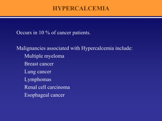
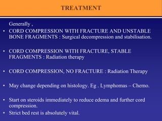
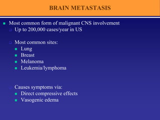
ONCOLOGIC
EMERGENCIES
2014
A PRIMER FOR NURSING STUDENTS
INTRODUCTION
 SVC SYNDROME
 FEBRILE NEUTROPENIA
 HYPERCALCEMIA OF MALIGNANCY
 HYPERLEUKOCYTOSIS
 SPINAL CORD COMPRESSION
 BRAIN METASTASIS
 TUMOR LYSIS SYNDROME
 PAIN
S V C SYNDROME
SVC SYNDROME
Superior vena cava syndrome (SVCS) is the mass effect in the mediastinum resulting
in obstruction of SVC and compression of other structures.
Obstruction may be :
INTERNAL : Thrombus
EXTERNAL : Mass effect
- malignant
- non-malignant
MALIGNANCIES ASSOCIATED WITH SVCS:
Lung Cancer – SCLC , NSCLC
N H L
Thymoma
Germ cell tumors of mediastinum
Metastatic tumors
Salsali M, Cliffton EE. N Y State J Med 1969 ; Bell DR Med J Aust 1986 ; Parish JM Mayo Clin Proc 1981
SVC SYNDROME
CLINICAL FEATURES
SYMPTOMS:
Early symptoms – Dyspnea and nonproductive cough , headache,
dysphagia, hoarseness, chest pain, facial puffiness
Late symptoms - Visual disturbances, dizziness, syncope, lethargy,
irritability and mental status changes
SVC SYNDROME
CLINICAL FEATURES
SIGNS :
Early signs - Edema of the face, neck, upper thorax, breasts, and
upper extremities (Stoke’s sign ) ,
Facial plethora & dilated veins of face, neck and thorax
( Pemberton’s sign )
Periorbital edema
Conjunctival edema & congestion
Compensatory tachycardia
Late signs - Cyanosis of the face or upper torso
Mental status changes
Tachypnea, orthopnea, stridor and respiratory distress
Seizures, stupor, coma
Haapoja & Blendowski, 1999 ;
SVC SYNDROME
EVALUATION - C X R
May show :
» Mediastinal widening
» Paratracheal shadow
» Pleural effusion
» Primary / Secondary
lung disease
SVC SYNDROME
MANAGEMENT
GENERAL MANAGEMENT:
Bed rest with the head elevated
Oxygen administration
Corticosteroids
Diuretic
No IV line in upper limbs.
SPECIFIC MANAGEMENT :
SCLC – Chemotherapy ± RT
NSCLC – RT + CT
NHL – CT ± RT
Catheter induced thrombosis - Thrombolysis
Stenting
FEBRILE NEUTROPENIA
FEBRILE NEUTROPENIA
DEFINITION :
FEVER : ~ single oral temperature of 101 º F (38.3° C)
OR
~ oral temp of 100.4 º F ( 38° C) lasting more than 1 hr
NEUTROPENIA :
~ ANC < 500 / mm3
OR
~ count of <1000 cells/mm3 with a predicted
decrease to <500 cells/mm3
FEBRILE NEUTROPENIA
FACTORS INFLUENCING RISK OF INFECTION
BREACH OF SKIN AND MUCOSAL BARRIERS :
IV access devices
Mucositis
Surgery
Tumor growth
DISEASE & THERAPY RELATED FACTORS :
CLL , MM  Hypogammaglobulinemia : Pneumococcus, H.influenza, N.meningitidis
ALL, HD, NHL  defective CMI : P.Carini, Cryptococcus, Salmonella
Steroids : Aspergillosis, Crytococcus, P.carini, Mycobacteria & atypical Mycobacteria
High dose Cytarabine  Mucositis : Streptococcal
FEBRILE NEUTROPENIA
INITIAL EVALUATION
HISTORY : Time since last chemotherapy administration, Major co-morbid illness,
Travel, Others at home with similar symptoms,
History of prior documented infections etc
PHYSICAL EXAMINATION : To find any focus of infection.
» IV access site
» Oropharynx
» Nasal cavity
» Skin including Perivaginal & Perineal regions
INVESTIGATIONS :
» CXR
» CBC, electrolytes, BUN, LFT
» Blood culture – 2 sets.
» Throat / wound swab as indicated
» Urine / Stool culture according to symptoms
FEBRILE NEUTROPENIA
CHARACTERISITCS OF HIGH RISK & LOW RISK PATIENTS
HIGH RISK
- Inpatients
- Associated co-morbidities
( hypotension, dehydration, hypoxia )
- Uncontrolled / progressive cancer
- Sr. Creatinine > 2 mg/dl
- LFT > 3 times normal
- HSCT / BMT recipient
- Prolonged severe neutropenia
anticipated
LOW RISK
- Outpatients
- No associated co-morbidities
- Good PS ( ECOG 0 –1 )
- Sr. Creatinine < 2 mg/dl
- LFT  3 times normal
- Non-transplant, solid tumor or
lymphoma patient
- Anticipated duration of neutropenia
< 7 days
I D S A RISK STRATIFICATION *
* Infectious Diseases Society of America guidelines , 2002.
FEBRILE NEUTROPENIA
MANAGEMENT ALGORITHM -1
Hughes et al. Comm & Inf Dis , 2002, 34: 730-51.
FEBRILE NEUTROPENIA
INDICATIONS FOR VANCOMYCIN
(1) Clinically suspected serious catheter-related infections
(e.g., bacteremia, cellulitis),
(2) Known colonization with penicillin- and cephalosporin-resistant
pneumococci or methicillin-resistant S. aureus,
(3) Positive results of blood culture for gram-positive bacteria before
final identification and susceptibility testing, or
(4) Hypotension or other evidence of cardiovascular impairment
IDSA guidelines , 2002
International Antimicrobial Therapy Co-operative Group of EORTC, NEJM , 1999.
FEBRILE NEUTROPENIA
INITIAL EMPIRICAL ANTIBIOTICS IN LOW RISK PATIENTS
Oral Ciprofloxacin + Amoxycillin / Clavulanate
For those allergic to Penicillins:
Ciprofloxacin + Clindamycin
IDSA guidelines , 2002
International Antimicrobial Therapy Co-operative Group of EORTC, NEJM , 1999.
FEBRILE NEUTROPENIA
MANAGEMENT ALGORITHM -2
IDSA guidelines , 2002
FEBRILE NEUTROPENIA
EMPIRICAL ANTIFUNGAL THERAPY
For patients with persistent fever after 3 days of antibiotics.
IDSA guidelines , 2002
FEBRILE NEUTROPENIA
DURATION OF ANTIBIOTIC THERAPY:
IDSA guidelines , 2002
FEBRILE NEUTROPENIA
COLONY STIMULATING FACTORS:
IDSA guidelines , 2002
• Can consistently shorten the duration of neutropenia
• Have not consistently and significantly reduced other measures of febrile
morbidity, including duration of fever, use of anti-infectives,
or costs of management of the febrile neutropenic episode.
• Possible use:
~ Conditions in which worsening of the course
is predicted and there is an expected long delay in recovery of the
marrow eg.pneumonia, hypotensive episodes, severe cellulitis or
sinusitis, systemic fungal infections, and multiorgan dysfunction
secondary to sepsis
~ For patients who remain severely neutropenic and have documented
infections that do not respond to appropriate antimicrobial therapy.
Not recommended for routine use to treat febrile or afebrile neutropenic
patients.
HYPERCALCEMIA OF
MALIGNANCY
HYPERCALCEMIA
Occurs in 10 % of cancer patients.
Malignancies associated with Hypercalcemia include:
Multiple myeloma
Breast cancer
Lung cancer
Lymphomas
Renal cell carcinoma
Esophageal cancer
HYPERCALCEMIA
TYPES OF HYPERCALCEMIA
HUMORAL HYPERCALCEMIA
~ PTHrP
LOCAL OSTEOLYTIC HYPERCALCEMIA
~ Osteoclast activation
~ IL- 1, IL – 6 , TNF
~ TGF α , PGE 2
~ RANKL ( receptor activator of nuclear factor kB ligand )
VITAMIN D LINKED HYPERCALCEMIA
~ activated mononuclear cells may secrete calcitriol
~ overexpression of RANKL has been suggested
HYPERCALCEMIA
CLINICAL FEATURES
GENERAL : Dehydration, Weight loss, Anorexia, Pruritus, Polydipsia
NEUROMUSCULAR : Fatigue, Lethargy, Muscle weakness, Seizure,
Hyporeflexia, Confusion, Psychosis, Coma.
GASTROINTESTINAL : Nausea, Vomiting, Constipation, Ileus .
GENITOURINARY : Polyuria
CARDIAC: Bradycardia, Prolonged PR interval, Shortened QT
interval, Wide T wave, Atrial or ventricular arrhythmias .
HYPERCALCEMIA - CLINICAL FEATURES
“stones, bones, groans and moans”
SPINAL CORD COMPRESSION
• Major emergency requiring radiation treatment
• Can lead to permanent neurologic dysfunction
– Ambulatory status is most important prognostic feature
• 80-90% of patients ambulatory at treatment retain function
SPINAL CORD COMPRESSION
• 2.5–5.0% of patients have spinal cord compression (SCC)
within the last 2 years of illness.
• Prostate, breast cancer, lung cancer most common
each ~15–20%
NHL, multiple myeloma, and renal cancer
~5–10% of patients
Men 40-60 years with prostate cancer = 17% incidence
• Thoracic spine affected in 60-80% of cases
• 50% present with disease in multiple spinal areas
SIGNS AND SYMPTOMS
• New onset back pain
– Initially localized, typically increasing in intensity
In particular:
– Pain that worsens when the patient is lying down
– Pain with percussion of vertebral bodies
• Weakness
– 60-85% of patients present with weakness
– ~2/3 are non-ambulatory at presentation
• Late neurologic signs are associated with permanent deficits such as
paraplegia
• Urinary retention
• Loss of sensory function
EVALUATION
• Non-contrast MRI of whole spine is best test
– If MRI not available, can use Myelography/CT
• MRI is better because
– Multiplanar imaging
– No radiation
– Contrast/needle not required to delineate lesions
– Can detect multiple lesion
– Should get whole spine MRI
• 97.6% sensitivity; 100.0% specificity
• Able to detect multi-level disease
• Biopsy if:
– metastatic disease not proven/documented
– no previous diagnosis of cancer
TREATMENT
Generally ,
• CORD COMPRESSION WITH FRACTURE AND UNSTABLE
BONE FRAGMENTS : Surgical decompression and stabilisation.
• CORD COMPRESSION WITH FRACTURE, STABLE
FRAGMENTS : Radiation therapy
• CORD COMPRESSION, NO FRACTURE : Radiation Therapy
• May change depending on histology. Eg . Lymphomas – Chemo.
• Start on steroids immediately to reduce edema and further cord
compression.
• Strict bed rest is absolutely vital.
BRAIN METASTASIS
BRAIN METASTASIS
 Most common form of malignant CNS involvement
 Up to 200,000 cases/year in US
 Most common sites:
 Lung
 Breast
 Melanoma
 Leukemia/lymphoma
 Causes symptoms via:
 Direct compressive effects
 Vasogenic edema
EVALUATION
 Signs/symptoms depend on location of mets
 Common:
 Headaches
 Seizures
 Focal deficits (e.g. weakness)
 Work up includes
 Physical Exam
 delineate neurologic deficits
 CT head
 MR head
 Can show lesions too small for CT
 Better tissue contrast
GENERAL MANAGEMENT
 Symptomatic treatment
 Anticonvulsants – ONLY IF SEIZURES OCCUR.
 Non-enzyme inducing anticonvulsants are preferred
 Phenytoin / Phosphenytoin
 Levetiracetam
 Hemorrhagic mets more likely cause seizures
 Prophylaxis may be indicated in these cases
 Dexamethasone
 For vasogenic edema
• Start with 16 mg IV bolus and switch to 8 mg BD.
 20% Mannitol – 100 ml / given over 15 min. TID.
 Check BP prior to infusion.
TREATMENT
• Solitary brain mets :
– Surgery  RT
– WBRT + Boost
– SRS / SRT
• Multiple brain mets :
– Palliative WBRT
– 30 Gy / 10 #
• No role of chemo.
HYPERLEUKOCYTOSIS
HYPERLEUKOCYTOSIS
A clinicopathologic syndrome caused by the sludging of circulating
leukemic blasts ( LEUKOSTASIS) in tissue microvasculature.
RISK FACTORS :
Younger age
Acute leukemias
Presence of certain cytogenetic abnormalities
- Philadelphia chromosome
- 11q23 translocation
Mortality rate approaches 40 %
HYPERLEUKOCYTOSIS
CLINICAL FEATURES
Symptoms arising from involvement of pulmonary and cerebral vasculature are more
common.
PULMONARY LEUKOSTASIS:
~ symptoms range from mild dyspnoea to respiratory distress
~ CXR  diffuse interstitial / alveolar infiltrate
~ ABG  pseudohypoxemia
INTRACRANIAL LEUKOSTASIS:
~ symptoms may range from confusion & somnolence to stupor & coma
~ may be preceded by focal CNS deficits
OTHER MANIFESTATIONS :
Retinal haemorrhage, Retinal vein thrombosis, Acute MI, Acute limb ischemia,
Renal vein thrombosis , Priapism and DIC.
HYPERLEUKOCYTOSIS
TREATMENT
GENERAL MEASURES:
~ Hydration
~ Alkalinisation of urine
~ Correction of thrombocytopenia / prevention of DIC
SPECIFIC MEASURES :
~ Leukapheresis – single session  WBC counts by 20 – 50 %
- also permits infusion of blood products.
~ Leukocytoreduction :Cytotoxic therapy - Hydroxyurea
~ Cranial radiation – has been used but not recommended routinely
PAIN
PAIN
• Moderate to severe pain experienced by 40% to 50% of
cancer patients.
• Very severe pain experienced by 25% to 30% of cancer
patients .
• 80% of terminal stage cancer experience moderate to
severe pain
OVERVIEW OF PAIN
• Causes –
– Infection
– Tumor related
–Nervous system, bone, visceral, mucosal
– Treatment Related
– surgery, radiation therapy, chemotherapy,
interventional procedures
• Types :
– Nociceptive : pain signals from nerve endings
– Neuropathic : damage to nerve fibres.
WHO LADDER OF PAIN MANAGEMENT
RADIATION & PAIN RELIEF
• Effective for Nociceptive and Neuropathic pain
• Effective for mild to moderate and severe pain
• Pain relief starting from within 24 hrs.
• Complete effects seen after 1 - 2 months.
• Brings about alleviation of other associated symptoms –
tumor swelling, anxiety and depression, appetite.
RT DOSE / FRACTIONATION
• 32.5 Gy / 13 #
• 30 Gy / 10 #
• 4 Gy / 5 #
• 5 Gy / 4 #
• 6 Gy / 2 #
• 8 Gy / 1 #
ALL ARE EQUAL AS FAR AS
PAIN RELIEF IS CONCERNED
Oncological emergencies - 2014

Oncological emergencies - 2014

  • 1.
  • 2.
    INTRODUCTION  SVC SYNDROME FEBRILE NEUTROPENIA  HYPERCALCEMIA OF MALIGNANCY  HYPERLEUKOCYTOSIS  SPINAL CORD COMPRESSION  BRAIN METASTASIS  TUMOR LYSIS SYNDROME  PAIN
  • 3.
    S V CSYNDROME
  • 4.
    SVC SYNDROME Superior venacava syndrome (SVCS) is the mass effect in the mediastinum resulting in obstruction of SVC and compression of other structures. Obstruction may be : INTERNAL : Thrombus EXTERNAL : Mass effect - malignant - non-malignant MALIGNANCIES ASSOCIATED WITH SVCS: Lung Cancer – SCLC , NSCLC N H L Thymoma Germ cell tumors of mediastinum Metastatic tumors Salsali M, Cliffton EE. N Y State J Med 1969 ; Bell DR Med J Aust 1986 ; Parish JM Mayo Clin Proc 1981
  • 6.
    SVC SYNDROME CLINICAL FEATURES SYMPTOMS: Earlysymptoms – Dyspnea and nonproductive cough , headache, dysphagia, hoarseness, chest pain, facial puffiness Late symptoms - Visual disturbances, dizziness, syncope, lethargy, irritability and mental status changes
  • 7.
    SVC SYNDROME CLINICAL FEATURES SIGNS: Early signs - Edema of the face, neck, upper thorax, breasts, and upper extremities (Stoke’s sign ) , Facial plethora & dilated veins of face, neck and thorax ( Pemberton’s sign ) Periorbital edema Conjunctival edema & congestion Compensatory tachycardia Late signs - Cyanosis of the face or upper torso Mental status changes Tachypnea, orthopnea, stridor and respiratory distress Seizures, stupor, coma Haapoja & Blendowski, 1999 ;
  • 8.
    SVC SYNDROME EVALUATION -C X R May show : » Mediastinal widening » Paratracheal shadow » Pleural effusion » Primary / Secondary lung disease
  • 9.
    SVC SYNDROME MANAGEMENT GENERAL MANAGEMENT: Bedrest with the head elevated Oxygen administration Corticosteroids Diuretic No IV line in upper limbs. SPECIFIC MANAGEMENT : SCLC – Chemotherapy ± RT NSCLC – RT + CT NHL – CT ± RT Catheter induced thrombosis - Thrombolysis Stenting
  • 10.
  • 11.
    FEBRILE NEUTROPENIA DEFINITION : FEVER: ~ single oral temperature of 101 º F (38.3° C) OR ~ oral temp of 100.4 º F ( 38° C) lasting more than 1 hr NEUTROPENIA : ~ ANC < 500 / mm3 OR ~ count of <1000 cells/mm3 with a predicted decrease to <500 cells/mm3
  • 12.
    FEBRILE NEUTROPENIA FACTORS INFLUENCINGRISK OF INFECTION BREACH OF SKIN AND MUCOSAL BARRIERS : IV access devices Mucositis Surgery Tumor growth DISEASE & THERAPY RELATED FACTORS : CLL , MM  Hypogammaglobulinemia : Pneumococcus, H.influenza, N.meningitidis ALL, HD, NHL  defective CMI : P.Carini, Cryptococcus, Salmonella Steroids : Aspergillosis, Crytococcus, P.carini, Mycobacteria & atypical Mycobacteria High dose Cytarabine  Mucositis : Streptococcal
  • 13.
    FEBRILE NEUTROPENIA INITIAL EVALUATION HISTORY: Time since last chemotherapy administration, Major co-morbid illness, Travel, Others at home with similar symptoms, History of prior documented infections etc PHYSICAL EXAMINATION : To find any focus of infection. » IV access site » Oropharynx » Nasal cavity » Skin including Perivaginal & Perineal regions INVESTIGATIONS : » CXR » CBC, electrolytes, BUN, LFT » Blood culture – 2 sets. » Throat / wound swab as indicated » Urine / Stool culture according to symptoms
  • 14.
    FEBRILE NEUTROPENIA CHARACTERISITCS OFHIGH RISK & LOW RISK PATIENTS HIGH RISK - Inpatients - Associated co-morbidities ( hypotension, dehydration, hypoxia ) - Uncontrolled / progressive cancer - Sr. Creatinine > 2 mg/dl - LFT > 3 times normal - HSCT / BMT recipient - Prolonged severe neutropenia anticipated LOW RISK - Outpatients - No associated co-morbidities - Good PS ( ECOG 0 –1 ) - Sr. Creatinine < 2 mg/dl - LFT  3 times normal - Non-transplant, solid tumor or lymphoma patient - Anticipated duration of neutropenia < 7 days I D S A RISK STRATIFICATION * * Infectious Diseases Society of America guidelines , 2002.
  • 15.
    FEBRILE NEUTROPENIA MANAGEMENT ALGORITHM-1 Hughes et al. Comm & Inf Dis , 2002, 34: 730-51.
  • 16.
    FEBRILE NEUTROPENIA INDICATIONS FORVANCOMYCIN (1) Clinically suspected serious catheter-related infections (e.g., bacteremia, cellulitis), (2) Known colonization with penicillin- and cephalosporin-resistant pneumococci or methicillin-resistant S. aureus, (3) Positive results of blood culture for gram-positive bacteria before final identification and susceptibility testing, or (4) Hypotension or other evidence of cardiovascular impairment IDSA guidelines , 2002 International Antimicrobial Therapy Co-operative Group of EORTC, NEJM , 1999.
  • 17.
    FEBRILE NEUTROPENIA INITIAL EMPIRICALANTIBIOTICS IN LOW RISK PATIENTS Oral Ciprofloxacin + Amoxycillin / Clavulanate For those allergic to Penicillins: Ciprofloxacin + Clindamycin IDSA guidelines , 2002 International Antimicrobial Therapy Co-operative Group of EORTC, NEJM , 1999.
  • 18.
  • 19.
    FEBRILE NEUTROPENIA EMPIRICAL ANTIFUNGALTHERAPY For patients with persistent fever after 3 days of antibiotics. IDSA guidelines , 2002
  • 20.
    FEBRILE NEUTROPENIA DURATION OFANTIBIOTIC THERAPY: IDSA guidelines , 2002
  • 21.
    FEBRILE NEUTROPENIA COLONY STIMULATINGFACTORS: IDSA guidelines , 2002 • Can consistently shorten the duration of neutropenia • Have not consistently and significantly reduced other measures of febrile morbidity, including duration of fever, use of anti-infectives, or costs of management of the febrile neutropenic episode. • Possible use: ~ Conditions in which worsening of the course is predicted and there is an expected long delay in recovery of the marrow eg.pneumonia, hypotensive episodes, severe cellulitis or sinusitis, systemic fungal infections, and multiorgan dysfunction secondary to sepsis ~ For patients who remain severely neutropenic and have documented infections that do not respond to appropriate antimicrobial therapy. Not recommended for routine use to treat febrile or afebrile neutropenic patients.
  • 22.
  • 23.
    HYPERCALCEMIA Occurs in 10% of cancer patients. Malignancies associated with Hypercalcemia include: Multiple myeloma Breast cancer Lung cancer Lymphomas Renal cell carcinoma Esophageal cancer
  • 24.
    HYPERCALCEMIA TYPES OF HYPERCALCEMIA HUMORALHYPERCALCEMIA ~ PTHrP LOCAL OSTEOLYTIC HYPERCALCEMIA ~ Osteoclast activation ~ IL- 1, IL – 6 , TNF ~ TGF α , PGE 2 ~ RANKL ( receptor activator of nuclear factor kB ligand ) VITAMIN D LINKED HYPERCALCEMIA ~ activated mononuclear cells may secrete calcitriol ~ overexpression of RANKL has been suggested
  • 25.
    HYPERCALCEMIA CLINICAL FEATURES GENERAL :Dehydration, Weight loss, Anorexia, Pruritus, Polydipsia NEUROMUSCULAR : Fatigue, Lethargy, Muscle weakness, Seizure, Hyporeflexia, Confusion, Psychosis, Coma. GASTROINTESTINAL : Nausea, Vomiting, Constipation, Ileus . GENITOURINARY : Polyuria CARDIAC: Bradycardia, Prolonged PR interval, Shortened QT interval, Wide T wave, Atrial or ventricular arrhythmias .
  • 26.
    HYPERCALCEMIA - CLINICALFEATURES “stones, bones, groans and moans”
  • 27.
  • 28.
  • 29.
    SPINAL CORD COMPRESSION •Major emergency requiring radiation treatment • Can lead to permanent neurologic dysfunction – Ambulatory status is most important prognostic feature • 80-90% of patients ambulatory at treatment retain function
  • 30.
    SPINAL CORD COMPRESSION •2.5–5.0% of patients have spinal cord compression (SCC) within the last 2 years of illness. • Prostate, breast cancer, lung cancer most common each ~15–20% NHL, multiple myeloma, and renal cancer ~5–10% of patients Men 40-60 years with prostate cancer = 17% incidence • Thoracic spine affected in 60-80% of cases • 50% present with disease in multiple spinal areas
  • 31.
    SIGNS AND SYMPTOMS •New onset back pain – Initially localized, typically increasing in intensity In particular: – Pain that worsens when the patient is lying down – Pain with percussion of vertebral bodies • Weakness – 60-85% of patients present with weakness – ~2/3 are non-ambulatory at presentation • Late neurologic signs are associated with permanent deficits such as paraplegia • Urinary retention • Loss of sensory function
  • 32.
    EVALUATION • Non-contrast MRIof whole spine is best test – If MRI not available, can use Myelography/CT • MRI is better because – Multiplanar imaging – No radiation – Contrast/needle not required to delineate lesions – Can detect multiple lesion – Should get whole spine MRI • 97.6% sensitivity; 100.0% specificity • Able to detect multi-level disease • Biopsy if: – metastatic disease not proven/documented – no previous diagnosis of cancer
  • 33.
    TREATMENT Generally , • CORDCOMPRESSION WITH FRACTURE AND UNSTABLE BONE FRAGMENTS : Surgical decompression and stabilisation. • CORD COMPRESSION WITH FRACTURE, STABLE FRAGMENTS : Radiation therapy • CORD COMPRESSION, NO FRACTURE : Radiation Therapy • May change depending on histology. Eg . Lymphomas – Chemo. • Start on steroids immediately to reduce edema and further cord compression. • Strict bed rest is absolutely vital.
  • 34.
  • 35.
    BRAIN METASTASIS  Mostcommon form of malignant CNS involvement  Up to 200,000 cases/year in US  Most common sites:  Lung  Breast  Melanoma  Leukemia/lymphoma  Causes symptoms via:  Direct compressive effects  Vasogenic edema
  • 36.
    EVALUATION  Signs/symptoms dependon location of mets  Common:  Headaches  Seizures  Focal deficits (e.g. weakness)  Work up includes  Physical Exam  delineate neurologic deficits  CT head  MR head  Can show lesions too small for CT  Better tissue contrast
  • 37.
    GENERAL MANAGEMENT  Symptomatictreatment  Anticonvulsants – ONLY IF SEIZURES OCCUR.  Non-enzyme inducing anticonvulsants are preferred  Phenytoin / Phosphenytoin  Levetiracetam  Hemorrhagic mets more likely cause seizures  Prophylaxis may be indicated in these cases  Dexamethasone  For vasogenic edema • Start with 16 mg IV bolus and switch to 8 mg BD.  20% Mannitol – 100 ml / given over 15 min. TID.  Check BP prior to infusion.
  • 38.
    TREATMENT • Solitary brainmets : – Surgery  RT – WBRT + Boost – SRS / SRT • Multiple brain mets : – Palliative WBRT – 30 Gy / 10 # • No role of chemo.
  • 39.
  • 40.
    HYPERLEUKOCYTOSIS A clinicopathologic syndromecaused by the sludging of circulating leukemic blasts ( LEUKOSTASIS) in tissue microvasculature. RISK FACTORS : Younger age Acute leukemias Presence of certain cytogenetic abnormalities - Philadelphia chromosome - 11q23 translocation Mortality rate approaches 40 %
  • 41.
    HYPERLEUKOCYTOSIS CLINICAL FEATURES Symptoms arisingfrom involvement of pulmonary and cerebral vasculature are more common. PULMONARY LEUKOSTASIS: ~ symptoms range from mild dyspnoea to respiratory distress ~ CXR  diffuse interstitial / alveolar infiltrate ~ ABG  pseudohypoxemia INTRACRANIAL LEUKOSTASIS: ~ symptoms may range from confusion & somnolence to stupor & coma ~ may be preceded by focal CNS deficits OTHER MANIFESTATIONS : Retinal haemorrhage, Retinal vein thrombosis, Acute MI, Acute limb ischemia, Renal vein thrombosis , Priapism and DIC.
  • 42.
    HYPERLEUKOCYTOSIS TREATMENT GENERAL MEASURES: ~ Hydration ~Alkalinisation of urine ~ Correction of thrombocytopenia / prevention of DIC SPECIFIC MEASURES : ~ Leukapheresis – single session  WBC counts by 20 – 50 % - also permits infusion of blood products. ~ Leukocytoreduction :Cytotoxic therapy - Hydroxyurea ~ Cranial radiation – has been used but not recommended routinely
  • 43.
  • 44.
    PAIN • Moderate tosevere pain experienced by 40% to 50% of cancer patients. • Very severe pain experienced by 25% to 30% of cancer patients . • 80% of terminal stage cancer experience moderate to severe pain
  • 45.
    OVERVIEW OF PAIN •Causes – – Infection – Tumor related –Nervous system, bone, visceral, mucosal – Treatment Related – surgery, radiation therapy, chemotherapy, interventional procedures • Types : – Nociceptive : pain signals from nerve endings – Neuropathic : damage to nerve fibres.
  • 46.
    WHO LADDER OFPAIN MANAGEMENT
  • 47.
    RADIATION & PAINRELIEF • Effective for Nociceptive and Neuropathic pain • Effective for mild to moderate and severe pain • Pain relief starting from within 24 hrs. • Complete effects seen after 1 - 2 months. • Brings about alleviation of other associated symptoms – tumor swelling, anxiety and depression, appetite.
  • 48.
    RT DOSE /FRACTIONATION • 32.5 Gy / 13 # • 30 Gy / 10 # • 4 Gy / 5 # • 5 Gy / 4 # • 6 Gy / 2 # • 8 Gy / 1 # ALL ARE EQUAL AS FAR AS PAIN RELIEF IS CONCERNED