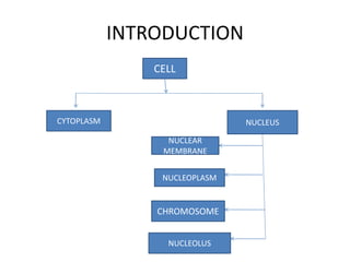
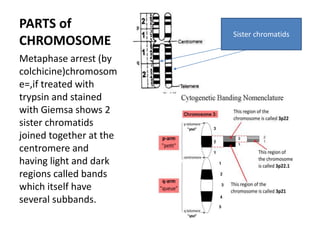
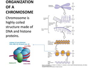
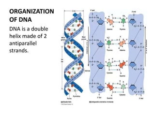
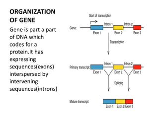
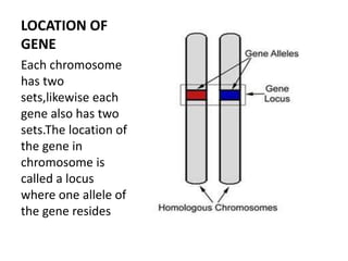
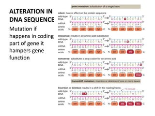
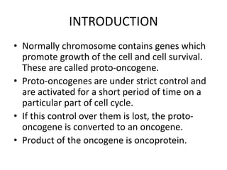
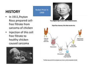
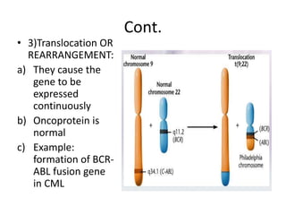
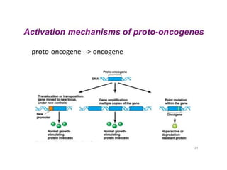
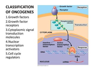
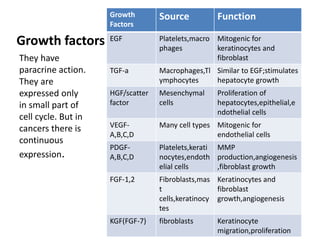
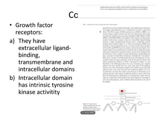
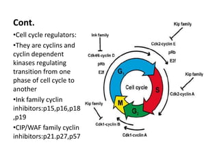
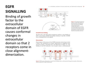
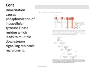
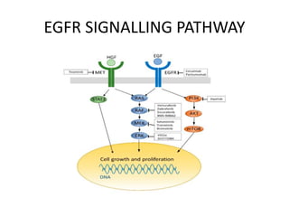
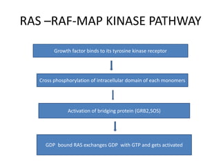
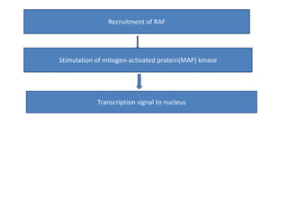
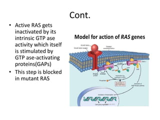
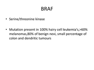
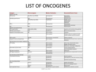
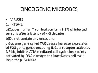
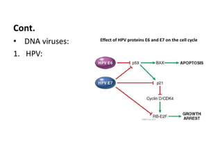
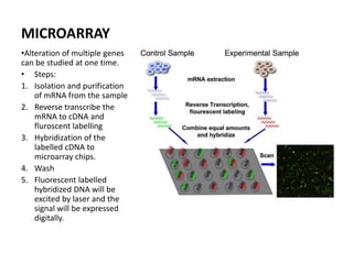
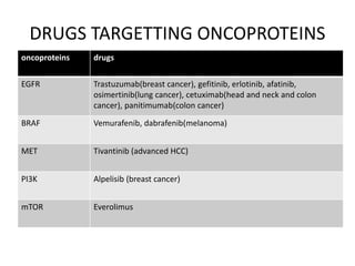
ONCOGENE
By SOUMYADIP ROY
Moderator: Dr. SHYAMA PREM
Additional Professor,
RADIATION ONCOLOGY, JIPMER
HEADINGS
• INTRODUCTION
• HISTORY
• MECHANISMS OF ACTIVATION
• CLASSIFICATION OF ONCOGENE
• LIST OF ONCOGENES
• ONCOGENIC MICROBES
• DETECTION OF ONCOGENES
• CLINICAL IMPLICATIONS
INTRODUCTION
CELL
CYTOPLASM NUCLEUS
NUCLEAR
MEMBRANE
NUCLEOPLASM
NUCLEOLUS
CHROMOSOME
PARTS of
CHROMOSOME
Metaphase arrest (by
colchicine)chromosom
e=,if treated with
trypsin and stained
with Giemsa shows 2
sister chromatids
joined together at the
centromere and
having light and dark
regions called bands
which itself have
several subbands.
Sister chromatids
ORGANIZATION
OF A
CHROMOSOME
Chromosome is
highly coiled
structure made of
DNA and histone
proteins.
ORGANIZATION
OF DNA
DNA is a double
helix made of 2
antiparallel
strands.
ORGANIZATION
OF GENE
Gene is part a part
of DNA which
codes for a
protein.It has
expressing
sequences(exons)
interspersed by
intervening
sequences(introns)
LOCATION OF
GENE
Each chromosome
has two
sets,likewise each
gene also has two
sets.The location of
the gene in
chromosome is
called a locus
where one allele of
the gene resides
ALTERATION IN
DNA SEQUENCE
Mutation if
happens in coding
part of gene it
hampers gene
function
INTRODUCTION
• Normally chromosome contains genes which
promote growth of the cell and cell survival.
These are called proto-oncogene.
• Proto-oncogenes are under strict control and
are activated for a short period of time on a
particular part of cell cycle.
• If this control over them is lost, the proto-
oncogene is converted to an oncogene.
• Product of the oncogene is oncoprotein.
HISTORY
• In 1911,Peyton
Rous prepared cell-
free filtrate from
sarcoma of chicken
• Injection of this cell
free filtrate to
healthy chicken
caused sarcoma
Nobel Prize in
1966
Cont.
• In 1958 Hary Rubin and
Howard Temin showed
morphological changes
in chicken embryo
fibroblast by infection
with RSV
Cont.
• C-src was found to
be the normal
counterpart of v-src
• Conversion of
normal
protooncogene c-
src to oncogene v-
src causes sarcoma.
• Transmission of v-
src from one host
to another is by the
process of
transduction
MECHANISMS OF ONCOGENE
ACTIVATIONS
• 3 mechanisms
1. Mutation:
a) It is insertion or deletion or missence
b) Affect only one allele; i.e.,heterozygous
c) Affects only one codon or clustered in
neighbouring codons
Cont.
2)Gene amplification:
a) Here the number of the copies of the gene
gets amplified
b) Oncoprotein is normal but present in
excessive amount.
c) Example is Her2/neu amplification in breast
cancer.
Cont.
• 3)Translocation OR
REARRANGEMENT:
a) They cause the
gene to be
expressed
continuously
b) Oncoprotein is
normal
c) Example:
formation of BCR-
ABL fusion gene
in CML
CLASSIFICATION
OF ONCOGENES
1.Growth factors
2.Growth factor
receptors
3.Cytoplasmic signal
transduction
molecules
4.Nuclear
transcription
activators
5.Cell cycle
regulators
Growth factors
Growth
Factors
Source Function
EGF Platelets,macro
phages
Mitogenic for
keratinocytes and
fibroblast
TGF-a Macrophages,Tl
ymphocytes
Similar to EGF;stimulates
hepatocyte growth
HGF/scatter
factor
Mesenchymal
cells
Proliferation of
hepatocytes,epithelial,e
ndothelial cells
VEGF-
A,B,C,D
Many cell types Mitogenic for
endothelial cells
PDGF-
A,B,C,D
Platelets,kerati
nocytes,endoth
elial cells
MMP
production,angiogenesis
,fibroblast growth
FGF-1,2 Fibroblasts,mas
t
cells,keratinocy
tes
Keratinocytes and
fibroblast
growth,angiogenesis
KGF(FGF-7) fibroblasts Keratinocyte
migration,proliferation
They have
paracrine action.
They are
expressed only
in small part of
cell cycle. But in
cancers there is
continuous
expression.
Cont.
• Growth factor
receptors:
a) They have
extracellular ligand-
binding,
transmembrane and
intracellular domains
b) Intracellular domain
has intrinsic tyrosine
kinase activitity
Cont.
d) Dimerization causes autophosphorylation
of intrinsic domain and recruitment of
other signalling proteins
e) Growth factor receptor oncoproteins are in
constitutively dimerized condition without
binding to ligand
Cont.
• Cytoplasmic signal transduction molecules:
a) These are serine/threonine or tyrosine
kinases
b) examples:RAS,RAF,MEK,PI3K,AKT
Cont.
• Nuclear transcription factors:
a)Growth factors ultimately induce synthesis or activity
of transcription factors
b)C-fos,N-myc
Cont.
•Cell cycle regulators:
•They are cyclins and
cyclin dependent
kinases regulating
transition from one
phase of cell cycle to
another
•Ink family cyclin
inhibitors:p15,p16,p18
,p19
•CIP/WAF family cyclin
inhibitors:p21.p27,p57
PROTOTYPE ONCOGENES
• EGFR:
a) Also called erbB or HER
b) 4 types:1,2,3,4
c) HER2 does not bind to a known ligand but
acts as co-factors for other members of the
family
d) HER3 does not have tyrosine kinase domain
Cont.
e)Ligand binding to extracellular domain causes
conformal changes and 2 receptors come in
close alignment-this is called dimerization.
F) ERBB1 is overexpressed in 80% of squamous
cell carcinomas of lung,80-100% of head and
neck malignancies and 50% glioblastomas
G) ERBB2 is overexpressed in 25% breast cancers
EGFR
SIGNALLING
Binding of growth
factor to the
extracellular
domain of EGFR
causes conformal
changes in
extracellular
domain so that 2
receptors come in
close alignment-
dimerization.
Cont
Dimerization
causes
phosphorylation of
intracellular
tyrosine kinase
residue which
leads to multiple
downstream
signalling molecule
recruitment.
EGFR SIGNALLING PATHWAY
RAS ONCOGENE
• Point mutation of RAS is single most common
abnormality of human tumours.
• Multiple growth factor(EGF,PDGF) signal
transduction pathways depend on RAS
• Mutated in 15-20% cancers
• 90% cholangiocarcinima,pancreatic
adenocarcinoma;50%colon,endometrial and
thyroid cancers and 30% lung and myeloid
leukemias have RAS mutation
Cont.
• 3 RAS proteins are: K-RAS,H-RAS and N-RAS
• K-RAS: colon and pancreas cancer
• H-RAS: bladder and kidney cancer
• N-RAS: melanoma and haematologic
malignancies
• Inactivated RAS in GDP bound state is anchored
to inner cell membrane by farnesyl group.
RAS –RAF-MAP KINASE PATHWAY
Growth factor binds to its tyrosine kinase receptor
Cross phosphorylation of intracellular domain of each monomers
Activation of bridging protein (GRB2,SOS)
GDP bound RAS exchanges GDP with GTP and gets activated
Recruitment of RAF
Stimulation of mitogen-activated protein(MAP) kinase
Transcription signal to nucleus
Cont.
• Active RAS gets
inactivated by its
intrinsic GTP ase
activity which itself
is stimulated by
GTP ase-activating
proteins(GAPs)
• This step is blocked
in mutant RAS
MAP KINASES
• They are serine/threonine kinases
• 3 parallel MAP kinase pathways are
there:MAPK,JNK,p38 pathways
• MAPK is activated by growth factors
• JNK and p38 are activated by environmental
stress signals,like ionizing radiations and UV
rays
• MAP enters nucleus to activate transcription
factors
BRAF
• Serine/threonine kinase
• Mutation present in 100% hairy cell leukemia's,>60%
melanomas,80% of benign nevi, small percentage of
colon and dendritic tumours
LIST OF ONCOGENES
ONCOGENIC MICROBES
• VIRUSES
1. HTLV-1:
a)Causes human T cell leukaemia in 3-5% of infected
persons after a latency of 4-5 decades
b)Do not contain any oncogene
c)But one gene called TAX causes increase expression
of FOS gene, genes encoding IL-2,its receptor activates
NF-Kb, inhibits ATM mediated cell cycle checkpoints
activated by DNA damage and inactivates cell cycle
inhibitor p16/INK4a
Cont.
• DNA viruses:
1. HPV:
Cont.
2)EBV:
a)African Burkitt lymphoma,nasopharyngeal
carcinoma,subset of Hodgkin lymphoma
b)LMP-1(latent membrane protein) acts as an
oncogene and behaves like constitutively active CD40
receptor which helper cell uses to activate B cells.
c)EBNA-2 activates NOTCH receptor, cyclin D and src
protooncogene
3)Hepatitis B and C :
a)Cause 70-85% of HCC
b)Chronic inflammation is the predominant etiology of
HCC which causes DNA damage.
c)NF-Kb pathway plays role
d)HBx gene of HBV activates several transcription
factors
Cont.
3)Hepatitis B and C :
a)Cause 70-85% of
HCC
b)Chronic
inflammation is the
predominant etiology
of HCC which causes
DNA damage.
c)NF-Kb pathway plays
role
d)HBx gene of HBV
activates several
transcription factors
Genome and transcribed component of HBV
Cont.
Bacteria
H.pylori:
3% of Chronically
infected patients
develop
adenocarcinoma after
decades
Pathogenic strains of
H.pylori contain
cytotoxin associated A
A(CagA) gene acting as
a growth promoting
gene
DETECTION OF
ONCOGENIC
ALTERATION
FISH:DNA probes
specific for a gene or
particular chromosome
region are labeled and
hybridized to denatured
metaphase
chromosomes or on
interphase nuclei on
paraffin embedded
tissue.After washing
fluorescent labeled
antibodies are added
and cells or tissues are
examined under
fluorescent microscopy
MICROARRAY
•Alteration of multiple genes
can be studied at one time.
• Steps:
1. Isolation and purification
of mRNA from the sample
2. Reverse transcribe the
mRNA to cDNA and
fluroscent labelling
3. Hybridization of the
labelled cDNA to
microarray chips.
4. Wash
5. Fluorescent labelled
hybridized DNA will be
excited by laser and the
signal will be expressed
digitally.
DRUGS TARGETTING ONCOPROTEINS
oncoproteins drugs
EGFR Trastuzumab(breast cancer), gefitinib, erlotinib, afatinib,
osimertinib(lung cancer), cetuximab(head and neck and colon
cancer), panitimumab(colon cancer)
BRAF Vemurafenib, dabrafenib(melanoma)
MET Tivantinib (advanced HCC)
PI3K Alpelisib (breast cancer)
mTOR Everolimus

Oncogene

  • 1.
    ONCOGENE By SOUMYADIP ROY Moderator:Dr. SHYAMA PREM Additional Professor, RADIATION ONCOLOGY, JIPMER
  • 2.
    HEADINGS • INTRODUCTION • HISTORY •MECHANISMS OF ACTIVATION • CLASSIFICATION OF ONCOGENE • LIST OF ONCOGENES • ONCOGENIC MICROBES • DETECTION OF ONCOGENES • CLINICAL IMPLICATIONS
  • 3.
  • 4.
    PARTS of CHROMOSOME Metaphase arrest(by colchicine)chromosom e=,if treated with trypsin and stained with Giemsa shows 2 sister chromatids joined together at the centromere and having light and dark regions called bands which itself have several subbands. Sister chromatids
  • 5.
    ORGANIZATION OF A CHROMOSOME Chromosome is highlycoiled structure made of DNA and histone proteins.
  • 6.
    ORGANIZATION OF DNA DNA isa double helix made of 2 antiparallel strands.
  • 7.
    ORGANIZATION OF GENE Gene ispart a part of DNA which codes for a protein.It has expressing sequences(exons) interspersed by intervening sequences(introns)
  • 8.
    LOCATION OF GENE Each chromosome hastwo sets,likewise each gene also has two sets.The location of the gene in chromosome is called a locus where one allele of the gene resides
  • 9.
    ALTERATION IN DNA SEQUENCE Mutationif happens in coding part of gene it hampers gene function
  • 10.
    INTRODUCTION • Normally chromosomecontains genes which promote growth of the cell and cell survival. These are called proto-oncogene. • Proto-oncogenes are under strict control and are activated for a short period of time on a particular part of cell cycle. • If this control over them is lost, the proto- oncogene is converted to an oncogene. • Product of the oncogene is oncoprotein.
  • 11.
    HISTORY • In 1911,Peyton Rousprepared cell- free filtrate from sarcoma of chicken • Injection of this cell free filtrate to healthy chicken caused sarcoma Nobel Prize in 1966
  • 12.
    Cont. • In 1958Hary Rubin and Howard Temin showed morphological changes in chicken embryo fibroblast by infection with RSV
  • 13.
    Cont. • C-src wasfound to be the normal counterpart of v-src • Conversion of normal protooncogene c- src to oncogene v- src causes sarcoma. • Transmission of v- src from one host to another is by the process of transduction
  • 14.
    MECHANISMS OF ONCOGENE ACTIVATIONS •3 mechanisms 1. Mutation: a) It is insertion or deletion or missence b) Affect only one allele; i.e.,heterozygous c) Affects only one codon or clustered in neighbouring codons
  • 15.
    Cont. 2)Gene amplification: a) Herethe number of the copies of the gene gets amplified b) Oncoprotein is normal but present in excessive amount. c) Example is Her2/neu amplification in breast cancer.
  • 16.
    Cont. • 3)Translocation OR REARRANGEMENT: a)They cause the gene to be expressed continuously b) Oncoprotein is normal c) Example: formation of BCR- ABL fusion gene in CML
  • 18.
    CLASSIFICATION OF ONCOGENES 1.Growth factors 2.Growthfactor receptors 3.Cytoplasmic signal transduction molecules 4.Nuclear transcription activators 5.Cell cycle regulators
  • 19.
    Growth factors Growth Factors Source Function EGFPlatelets,macro phages Mitogenic for keratinocytes and fibroblast TGF-a Macrophages,Tl ymphocytes Similar to EGF;stimulates hepatocyte growth HGF/scatter factor Mesenchymal cells Proliferation of hepatocytes,epithelial,e ndothelial cells VEGF- A,B,C,D Many cell types Mitogenic for endothelial cells PDGF- A,B,C,D Platelets,kerati nocytes,endoth elial cells MMP production,angiogenesis ,fibroblast growth FGF-1,2 Fibroblasts,mas t cells,keratinocy tes Keratinocytes and fibroblast growth,angiogenesis KGF(FGF-7) fibroblasts Keratinocyte migration,proliferation They have paracrine action. They are expressed only in small part of cell cycle. But in cancers there is continuous expression.
  • 20.
    Cont. • Growth factor receptors: a)They have extracellular ligand- binding, transmembrane and intracellular domains b) Intracellular domain has intrinsic tyrosine kinase activitity
  • 21.
    Cont. d) Dimerization causesautophosphorylation of intrinsic domain and recruitment of other signalling proteins e) Growth factor receptor oncoproteins are in constitutively dimerized condition without binding to ligand
  • 22.
    Cont. • Cytoplasmic signaltransduction molecules: a) These are serine/threonine or tyrosine kinases b) examples:RAS,RAF,MEK,PI3K,AKT
  • 23.
    Cont. • Nuclear transcriptionfactors: a)Growth factors ultimately induce synthesis or activity of transcription factors b)C-fos,N-myc
  • 24.
    Cont. •Cell cycle regulators: •Theyare cyclins and cyclin dependent kinases regulating transition from one phase of cell cycle to another •Ink family cyclin inhibitors:p15,p16,p18 ,p19 •CIP/WAF family cyclin inhibitors:p21.p27,p57
  • 25.
    PROTOTYPE ONCOGENES • EGFR: a)Also called erbB or HER b) 4 types:1,2,3,4 c) HER2 does not bind to a known ligand but acts as co-factors for other members of the family d) HER3 does not have tyrosine kinase domain
  • 26.
    Cont. e)Ligand binding toextracellular domain causes conformal changes and 2 receptors come in close alignment-this is called dimerization. F) ERBB1 is overexpressed in 80% of squamous cell carcinomas of lung,80-100% of head and neck malignancies and 50% glioblastomas G) ERBB2 is overexpressed in 25% breast cancers
  • 27.
    EGFR SIGNALLING Binding of growth factorto the extracellular domain of EGFR causes conformal changes in extracellular domain so that 2 receptors come in close alignment- dimerization.
  • 28.
    Cont Dimerization causes phosphorylation of intracellular tyrosine kinase residuewhich leads to multiple downstream signalling molecule recruitment.
  • 29.
  • 30.
    RAS ONCOGENE • Pointmutation of RAS is single most common abnormality of human tumours. • Multiple growth factor(EGF,PDGF) signal transduction pathways depend on RAS • Mutated in 15-20% cancers • 90% cholangiocarcinima,pancreatic adenocarcinoma;50%colon,endometrial and thyroid cancers and 30% lung and myeloid leukemias have RAS mutation
  • 31.
    Cont. • 3 RASproteins are: K-RAS,H-RAS and N-RAS • K-RAS: colon and pancreas cancer • H-RAS: bladder and kidney cancer • N-RAS: melanoma and haematologic malignancies • Inactivated RAS in GDP bound state is anchored to inner cell membrane by farnesyl group.
  • 32.
    RAS –RAF-MAP KINASEPATHWAY Growth factor binds to its tyrosine kinase receptor Cross phosphorylation of intracellular domain of each monomers Activation of bridging protein (GRB2,SOS) GDP bound RAS exchanges GDP with GTP and gets activated
  • 33.
    Recruitment of RAF Stimulationof mitogen-activated protein(MAP) kinase Transcription signal to nucleus
  • 34.
    Cont. • Active RASgets inactivated by its intrinsic GTP ase activity which itself is stimulated by GTP ase-activating proteins(GAPs) • This step is blocked in mutant RAS
  • 35.
    MAP KINASES • Theyare serine/threonine kinases • 3 parallel MAP kinase pathways are there:MAPK,JNK,p38 pathways • MAPK is activated by growth factors • JNK and p38 are activated by environmental stress signals,like ionizing radiations and UV rays • MAP enters nucleus to activate transcription factors
  • 36.
    BRAF • Serine/threonine kinase •Mutation present in 100% hairy cell leukemia's,>60% melanomas,80% of benign nevi, small percentage of colon and dendritic tumours
  • 37.
  • 38.
    ONCOGENIC MICROBES • VIRUSES 1.HTLV-1: a)Causes human T cell leukaemia in 3-5% of infected persons after a latency of 4-5 decades b)Do not contain any oncogene c)But one gene called TAX causes increase expression of FOS gene, genes encoding IL-2,its receptor activates NF-Kb, inhibits ATM mediated cell cycle checkpoints activated by DNA damage and inactivates cell cycle inhibitor p16/INK4a
  • 39.
  • 40.
    Cont. 2)EBV: a)African Burkitt lymphoma,nasopharyngeal carcinoma,subsetof Hodgkin lymphoma b)LMP-1(latent membrane protein) acts as an oncogene and behaves like constitutively active CD40 receptor which helper cell uses to activate B cells. c)EBNA-2 activates NOTCH receptor, cyclin D and src protooncogene
  • 41.
    3)Hepatitis B andC : a)Cause 70-85% of HCC b)Chronic inflammation is the predominant etiology of HCC which causes DNA damage. c)NF-Kb pathway plays role d)HBx gene of HBV activates several transcription factors
  • 42.
    Cont. 3)Hepatitis B andC : a)Cause 70-85% of HCC b)Chronic inflammation is the predominant etiology of HCC which causes DNA damage. c)NF-Kb pathway plays role d)HBx gene of HBV activates several transcription factors Genome and transcribed component of HBV
  • 43.
    Cont. Bacteria H.pylori: 3% of Chronically infectedpatients develop adenocarcinoma after decades Pathogenic strains of H.pylori contain cytotoxin associated A A(CagA) gene acting as a growth promoting gene
  • 44.
    DETECTION OF ONCOGENIC ALTERATION FISH:DNA probes specificfor a gene or particular chromosome region are labeled and hybridized to denatured metaphase chromosomes or on interphase nuclei on paraffin embedded tissue.After washing fluorescent labeled antibodies are added and cells or tissues are examined under fluorescent microscopy
  • 45.
    MICROARRAY •Alteration of multiplegenes can be studied at one time. • Steps: 1. Isolation and purification of mRNA from the sample 2. Reverse transcribe the mRNA to cDNA and fluroscent labelling 3. Hybridization of the labelled cDNA to microarray chips. 4. Wash 5. Fluorescent labelled hybridized DNA will be excited by laser and the signal will be expressed digitally.
  • 46.
    DRUGS TARGETTING ONCOPROTEINS oncoproteinsdrugs EGFR Trastuzumab(breast cancer), gefitinib, erlotinib, afatinib, osimertinib(lung cancer), cetuximab(head and neck and colon cancer), panitimumab(colon cancer) BRAF Vemurafenib, dabrafenib(melanoma) MET Tivantinib (advanced HCC) PI3K Alpelisib (breast cancer) mTOR Everolimus