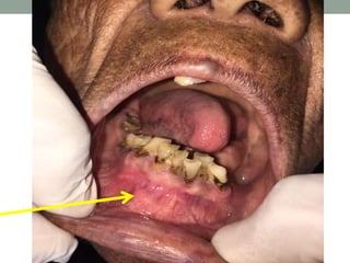
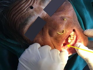
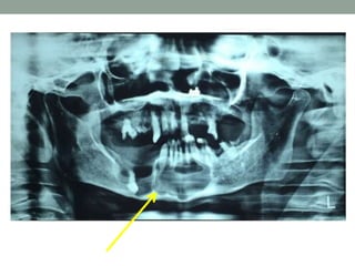
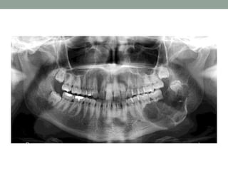
Odontogenic keratocyst (OKC) is the cyst arising from the cell rests of dental lamina. It can occur anywhere in the jaw, but commonly seen in the posterior part of the mandible. Radiographically, most OKCs are unilocular when presented at the periapex and can be mistaken for radicular or lateral periodontal cyst.
Fibro-osseous lesions of the jaws
Fibrous dysplasia
Cemento-osseous dysplasia
Focal cemento-osseous dysplasia
Periapical cemento-osseous dysplasia
Florid cemento-osseous dysplasia
Ossifying fibroma
Juvenile aggressive ossifying fibroma
Cherubism
Fibro-osseous lesions (FOL) are characterized by replacement of normal bone architecture by collagen fibers and fibroblasts containing calcified tissue.
They include a wide variety of lesions of developmental, dysplastic and neoplastic origins with clinical and radiographic presentation and behavior.
Because of the histological similarities between diverse diseases, proper diagnosis requires correlation of history, clinical and radiographic findings.Fibrous Dysplasia
2. Reactive (dysplastic lesions arising in the tooth-bearing area (presumably of periodontal origin).
a. Periapical cemento-osseous dysplasia
b. Focal cemento-osseous dysplasia
c. Florid cemento-osseous dysplasia
3. Fibro-osseous neoplasms (widely designated as cementifying fibroma, ossifying fibroma or cemento-ossifying fibroma.Bone dysplasias
a. Fibrous dyspla i. Monostoticii. Polyostotic
iii. Polyostotic with endocrinopathy (McCune-Albright)
iv Osteofibrous dysplasia
b. Osteitis deformansc. Pagetoid heritable bone dysplasias of childhood
d. Segmental odontomaxillary dysplasia
2. Cemento-osseous dysplasias
a. Focal cemento-osseous dysplasia b. Florid cemento-osseous dysplasia
3.Inflammatory/reactive processes
a. Focal sclerosing osteomyelitisb. Diffuse sclerosing osteomyelitis
c. Proliferative periostitis
4. Metabolic Disease: hyperparathyroidism
5. Neoplastic lesions (Ossifying fibromas)
a. Ossifying fibromab. Hyperparathyroidism jaw lesion syndrome
c. Juvenile ossifying fibroma i. Trabecular typeii. Psammomatoid type
d. Gigantiform cementomas
Dr. Ahmed M. Adawy, Professor Emeritus, Dep. Oral & Maxillofacial Surgery. Former Dean, Faculty of Dental Medicine, Al-Azhar University. Mandibular angle fractures account for 23% to 42% of all facial fractures. Fracture of mandibular angle can be classified as (A) Vertical favorable or unfavorable, (B) Horizontally favorable of unfavorable. Traditionally, mandibular angle fractures have been treated with either closed reduction and inter-maxillary fixation or open reduction and internal fixation with or without inter-maxillary fixation. Patients treated with inter-maxillary fixation have a restricted airway and loose excess weight. Rigid internal fixation and early return to function have eliminated the use of wire osteosenthysis and prolonged use of inter-maxillary fixation. The principal of rigid fixation, however, have inherent set of disadvantages including damage to the inferior alveolar nerve and the marginal mandibular branch of facial nerve. Postoperative malocclusion rates are also high. With the introduction of semi-rigid technique fracture of the mandibular angle could be treated according to Champy’s Ideal lines of osteosenthysis. The technique involves placement of a single monocortial miniplate on the superior border of the mandible. However, some studies suggested using a second miniplate along the inferior border. Wether one or two miniplates should be used is still debatable. The application of 3D plates may provide additional stability in 3 dimension and good resistance against torque forces.
ORN is an inflammatory condition of bone that occurs after the bone has been exposed to therapeutic doses of radiation usually given for a malignancies.
Gingival recession is a problem affecting almost all middle and older aged to some degree. It can be assessed by an appearance of a long clinical tooth and varied proportion of the teeth when compared with adjacent teeth...
The Indian Dental Academy is the Leader in continuing dental education , training dentists in all aspects of dentistry and
offering a wide range of dental certified courses in different formats.for more details please visit
www.indiandentalacademy.com
Includes most common tumors of oral cavity including scc,bcc, melanoma, ameloblastoma, odontoma, fibromas, pindborg tumors etc.
Presented by Dr. Binaya Subedi
mucogingival surgery or plastic surgery of muco-gingival tissue is a surgical procedure targeted to correct and eliminate anatomic, developmental and traumatic alterations of gingiva.
Erythema multiforme, Steven-Johnson syndrome and Toxic Epidermal NecrolysisBinaya Subedi
Erythema Multiforme is a common Vesiculobullous deramtological condition with mucosal manifestations trigged by Herpes virus infection and certain sulpha containing drugs.
Title: Sense of Smell
Presenter: Dr. Faiza, Assistant Professor of Physiology
Qualifications:
MBBS (Best Graduate, AIMC Lahore)
FCPS Physiology
ICMT, CHPE, DHPE (STMU)
MPH (GC University, Faisalabad)
MBA (Virtual University of Pakistan)
Learning Objectives:
Describe the primary categories of smells and the concept of odor blindness.
Explain the structure and location of the olfactory membrane and mucosa, including the types and roles of cells involved in olfaction.
Describe the pathway and mechanisms of olfactory signal transmission from the olfactory receptors to the brain.
Illustrate the biochemical cascade triggered by odorant binding to olfactory receptors, including the role of G-proteins and second messengers in generating an action potential.
Identify different types of olfactory disorders such as anosmia, hyposmia, hyperosmia, and dysosmia, including their potential causes.
Key Topics:
Olfactory Genes:
3% of the human genome accounts for olfactory genes.
400 genes for odorant receptors.
Olfactory Membrane:
Located in the superior part of the nasal cavity.
Medially: Folds downward along the superior septum.
Laterally: Folds over the superior turbinate and upper surface of the middle turbinate.
Total surface area: 5-10 square centimeters.
Olfactory Mucosa:
Olfactory Cells: Bipolar nerve cells derived from the CNS (100 million), with 4-25 olfactory cilia per cell.
Sustentacular Cells: Produce mucus and maintain ionic and molecular environment.
Basal Cells: Replace worn-out olfactory cells with an average lifespan of 1-2 months.
Bowman’s Gland: Secretes mucus.
Stimulation of Olfactory Cells:
Odorant dissolves in mucus and attaches to receptors on olfactory cilia.
Involves a cascade effect through G-proteins and second messengers, leading to depolarization and action potential generation in the olfactory nerve.
Quality of a Good Odorant:
Small (3-20 Carbon atoms), volatile, water-soluble, and lipid-soluble.
Facilitated by odorant-binding proteins in mucus.
Membrane Potential and Action Potential:
Resting membrane potential: -55mV.
Action potential frequency in the olfactory nerve increases with odorant strength.
Adaptation Towards the Sense of Smell:
Rapid adaptation within the first second, with further slow adaptation.
Psychological adaptation greater than receptor adaptation, involving feedback inhibition from the central nervous system.
Primary Sensations of Smell:
Camphoraceous, Musky, Floral, Pepperminty, Ethereal, Pungent, Putrid.
Odor Detection Threshold:
Examples: Hydrogen sulfide (0.0005 ppm), Methyl-mercaptan (0.002 ppm).
Some toxic substances are odorless at lethal concentrations.
Characteristics of Smell:
Odor blindness for single substances due to lack of appropriate receptor protein.
Behavioral and emotional influences of smell.
Transmission of Olfactory Signals:
From olfactory cells to glomeruli in the olfactory bulb, involving lateral inhibition.
Primitive, less old, and new olfactory systems with different path
Ozempic: Preoperative Management of Patients on GLP-1 Receptor Agonists Saeid Safari
Preoperative Management of Patients on GLP-1 Receptor Agonists like Ozempic and Semiglutide
ASA GUIDELINE
NYSORA Guideline
2 Case Reports of Gastric Ultrasound
New Directions in Targeted Therapeutic Approaches for Older Adults With Mantl...i3 Health
i3 Health is pleased to make the speaker slides from this activity available for use as a non-accredited self-study or teaching resource.
This slide deck presented by Dr. Kami Maddocks, Professor-Clinical in the Division of Hematology and
Associate Division Director for Ambulatory Operations
The Ohio State University Comprehensive Cancer Center, will provide insight into new directions in targeted therapeutic approaches for older adults with mantle cell lymphoma.
STATEMENT OF NEED
Mantle cell lymphoma (MCL) is a rare, aggressive B-cell non-Hodgkin lymphoma (NHL) accounting for 5% to 7% of all lymphomas. Its prognosis ranges from indolent disease that does not require treatment for years to very aggressive disease, which is associated with poor survival (Silkenstedt et al, 2021). Typically, MCL is diagnosed at advanced stage and in older patients who cannot tolerate intensive therapy (NCCN, 2022). Although recent advances have slightly increased remission rates, recurrence and relapse remain very common, leading to a median overall survival between 3 and 6 years (LLS, 2021). Though there are several effective options, progress is still needed towards establishing an accepted frontline approach for MCL (Castellino et al, 2022). Treatment selection and management of MCL are complicated by the heterogeneity of prognosis, advanced age and comorbidities of patients, and lack of an established standard approach for treatment, making it vital that clinicians be familiar with the latest research and advances in this area. In this activity chaired by Michael Wang, MD, Professor in the Department of Lymphoma & Myeloma at MD Anderson Cancer Center, expert faculty will discuss prognostic factors informing treatment, the promising results of recent trials in new therapeutic approaches, and the implications of treatment resistance in therapeutic selection for MCL.
Target Audience
Hematology/oncology fellows, attending faculty, and other health care professionals involved in the treatment of patients with mantle cell lymphoma (MCL).
Learning Objectives
1.) Identify clinical and biological prognostic factors that can guide treatment decision making for older adults with MCL
2.) Evaluate emerging data on targeted therapeutic approaches for treatment-naive and relapsed/refractory MCL and their applicability to older adults
3.) Assess mechanisms of resistance to targeted therapies for MCL and their implications for treatment selection
Lung Cancer: Artificial Intelligence, Synergetics, Complex System Analysis, S...Oleg Kshivets
RESULTS: Overall life span (LS) was 2252.1±1742.5 days and cumulative 5-year survival (5YS) reached 73.2%, 10 years – 64.8%, 20 years – 42.5%. 513 LCP lived more than 5 years (LS=3124.6±1525.6 days), 148 LCP – more than 10 years (LS=5054.4±1504.1 days).199 LCP died because of LC (LS=562.7±374.5 days). 5YS of LCP after bi/lobectomies was significantly superior in comparison with LCP after pneumonectomies (78.1% vs.63.7%, P=0.00001 by log-rank test). AT significantly improved 5YS (66.3% vs. 34.8%) (P=0.00000 by log-rank test) only for LCP with N1-2. Cox modeling displayed that 5YS of LCP significantly depended on: phase transition (PT) early-invasive LC in terms of synergetics, PT N0—N12, cell ratio factors (ratio between cancer cells- CC and blood cells subpopulations), G1-3, histology, glucose, AT, blood cell circuit, prothrombin index, heparin tolerance, recalcification time (P=0.000-0.038). Neural networks, genetic algorithm selection and bootstrap simulation revealed relationships between 5YS and PT early-invasive LC (rank=1), PT N0—N12 (rank=2), thrombocytes/CC (3), erythrocytes/CC (4), eosinophils/CC (5), healthy cells/CC (6), lymphocytes/CC (7), segmented neutrophils/CC (8), stick neutrophils/CC (9), monocytes/CC (10); leucocytes/CC (11). Correct prediction of 5YS was 100% by neural networks computing (area under ROC curve=1.0; error=0.0).
CONCLUSIONS: 5YS of LCP after radical procedures significantly depended on: 1) PT early-invasive cancer; 2) PT N0--N12; 3) cell ratio factors; 4) blood cell circuit; 5) biochemical factors; 6) hemostasis system; 7) AT; 8) LC characteristics; 9) LC cell dynamics; 10) surgery type: lobectomy/pneumonectomy; 11) anthropometric data. Optimal diagnosis and treatment strategies for LC are: 1) screening and early detection of LC; 2) availability of experienced thoracic surgeons because of complexity of radical procedures; 3) aggressive en block surgery and adequate lymph node dissection for completeness; 4) precise prediction; 5) adjuvant chemoimmunoradiotherapy for LCP with unfavorable prognosis.
Factory Supply Best Quality Pmk Oil CAS 28578–16–7 PMK Powder in Stockrebeccabio
Factory Supply Best Quality Pmk Oil CAS 28578–16–7 PMK Powder in Stock
Telegram: bmksupplier
signal: +85264872720
threema: TUD4A6YC
You can contact me on Telegram or Threema
Communicate promptly and reply
Free of customs clearance, Double Clearance 100% pass delivery to USA, Canada, Spain, Germany, Netherland, Poland, Italy, Sweden, UK, Czech Republic, Australia, Mexico, Russia, Ukraine, Kazakhstan.Door to door service
Hot Selling Organic intermediates
micro teaching on communication m.sc nursing.pdfAnurag Sharma
Microteaching is a unique model of practice teaching. It is a viable instrument for the. desired change in the teaching behavior or the behavior potential which, in specified types of real. classroom situations, tends to facilitate the achievement of specified types of objectives.
Tom Selleck Health: A Comprehensive Look at the Iconic Actor’s Wellness Journeygreendigital
Tom Selleck, an enduring figure in Hollywood. has captivated audiences for decades with his rugged charm, iconic moustache. and memorable roles in television and film. From his breakout role as Thomas Magnum in Magnum P.I. to his current portrayal of Frank Reagan in Blue Bloods. Selleck's career has spanned over 50 years. But beyond his professional achievements. fans have often been curious about Tom Selleck Health. especially as he has aged in the public eye.
Follow us on: Pinterest
Introduction
Many have been interested in Tom Selleck health. not only because of his enduring presence on screen but also because of the challenges. and lifestyle choices he has faced and made over the years. This article delves into the various aspects of Tom Selleck health. exploring his fitness regimen, diet, mental health. and the challenges he has encountered as he ages. We'll look at how he maintains his well-being. the health issues he has faced, and his approach to ageing .
Early Life and Career
Childhood and Athletic Beginnings
Tom Selleck was born on January 29, 1945, in Detroit, Michigan, and grew up in Sherman Oaks, California. From an early age, he was involved in sports, particularly basketball. which played a significant role in his physical development. His athletic pursuits continued into college. where he attended the University of Southern California (USC) on a basketball scholarship. This early involvement in sports laid a strong foundation for his physical health and disciplined lifestyle.
Transition to Acting
Selleck's transition from an athlete to an actor came with its physical demands. His first significant role in "Magnum P.I." required him to perform various stunts and maintain a fit appearance. This role, which he played from 1980 to 1988. necessitated a rigorous fitness routine to meet the show's demands. setting the stage for his long-term commitment to health and wellness.
Fitness Regimen
Workout Routine
Tom Selleck health and fitness regimen has evolved. adapting to his changing roles and age. During his "Magnum, P.I." days. Selleck's workouts were intense and focused on building and maintaining muscle mass. His routine included weightlifting, cardiovascular exercises. and specific training for the stunts he performed on the show.
Selleck adjusted his fitness routine as he aged to suit his body's needs. Today, his workouts focus on maintaining flexibility, strength, and cardiovascular health. He incorporates low-impact exercises such as swimming, walking, and light weightlifting. This balanced approach helps him stay fit without putting undue strain on his joints and muscles.
Importance of Flexibility and Mobility
In recent years, Selleck has emphasized the importance of flexibility and mobility in his fitness regimen. Understanding the natural decline in muscle mass and joint flexibility with age. he includes stretching and yoga in his routine. These practices help prevent injuries, improve posture, and maintain mobilit
Title: Sense of Taste
Presenter: Dr. Faiza, Assistant Professor of Physiology
Qualifications:
MBBS (Best Graduate, AIMC Lahore)
FCPS Physiology
ICMT, CHPE, DHPE (STMU)
MPH (GC University, Faisalabad)
MBA (Virtual University of Pakistan)
Learning Objectives:
Describe the structure and function of taste buds.
Describe the relationship between the taste threshold and taste index of common substances.
Explain the chemical basis and signal transduction of taste perception for each type of primary taste sensation.
Recognize different abnormalities of taste perception and their causes.
Key Topics:
Significance of Taste Sensation:
Differentiation between pleasant and harmful food
Influence on behavior
Selection of food based on metabolic needs
Receptors of Taste:
Taste buds on the tongue
Influence of sense of smell, texture of food, and pain stimulation (e.g., by pepper)
Primary and Secondary Taste Sensations:
Primary taste sensations: Sweet, Sour, Salty, Bitter, Umami
Chemical basis and signal transduction mechanisms for each taste
Taste Threshold and Index:
Taste threshold values for Sweet (sucrose), Salty (NaCl), Sour (HCl), and Bitter (Quinine)
Taste index relationship: Inversely proportional to taste threshold
Taste Blindness:
Inability to taste certain substances, particularly thiourea compounds
Example: Phenylthiocarbamide
Structure and Function of Taste Buds:
Composition: Epithelial cells, Sustentacular/Supporting cells, Taste cells, Basal cells
Features: Taste pores, Taste hairs/microvilli, and Taste nerve fibers
Location of Taste Buds:
Found in papillae of the tongue (Fungiform, Circumvallate, Foliate)
Also present on the palate, tonsillar pillars, epiglottis, and proximal esophagus
Mechanism of Taste Stimulation:
Interaction of taste substances with receptors on microvilli
Signal transduction pathways for Umami, Sweet, Bitter, Sour, and Salty tastes
Taste Sensitivity and Adaptation:
Decrease in sensitivity with age
Rapid adaptation of taste sensation
Role of Saliva in Taste:
Dissolution of tastants to reach receptors
Washing away the stimulus
Taste Preferences and Aversions:
Mechanisms behind taste preference and aversion
Influence of receptors and neural pathways
Impact of Sensory Nerve Damage:
Degeneration of taste buds if the sensory nerve fiber is cut
Abnormalities of Taste Detection:
Conditions: Ageusia, Hypogeusia, Dysgeusia (parageusia)
Causes: Nerve damage, neurological disorders, infections, poor oral hygiene, adverse drug effects, deficiencies, aging, tobacco use, altered neurotransmitter levels
Neurotransmitters and Taste Threshold:
Effects of serotonin (5-HT) and norepinephrine (NE) on taste sensitivity
Supertasters:
25% of the population with heightened sensitivity to taste, especially bitterness
Increased number of fungiform papillae
- Video recording of this lecture in English language: https://youtu.be/lK81BzxMqdo
- Video recording of this lecture in Arabic language: https://youtu.be/Ve4P0COk9OI
- Link to download the book free: https://nephrotube.blogspot.com/p/nephrotube-nephrology-books.html
- Link to NephroTube website: www.NephroTube.com
- Link to NephroTube social media accounts: https://nephrotube.blogspot.com/p/join-nephrotube-on-social-media.html
TEST BANK for Operations Management, 14th Edition by William J. Stevenson, Ve...kevinkariuki227
TEST BANK for Operations Management, 14th Edition by William J. Stevenson, Verified Chapters 1 - 19, Complete Newest Version.pdf
Explore natural remedies for syphilis treatment in Singapore. Discover alternative therapies, herbal remedies, and lifestyle changes that may complement conventional treatments. Learn about holistic approaches to managing syphilis symptoms and supporting overall health.
4. History of Presenting illness
• She was in her usual state of health, when she noticed a
hard swelling in lower front region of jaw 3 months back,
which was gradual on onset, progressive associated with
pain, looseness of teeth, foul taste and breath.
• Pain on lower back and front region of jaw which was
gradual on onset, throbbing type, radiating towards ears
and neck, mildly relieved on medications (painkillers),
aggravated during eating.
• There was no history of fever, vomiting or trauma.
6. Past Dental History
• She had undergone removal of teeth in lower right back
region of jaw which was eventful (unhealed socket).
7. Personal History
• She consumes tobacco in chewable form since past 15
years and places it in lower right vestibular area.
• She brushes randomly with neem stick (twig-datun)
9. Extra-oral Examination
Facial Symmetry:
• Asymmetrical
• Swelling present over right mandibular posterior region and
anterior region of around 3×3 cm in size, without any changes
or ulcerations on overlying skin.
TMJ:
• Deviated towards right on opening and closing. Non-tender
Lymph Nodes:
• Multiple palpable right submandibular nodes, oval in shape,
around 0.5-1 cm in diameter, soft, freely mobile and non-
tender.
10. Intra-oral Examinations
• A swelling extending from distal of right mandibular canine to mesial
to left mandibular 1st premolar, of around 3×3 cm in size, without any
surface changes. On palpation, it was non-tender, bony hard with
egg-shell cracking present.
• A bony defect was present distal to right mandibular canine of around
1cm in diameter, with pus like discharge. On palpation it was tender.
• A fluctuant swelling present over right alveo-lingual sulcus of around
5×1 cm in dimension with no surface alterations and was fluctuant on
palpation.
• Generalized gingival recession present.
14. Investigations
• Aspiration of cystic contents:
Showed dirty pus colored fluid, contained shredded and
fragmented cells of cystic lining.
• Protein analysis:
Toller postulated that a protein level of less than 4.0 gm/100ml
indicated a diagnosis of OKC.
• Radiography:
OPG was advised which showed, multilocular radiolucency
extending from right rams-angle region to mandibular anterior
region with well defined sclerotic border without roots resorption
and thinning of cortical plates and only few mm of mandibular lower
border remaining.
17. Treatment Options:
• Curettage with peripheral osteotomy keeping a safety
margin of 1mm
• Curettage followed by Chemical cauterization with
carnoy’s solution
• Marginal mandibulectomy leaving lower border intact
• Segmental hemimandibulectomy with left anterior
sectional mandibulectomy.
Prognosis:
• Poor, depending upon the patient’s condition, severity of
lesion and amount of mandible involved.
• Recurrence is most likely.
20. • The odontogenic keratocyst is a distinctive form of developmental
odontogenic cyst that deserves special consideration because of
its specific histopathologic features and clinical behavior.
• There is general agreement that the odontogenic keratocyst
arises from cell rests of the dental lamina.
• 3%-11% of odontogenic cyst
• Classified under benign odontogenic tumors by WHO because;
• Behaviour: Locally destructive and high recurrence rate
• Histopathology: basal epithelial layer shows proliferation and
budding into the underlying CT
• Genetics: PTCH gene mutation
• Possible association with Nevoid basal cell carcinoma
• Reclassified under developmental odontogenic cyst by WHO in
2017
21. • Age:
• Occurs over a wide range
of age groups from first to
ninth decades.
• Bimodal distribution with
peak incidence: 2nd-3rd
decade & 5th- later
• Sex: Male predilection
• Site:
• Mandible (65%) ramus
3rd molar area > 1st & 2nd
molar area > anterior
maxilla (25%)
• In most of cases, more
than one OKC are
present.
Clinical Features:
22. • OKC grows within medullary cavity of the bone without causing
obvious bone expansion in anteroposterior direction which is
less compared to other cysts of comparable size.
• Small OKC are asymptomatic and are discovered only during
radiographic examination.
• Larger lesions produce swellings involving the maxillary sinus and the
entire ascending ramus, including the condylar and coronoid
processes causing apparent facial asymmetry, pain, paraesthesia of
lips and mobility of non-periodontal origin.
• Displacement of tooth is common that root resorption but both can
occur concomittently.
• When maxillary anterior region is involved, tends to get infected
due to vicinity to maxillary sinus.
23. • OKC is associated with GORLIN-GOLTZ (NBSCS)
syndrome characterized by;
• Basal cell carcinoma
• Odontogenic Keratocyst of the jaws
• Bifid ribs– sixth rib
• Plantar & palmar pits
• Occular hypertelorism
• Ectopic calcifications
24. Radiographic Features
• OKC demonstrate a well-defined radiolucent area with
smooth and often corticated margins. Larger lesions tends
to be multilocular.
• Sometimes associated with unerupted tooth mimicking
dentigerous cyst.
• Root resorption is common.
25.
26. • Radiographic classification (Toller, 1970)
• Replacemental type: develops in place of normal tooth
• Envelopemental type: embraces an adjacent unerupted
tooth
• Extraneous type: develops in ascending ramus away
from teeth
• Collateral type: adjacent to root of teeth
27. Treatment and Prognosis
• Enucleation with curettage
• Enucleation with peripheral osteotomy with safety
margins.
• Chemical cauterization with Carnoys solution after
enucleation
• Surgical resection of a marginal bone, part of, half of or
whole mandible depending upon severity.
Prognosis:
• Potential malignant transformation but rare
• Recurrence is high (15-20%)
28. Recurrence is due to;
• Thin, fragile lining is very difficult to remove completely.
• New cysts develop from satellite cysts left behind.
• Some cysts may be left behind in cases of Gorlin – Gotz
syndrome.
• New cysts can also develop from basal cells of overlying
oral epithelium, especially in ramus – 3rd molar region.