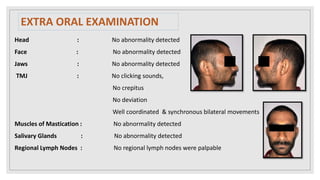
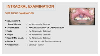
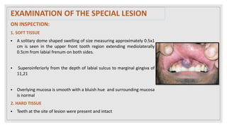
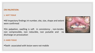
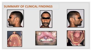
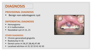
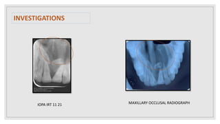
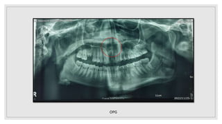
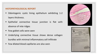
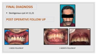
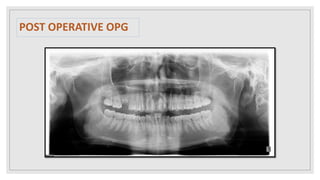
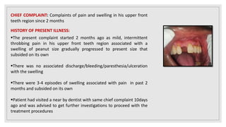
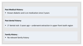
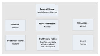
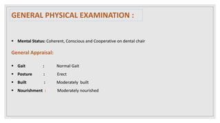
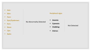
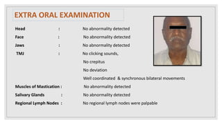
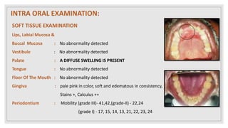
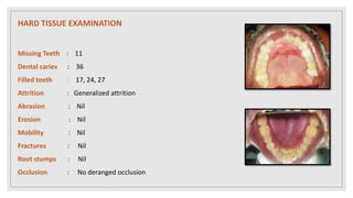
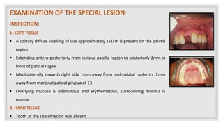
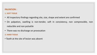
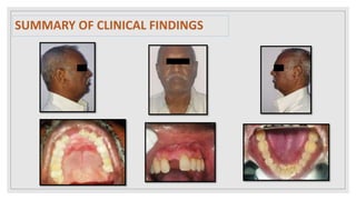
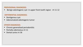
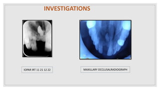
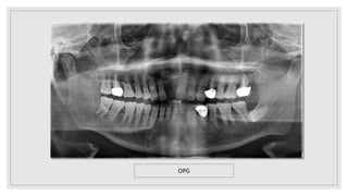
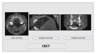
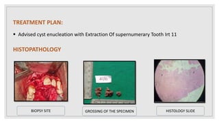
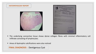
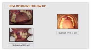
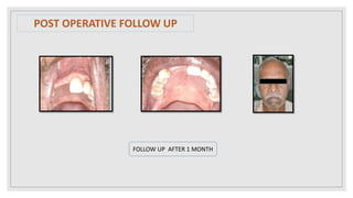
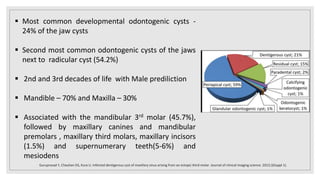
DENTIGEROUS CYST IN ANTERIOR
MAXILLA: A RARE ENTITY
-CASE SERIES
Presenting by :
Anitha Chesetti
Oral Medicine And Radiology
GSL Dental College And Hospital
CASE- 1
OP No : 2212030009
Age : 26 years
Name : P Suresh
Gender : Male
Address : Yanam
Chief complaint:
 Complaints of swelling in upper front tooth region since 1 month
History of Present Illness:
 The present complaint started 1 month ago as a small swelling in the
upper front tooth region which gradually progressed to present size,
with no associated pain and secondary changes.
 Patient gives history of same swelling in the upper front tooth region
1 year ago and visited nearby dentist and he was advised biopsy and
underwent biopsy (Biopsy report revealed benign cystic lesion) &
swelling has not subsided.
 Patient again visited a nearby hospital 1 month ago, advised root canal
treatment and patient didn’t undergo treatment, swelling has not
subsided and he further visited GSL dental hospital for further
treatment.
Past Medical History:
 No relevant past medical history
Past dental history:
 1st dental visit 1 and half year ago – swelling in upper front tooth region
Family History :
 No relevant family history
Appetite:
Normal
Bowel and bladder:
Normal
Micturition :
Normal
Oral Hygiene Habits:
Brushes once daily
with tooth brush
and tooth paste
Deleterious habits :
No H/O
Sleep :
Normal
Personal history;
Marital status: Married
 Mental Status: Coherent, Conscious and Cooperative on dental chair
General Appraisal:
 Gait : Normal Gait
 Posture : Erect
 Built : Moderately built
 Nourishment : Moderately nourished
GENERAL PHYSICAL EXAMINATION :
 Hair:
 Skin:
 Face:
 Eyes/Eyebrows:
 Ears:
 Nose:
 Lips:
 Nails:
No Abnormality Detected
Peripheral signs
 Anemia
 Cyanosis:
 Clubbing:
 Icterus:
Not Detected
Vital signs
 Pulse rate : 68 beats/min
 B.P : 120/70 mmHg
 Respiratory rate : 18 breaths / min
 Temperature : 98.3 ⁰ F
 Height : 5’6”
 Weight : 53kgs
EXTRA ORAL EXAMINATION
Head : No abnormality detected
Face : No abnormality detected
Jaws : No abnormality detected
TMJ : No clicking sounds,
No crepitus
No deviation
Well coordinated & synchronous bilateral movements
Muscles of Mastication : No abnormality detected
Salivary Glands : No abnormality detected
Regional Lymph Nodes : No regional lymph nodes were palpable
INTRAORAL EXAMINATION
SOFT TISSUE EXAMINATION
 Lips , Alveolar &
Buccal Mucosa : No Abnormality Detected
 Labial Mucosa : NODULAR GROWTH ON LABIAL FRENUM
 Palate : No Abnormality Detected
 Tongue : No Abnormality Detected
 Floor Of The Mouth : No Abnormality Detected
 Gingiva & Coral pink in color, firm in consistency
 Periodontium : Calculus + stains +
HARD TISSUE EXAMINATION
Dental caries : 36,37,38,46,47,48
Filled teeth : Nil
Attrition : 31,32,41,42,43
Abrasion : Nil
Erosion : Nil
Mobility : Nil
Fractures : Nil
Root stumps : 16
Occlusion : No Derranged occlusion
ON INSPECTION:
1. SOFT TISSUE
 A solitary dome shaped swelling of size measuring approximately 0.5x1
cm is seen in the upper front tooth region extending mediolaterally
0.5cm from labial frenum on both sides.
 Superoinferiorly from the depth of labial sulcus to marginal gingiva of
11,21
 Overlying mucosa is smooth with a bluish hue and surrounding mucosa
is normal
2. HARD TISSUE
 Teeth at the site of lesion were present and intact
EXAMINATION OF THE SPECIAL LESION
ON PALPATION:
1. SOFT TISSUE
All inspectory findings irt number, site, size, shape and extent
were confirmed
On palpation, swelling is soft in consistency , non-tender,
non compressible, non reducible, non pulsatile and no
discharge on provocation
2. HARD TISSUE
Teeth associated with lesion were not mobile
SUMMARY OF CLINICAL FINDINGS
DIAGNOSIS
PROVISIONAL DIAGNOSIS
 Benign non-odontogenic cyst
DIFFERENTIAL DIAGNOSIS
 Hemangioma
 A V malformation
 Nasolabial cyst irt 11, 21
OTHER DIAGNOSIS:
 Chronic generalized gingivitis
 Rootstump irt 16
 Dental caries irt 36,37,38,46,47,48
 Localised attrition irt 31 32 33 41 42 43
INVESTIGATIONS
IOPA IRT 11 21 MAXILLARY OCCLUSAL RADIOGRAPH
OPG
 Dentigerous cyst irt 11, 21
DIFFERENTIAL DIAGNOSIS
 Adenamatoid odontogenic tumour
 CEOC
RADIOLOGICAL DIAGNOSIS
TREATMENT PLAN
 Cyst enucleation along with
extraction of supernumerary
tooth
 Endodontic treatment irt 11 12
13 21 22 23
HISTOPATHOLOGICAL REPORT
 Odontogenic cystic lining epithelium exhibiting 1-2
layers thickness.
 Epithelial connective tissue junction is flat with
absence of rete ridges
 Few goblet cells were seen
 Underlying connective tissue shows dense collagen
bundles with minimal inflammatory cell infiltrate
 Few dilated blood capillaries are also seen
FINAL DIAGNOSIS
 Dentigerous cyst irt 11,21
POST OPERATIVE FOLLOW UP
1 WEEK FOLLOWUP 1 MONTH FOLLOWUP
POST OPERATIVE OPG
CASE- 2
OP No : 2104100002
Age : 73 years
Name : B Satya Prasad
Gender : Male
Address : Rajahmundry
CHIEF COMPLAINT: Complaints of pain and swelling in his upper front
teeth region since 2 months
HISTORY OF PRESENT ILLNESS:
The present complaint started 2 months ago as mild, intermittent
throbbing pain in his upper front teeth region associated with a
swelling of peanut size gradually progressed to present size that
subsided on its own
There was no associated discharge/bleeding/paresthesia/ulceration
with the swelling
There were 3-4 episodes of swelling associated with pain in past 2
months and subsided on its own
Patient had visited a near by dentist with same chief complaint 10days
ago and was advised to get further investigations to proceed with the
treatment procedures
Past Medical History:
 Known diabetic and is on medication since 3 years
Past dental history:
 1st dental visit- 3 years ago – underwent extraction in upper front tooth region
Family History :
 No relevant family history
Appetite:
Normal
Bowel and bladder:
Normal
Micturition :
Normal
Oral Hygiene Habits:
Brushes once daily
with tooth brush
and tooth paste
Deleterious habits :
No H/O
Sleep :
Normal
Personal history;
Marital status: Married
 Mental Status: Coherent, Conscious and Cooperative on dental chair
General Appraisal:
 Gait : Normal Gait
 Posture : Erect
 Built : Moderately built
 Nourishment : Moderately nourished
GENERAL PHYSICAL EXAMINATION :
 Hair:
 Skin:
 Face:
 Eyes/Eyebrows:
 Ears:
 Nose:
 Lips:
 Nails:
No Abnormality Detected
Peripheral signs
 Anemia
 Cyanosis:
 Clubbing:
 Icterus:
Not Detected
Vital signs
 Pulse rate : 74 beats/min
 B.P : 140/70 mmHg
 Respiratory rate : 17 breaths / min
 Temperature : 98.6 ⁰ F
 Height : 5’10”
 Weight : 54kgs
EXTRA ORAL EXAMINATION
Head : No abnormality detected
Face : No abnormality detected
Jaws : No abnormality detected
TMJ : No clicking sounds,
No crepitus
No deviation
Well coordinated & synchronous bilateral movements
Muscles of Mastication : No abnormality detected
Salivary Glands : No abnormality detected
Regional Lymph Nodes : No regional lymph nodes were palpable
SOFT TISSUE EXAMINATION
Lips, Labial Mucosa &
Buccal Mucosa : No abnormality detected
Vestibule : No abnormality detected
Palate : A DIFFUSE SWELLING IS PRESENT
Tongue : No abnormality detected
Floor Of The Mouth : No abnormality detected
Gingiva : pale pink in color, soft and edematous in consistency,
Stains +, Calculus ++
Periodontium : Mobility (grade III)- 41,42,(grade-II) - 22,24
(grade I) - 17, 15, 14, 13, 21, 22, 23, 24
INTRA ORAL EXAMINATION:
HARD TISSUE EXAMINATION
Missing Teeth : 11
Dental caries : 36
Filled teeth : 17, 24, 27
Attrition : Generalized attrition
Abrasion : Nil
Erosion : Nil
Mobility : Nil
Fractures : Nil
Root stumps : Nil
Occlusion : No deranged occlusion
EXAMINATION OF THE SPECIAL LESION:
INSPECTION:
1. SOFT TISSUE
 A solitary diffuse swelling of size approximately 1x1cm is present on the palatal
region.
 Extending antero-posteriorly from incisive papilla region to posteriorly 2mm in
front of palatal rugae
 Mediolaterally towards right side 1mm away from mid-palatal raphe to 2mm
away from marginal palatal gingiva of 13
 Overlying mucosa is edematous and erythematous, surrounding mucosa is
normal
2. HARD TISSUE
 Tooth at the site of lesion was absent
PALPATION :
1. SOFT TISSUE
• All inspectory findings regarding site, size, shape and extent are confirmed
• On palpation, swelling is non-tender, soft in consistency, non compressible, non
reducible and non pulsatile
• There was no discharge on provocation
2. HARD TISSUE
• Tooth at the site of lesion was absent
SUMMARY OF CLINICAL FINDINGS
PROVISIONAL DIAGNOSIS:
 Benign odontogenic cyst in upper front tooth region irt 11 12
DIFFERENTIAL DIAGNOSIS:
 Dentigerous cyst
 Adenomatoid odontogenic tumor
OTHER DIAGNOSIS:
 Chronic generalized periodontitis
 Partially edentulous irt 11
 Dental caries irt 36
INVESTIGATIONS
IOPAR IRT 11 21 12 22 MAXILLARY OCCLUSALRADIOGRAPH
OPG
AXIAL SECTION SAGGITAL SECTION CORONAL SECTION
CBCT
TREATMENT PLAN:
 Advised cyst enucleation with Extraction Of supernumerary Tooth Irt 11
HISTOPATHOLOGY
GROSSING OF THE SPECIMEN HISTOLOGY SLIDE
BIOPSY SITE
HISTOPATHOLOGY REPORT
 The underlying connective tissue shows dense collagen fibres with minimal inflammatory cell
infiltrate consisting of lymphocytes.
 Areas of dystrophic calcifications were also noticed
FINAL DIAGNOSIS : Dentigerous Cyst
FOLLOW UP AFTER 7 DAYS
FOLLOW UP AFTER 15 DAYS
POST OPERATIVE FOLLOW UP
FOLLOW UP AFTER 1 MONTH
DISCUSSION
 Cyst is defined ‘a pathological cavity having fluid, semifluid or gaseous
contents and which is not created by the accumulation of pus’
(Kramer (1974) )
 Odontogenic cysts - arising from the odontogenic epithelium and
Non odontogenic(Fissural cysts) - arising from the oral epithelium
trapped between fusing processes during embryogenesis
 Dentigenous cyst (follicular, pericoronal) –
“An odontogenic cyst that surrounds the crown of an impacted tooth;
caused by fluid accumulation between the reduced enamel
epithelium and the enamel surface, resulting in a cyst in which the
crown is located within the lumen”
Rajendran, A., & Sundaram, S. (2014). Shafer’s textbook of oral pathology. Elsevier Health Sciences Apac.
 Dentigerous cyst develops around the crown of an unerupted tooth
by accumulation of fluid either between the reduced enamel
epithelium and enamel
 This fluid accumulation occurs as a result of the pressure exerted by
an erupting tooth on an impacted follicle, which obstructs the
venous outflow and thereby induces rapid transudation of serum
across the capillary wall.
 Two types of dentigerous cysts occur( Ben and Altini)
First type - Developmental in origin,
Occurs in mature teeth
Result of impaction
Second type - Inflammatory in origin
Occurs in immature teeth
Result of inflammation from a non-vital deciduous
tooth or some other source spreading to involve the
tooth follicle
Önay Ö, Süslü AE, Yılmaz T. Huge dentigerous cyst in the maxillary sinus: a rare case in childhood. Turkish archives of otorhinolaryngology. 2019 Mar;57(1):54.
 Most common developmental odontogenic cysts -
24% of the jaw cysts
 Second most common odontogenic cysts of the jaws
next to radicular cyst (54.2%)
 2nd and 3rd decades of life with Male prediliction
 Mandible – 70% and Maxilla – 30%
 Associated with the mandibular 3rd molar (45.7%),
followed by maxillary canines and mandibular
premolars , maxillary third molars, maxillary incisors
(1.5%) and supernumerary teeth(5-6%) and
mesiodens
Guruprasad Y, Chauhan DS, Kura U. Infected dentigerous cyst of maxillary sinus arising from an ectopic third molar. Journal of clinical imaging science. 2013;3(Suppl 1).
 Mourshed stated that only 1.44% of impacted teeth undergo
dentigerous cyst transformation
 So dentigerous cysts involving the permanent central incisor
are rare( 44 Cases)
 Clinically, it is often asymptomatic; it is discovered as incidental
radiographic finding or when acute inflammation, or swelling
develops but have the potential to become extremely large
and cause cortical expansion and erosion
 Expansion of the bone, facial asymmetry, extreme
displacement of teeth, severe root resorption of adjacent teeth
and pain are all possible sequelae of continuous enlargement
of the cyst
Arakeri G, Rai KK, Shivakumar HR, Khaji SI. A massive dentigerous cyst of the mandible in a young patient: a case report. Plastic and Aesthetic Research. 2015 Sep 15;2:294-8.
 Cystic involvement of an unerupted mandibular third
molar - ‘hollowing-out’ of the entire ramus extending
up to the coronoid process and condyle as well as in
expansion of the cortical plate due to the pressure
exerted by the lesion and sometimes comes to lie
compressed against the inferior border of the
mandible.
 Cyst associated with a maxillary cuspid - expansion
of the anterior maxilla and resemble an acute
sinusitis or cellulitis
Nagarajan N, Jayachandran S, Jayaram V, Nisha A. Dentigerous Cyst in the Maxillary Anterior Region of a Pediatric Patient. Annals of the National Academy of Medical Sciences (India). 2021 Jan;57(01):58-61.
 Radiographically, If the follicular space on radiograph is more than 5 mm, an
odontogenic cyst can be suspected
 It usually occurs as a well-defined unilocular radiolucency, often with a
sclerotic border around the impacted crown of the tooth
 The cyst to crown relationship in a dentigerous cyst can show different
variations (Thoma-Robinson-Bernier)
• Central variety - The cyst surrounds the crown of the tooth with the
crown projecting into the cyst(mandibular 3rd molar, maxillary canine)
• Lateral variety - When the cyst grows laterally along the root surface
surrounding the crown partially (mandibular 3rd molar)
• Circumferential variety - a considerable amount of root appears to lie
within the cyst along with the crown that is surrounded by the cyst
Nagarajan N, Jayachandran S, Jayaram V, Nisha A. Dentigerous Cyst in the Maxillary Anterior Region of a Pediatric Patient. Annals of the National Academy of Medical Sciences (India). 2021 Jan;57(01):58-61.
 The content of the cyst lumen is usually a thin,
watery yellow fluid, occasionally blood tinged.
 Histologically, the dentigerous cyst is usually
composed of a thin fibrous connective tissue
wall lined by nonkeratinized stratified
squamous epithelium consisting of myxoid
tissue, odontogenic remnants and rarely
sebaceous cells.
 Rete peg formation is generally absent except
in cases that are secondarily infected.
Rohilla M, Marwah N, Tyagi R. Anterior maxillary dentigerous cyst. International journal of clinical pediatric dentistry. 2009 Jan;2(1):42.
Differential diagnosis
1.Unicystic Ameloblastoma - They will grow laterally away from the tooth in comparison to dentigerous
cyst, which envelop the tooth symmetrically and is more common in the premolar-molar area
2.Adenomatoid odontogenic tumor- rare and usually occur in the maxillary anterior region, Cystic lining is
not attached to CEJ, it involves the uneruptedtooth
3.Calcifying odontogenic Cyst - it may occur as a pericoronal radiolucency and may contain evidences of
calcification, Cystic lining is not attached to CEJ, it involves the unerupted tooth
4.Odontogenic keratocyst - does not expand the bone to the same extent, is less likely to resorb teeth
and may attach further apically on the root instead at the cementoenamel junction
5.Hyper plastic follicle - if the follicular space is more than 5 mm, a dentigerous cyst is suspected
 These are usually solitary however bilateral and multiple
cysts - syndromes such as basal cell nevus syndrome,
mucopolysaccharidosis, Gardner’s syndrome, cleidocranial
dysplasia, bifid rib syndrome and prolonged concurrent use
of cyclosporine & calcium channel blockers
 Various treatment plans proposed for dentigerous cysts are:
(a) Cyst enucleation along with extraction of the
involved tooth.
(b) Marsupialization technique - involves removal of
the cyst, however, the developing tooth is
preserved.
 Potential Complications : untreated Dentigerous cyst may
transform into tumours like mural ameloblastoma, and has
the potential of developing into malignancies like squamous
cell carcinoma and mucoepidermoid carcinoma
Nagarajan N, Jayachandran S, Jayaram V, Nisha A. Dentigerous Cyst in the Maxillary Anterior Region of a Pediatric Patient. Annals of the National Academy of Medical Sciences (India). 2021 Jan;57(01):58-61.
 Dentigerous cyst is a common developmental odontogenic
cyst, mostly seen in relation with impacted mandibular third
molars and maxillary canines
 Usually, they are asymptomatic and this delays the diagnosis
 Prompt diagnosis and treatment is mandatory to prevent
dreadful complications
 A dentigerous cyst associated with an anterior tooth will
result in failure or eruption of the tooth and therefore lead to
esthetic and orthodontic problems
CONCLUSION
REFERENCES
1. Shear, M., & Speight, P. (2008). Cysts of the oral and maxillofacial regions (4th ed.). Wiley-Blackwell.
2. Rajendran, A., & Sundaram, S. (2014). Shafer’s textbook of oral pathology. Elsevier Health Sciences Apac.
3. Nagarajan N, Jayachandran S, Jayaram V, Nisha A. Dentigerous Cyst in the Maxillary Anterior Region of a Pediatric
Patient. Annals of the National Academy of Medical Sciences (India). 2021 Jan;57(01):58-61.
4. Rohilla M, Marwah N, Tyagi R. Anterior maxillary dentigerous cyst. International journal of clinical pediatric dentistry.
2009 Jan;2(1):42.
5. Cura N, Hanttash A, Inceoglu B, Orhan K, Oncul AM. Dentigerous cysts in four quadrants of a nonsyndromic patient:
case report and literature review. Oral Radiology. 2015 Jan;31:49-58.
6. Arakeri G, Rai KK, Shivakumar HR, Khaji SI. A massive dentigerous cyst of the mandible in a young patient: a case
report. Plastic and Aesthetic Research. 2015 Sep 15;2:294-8.
7. Guruprasad Y, Chauhan DS, Kura U. Infected dentigerous cyst of maxillary sinus arising from an ectopic third molar.
Journal of clinical imaging science. 2013;3(Suppl 1).
8. Önay Ö, Süslü AE, Yılmaz T. Huge dentigerous cyst in the maxillary sinus: a rare case in childhood. Turkish archives of
otorhinolaryngology.2019Mar;57(1):54
9. Wood NK, goaz PW. Differential diagnosis of oral and maxillofacial lesions. 5th ed. St. Louis, MO: mosby; 1997
10.White SC, pharoah MJ. White and pharoah’s oral radiology: principles and interpretation. 8th ed. London, england:
mosby; 2018.

DENTIGEROUS CYST - a case presentation with review

  • 1.
    DENTIGEROUS CYST INANTERIOR MAXILLA: A RARE ENTITY -CASE SERIES Presenting by : Anitha Chesetti Oral Medicine And Radiology GSL Dental College And Hospital
  • 2.
  • 3.
    OP No :2212030009 Age : 26 years Name : P Suresh Gender : Male Address : Yanam
  • 4.
    Chief complaint:  Complaintsof swelling in upper front tooth region since 1 month History of Present Illness:  The present complaint started 1 month ago as a small swelling in the upper front tooth region which gradually progressed to present size, with no associated pain and secondary changes.  Patient gives history of same swelling in the upper front tooth region 1 year ago and visited nearby dentist and he was advised biopsy and underwent biopsy (Biopsy report revealed benign cystic lesion) & swelling has not subsided.  Patient again visited a nearby hospital 1 month ago, advised root canal treatment and patient didn’t undergo treatment, swelling has not subsided and he further visited GSL dental hospital for further treatment.
  • 5.
    Past Medical History: No relevant past medical history Past dental history:  1st dental visit 1 and half year ago – swelling in upper front tooth region Family History :  No relevant family history
  • 6.
    Appetite: Normal Bowel and bladder: Normal Micturition: Normal Oral Hygiene Habits: Brushes once daily with tooth brush and tooth paste Deleterious habits : No H/O Sleep : Normal Personal history; Marital status: Married
  • 7.
     Mental Status:Coherent, Conscious and Cooperative on dental chair General Appraisal:  Gait : Normal Gait  Posture : Erect  Built : Moderately built  Nourishment : Moderately nourished GENERAL PHYSICAL EXAMINATION :
  • 8.
     Hair:  Skin: Face:  Eyes/Eyebrows:  Ears:  Nose:  Lips:  Nails: No Abnormality Detected Peripheral signs  Anemia  Cyanosis:  Clubbing:  Icterus: Not Detected
  • 9.
    Vital signs  Pulserate : 68 beats/min  B.P : 120/70 mmHg  Respiratory rate : 18 breaths / min  Temperature : 98.3 ⁰ F  Height : 5’6”  Weight : 53kgs
  • 10.
    EXTRA ORAL EXAMINATION Head: No abnormality detected Face : No abnormality detected Jaws : No abnormality detected TMJ : No clicking sounds, No crepitus No deviation Well coordinated & synchronous bilateral movements Muscles of Mastication : No abnormality detected Salivary Glands : No abnormality detected Regional Lymph Nodes : No regional lymph nodes were palpable
  • 11.
    INTRAORAL EXAMINATION SOFT TISSUEEXAMINATION  Lips , Alveolar & Buccal Mucosa : No Abnormality Detected  Labial Mucosa : NODULAR GROWTH ON LABIAL FRENUM  Palate : No Abnormality Detected  Tongue : No Abnormality Detected  Floor Of The Mouth : No Abnormality Detected  Gingiva & Coral pink in color, firm in consistency  Periodontium : Calculus + stains +
  • 12.
    HARD TISSUE EXAMINATION Dentalcaries : 36,37,38,46,47,48 Filled teeth : Nil Attrition : 31,32,41,42,43 Abrasion : Nil Erosion : Nil Mobility : Nil Fractures : Nil Root stumps : 16 Occlusion : No Derranged occlusion
  • 13.
    ON INSPECTION: 1. SOFTTISSUE  A solitary dome shaped swelling of size measuring approximately 0.5x1 cm is seen in the upper front tooth region extending mediolaterally 0.5cm from labial frenum on both sides.  Superoinferiorly from the depth of labial sulcus to marginal gingiva of 11,21  Overlying mucosa is smooth with a bluish hue and surrounding mucosa is normal 2. HARD TISSUE  Teeth at the site of lesion were present and intact EXAMINATION OF THE SPECIAL LESION
  • 14.
    ON PALPATION: 1. SOFTTISSUE All inspectory findings irt number, site, size, shape and extent were confirmed On palpation, swelling is soft in consistency , non-tender, non compressible, non reducible, non pulsatile and no discharge on provocation 2. HARD TISSUE Teeth associated with lesion were not mobile
  • 15.
  • 16.
    DIAGNOSIS PROVISIONAL DIAGNOSIS  Benignnon-odontogenic cyst DIFFERENTIAL DIAGNOSIS  Hemangioma  A V malformation  Nasolabial cyst irt 11, 21 OTHER DIAGNOSIS:  Chronic generalized gingivitis  Rootstump irt 16  Dental caries irt 36,37,38,46,47,48  Localised attrition irt 31 32 33 41 42 43
  • 17.
    INVESTIGATIONS IOPA IRT 1121 MAXILLARY OCCLUSAL RADIOGRAPH
  • 18.
  • 19.
     Dentigerous cystirt 11, 21 DIFFERENTIAL DIAGNOSIS  Adenamatoid odontogenic tumour  CEOC RADIOLOGICAL DIAGNOSIS
  • 20.
    TREATMENT PLAN  Cystenucleation along with extraction of supernumerary tooth  Endodontic treatment irt 11 12 13 21 22 23
  • 21.
    HISTOPATHOLOGICAL REPORT  Odontogeniccystic lining epithelium exhibiting 1-2 layers thickness.  Epithelial connective tissue junction is flat with absence of rete ridges  Few goblet cells were seen  Underlying connective tissue shows dense collagen bundles with minimal inflammatory cell infiltrate  Few dilated blood capillaries are also seen
  • 22.
    FINAL DIAGNOSIS  Dentigerouscyst irt 11,21 POST OPERATIVE FOLLOW UP 1 WEEK FOLLOWUP 1 MONTH FOLLOWUP
  • 23.
  • 24.
  • 25.
    OP No :2104100002 Age : 73 years Name : B Satya Prasad Gender : Male Address : Rajahmundry
  • 26.
    CHIEF COMPLAINT: Complaintsof pain and swelling in his upper front teeth region since 2 months HISTORY OF PRESENT ILLNESS: The present complaint started 2 months ago as mild, intermittent throbbing pain in his upper front teeth region associated with a swelling of peanut size gradually progressed to present size that subsided on its own There was no associated discharge/bleeding/paresthesia/ulceration with the swelling There were 3-4 episodes of swelling associated with pain in past 2 months and subsided on its own Patient had visited a near by dentist with same chief complaint 10days ago and was advised to get further investigations to proceed with the treatment procedures
  • 27.
    Past Medical History: Known diabetic and is on medication since 3 years Past dental history:  1st dental visit- 3 years ago – underwent extraction in upper front tooth region Family History :  No relevant family history
  • 28.
    Appetite: Normal Bowel and bladder: Normal Micturition: Normal Oral Hygiene Habits: Brushes once daily with tooth brush and tooth paste Deleterious habits : No H/O Sleep : Normal Personal history; Marital status: Married
  • 29.
     Mental Status:Coherent, Conscious and Cooperative on dental chair General Appraisal:  Gait : Normal Gait  Posture : Erect  Built : Moderately built  Nourishment : Moderately nourished GENERAL PHYSICAL EXAMINATION :
  • 30.
     Hair:  Skin: Face:  Eyes/Eyebrows:  Ears:  Nose:  Lips:  Nails: No Abnormality Detected Peripheral signs  Anemia  Cyanosis:  Clubbing:  Icterus: Not Detected
  • 31.
    Vital signs  Pulserate : 74 beats/min  B.P : 140/70 mmHg  Respiratory rate : 17 breaths / min  Temperature : 98.6 ⁰ F  Height : 5’10”  Weight : 54kgs
  • 32.
    EXTRA ORAL EXAMINATION Head: No abnormality detected Face : No abnormality detected Jaws : No abnormality detected TMJ : No clicking sounds, No crepitus No deviation Well coordinated & synchronous bilateral movements Muscles of Mastication : No abnormality detected Salivary Glands : No abnormality detected Regional Lymph Nodes : No regional lymph nodes were palpable
  • 33.
    SOFT TISSUE EXAMINATION Lips,Labial Mucosa & Buccal Mucosa : No abnormality detected Vestibule : No abnormality detected Palate : A DIFFUSE SWELLING IS PRESENT Tongue : No abnormality detected Floor Of The Mouth : No abnormality detected Gingiva : pale pink in color, soft and edematous in consistency, Stains +, Calculus ++ Periodontium : Mobility (grade III)- 41,42,(grade-II) - 22,24 (grade I) - 17, 15, 14, 13, 21, 22, 23, 24 INTRA ORAL EXAMINATION:
  • 34.
    HARD TISSUE EXAMINATION MissingTeeth : 11 Dental caries : 36 Filled teeth : 17, 24, 27 Attrition : Generalized attrition Abrasion : Nil Erosion : Nil Mobility : Nil Fractures : Nil Root stumps : Nil Occlusion : No deranged occlusion
  • 35.
    EXAMINATION OF THESPECIAL LESION: INSPECTION: 1. SOFT TISSUE  A solitary diffuse swelling of size approximately 1x1cm is present on the palatal region.  Extending antero-posteriorly from incisive papilla region to posteriorly 2mm in front of palatal rugae  Mediolaterally towards right side 1mm away from mid-palatal raphe to 2mm away from marginal palatal gingiva of 13  Overlying mucosa is edematous and erythematous, surrounding mucosa is normal 2. HARD TISSUE  Tooth at the site of lesion was absent
  • 36.
    PALPATION : 1. SOFTTISSUE • All inspectory findings regarding site, size, shape and extent are confirmed • On palpation, swelling is non-tender, soft in consistency, non compressible, non reducible and non pulsatile • There was no discharge on provocation 2. HARD TISSUE • Tooth at the site of lesion was absent
  • 37.
  • 38.
    PROVISIONAL DIAGNOSIS:  Benignodontogenic cyst in upper front tooth region irt 11 12 DIFFERENTIAL DIAGNOSIS:  Dentigerous cyst  Adenomatoid odontogenic tumor OTHER DIAGNOSIS:  Chronic generalized periodontitis  Partially edentulous irt 11  Dental caries irt 36
  • 39.
    INVESTIGATIONS IOPAR IRT 1121 12 22 MAXILLARY OCCLUSALRADIOGRAPH
  • 40.
  • 41.
    AXIAL SECTION SAGGITALSECTION CORONAL SECTION CBCT
  • 42.
    TREATMENT PLAN:  Advisedcyst enucleation with Extraction Of supernumerary Tooth Irt 11 HISTOPATHOLOGY GROSSING OF THE SPECIMEN HISTOLOGY SLIDE BIOPSY SITE
  • 43.
    HISTOPATHOLOGY REPORT  Theunderlying connective tissue shows dense collagen fibres with minimal inflammatory cell infiltrate consisting of lymphocytes.  Areas of dystrophic calcifications were also noticed FINAL DIAGNOSIS : Dentigerous Cyst
  • 44.
    FOLLOW UP AFTER7 DAYS FOLLOW UP AFTER 15 DAYS POST OPERATIVE FOLLOW UP
  • 45.
    POST OPERATIVE FOLLOWUP FOLLOW UP AFTER 1 MONTH
  • 46.
    DISCUSSION  Cyst isdefined ‘a pathological cavity having fluid, semifluid or gaseous contents and which is not created by the accumulation of pus’ (Kramer (1974) )  Odontogenic cysts - arising from the odontogenic epithelium and Non odontogenic(Fissural cysts) - arising from the oral epithelium trapped between fusing processes during embryogenesis  Dentigenous cyst (follicular, pericoronal) – “An odontogenic cyst that surrounds the crown of an impacted tooth; caused by fluid accumulation between the reduced enamel epithelium and the enamel surface, resulting in a cyst in which the crown is located within the lumen” Rajendran, A., & Sundaram, S. (2014). Shafer’s textbook of oral pathology. Elsevier Health Sciences Apac.
  • 47.
     Dentigerous cystdevelops around the crown of an unerupted tooth by accumulation of fluid either between the reduced enamel epithelium and enamel  This fluid accumulation occurs as a result of the pressure exerted by an erupting tooth on an impacted follicle, which obstructs the venous outflow and thereby induces rapid transudation of serum across the capillary wall.  Two types of dentigerous cysts occur( Ben and Altini) First type - Developmental in origin, Occurs in mature teeth Result of impaction Second type - Inflammatory in origin Occurs in immature teeth Result of inflammation from a non-vital deciduous tooth or some other source spreading to involve the tooth follicle Önay Ö, Süslü AE, Yılmaz T. Huge dentigerous cyst in the maxillary sinus: a rare case in childhood. Turkish archives of otorhinolaryngology. 2019 Mar;57(1):54.
  • 48.
     Most commondevelopmental odontogenic cysts - 24% of the jaw cysts  Second most common odontogenic cysts of the jaws next to radicular cyst (54.2%)  2nd and 3rd decades of life with Male prediliction  Mandible – 70% and Maxilla – 30%  Associated with the mandibular 3rd molar (45.7%), followed by maxillary canines and mandibular premolars , maxillary third molars, maxillary incisors (1.5%) and supernumerary teeth(5-6%) and mesiodens Guruprasad Y, Chauhan DS, Kura U. Infected dentigerous cyst of maxillary sinus arising from an ectopic third molar. Journal of clinical imaging science. 2013;3(Suppl 1).
  • 49.
     Mourshed statedthat only 1.44% of impacted teeth undergo dentigerous cyst transformation  So dentigerous cysts involving the permanent central incisor are rare( 44 Cases)  Clinically, it is often asymptomatic; it is discovered as incidental radiographic finding or when acute inflammation, or swelling develops but have the potential to become extremely large and cause cortical expansion and erosion  Expansion of the bone, facial asymmetry, extreme displacement of teeth, severe root resorption of adjacent teeth and pain are all possible sequelae of continuous enlargement of the cyst Arakeri G, Rai KK, Shivakumar HR, Khaji SI. A massive dentigerous cyst of the mandible in a young patient: a case report. Plastic and Aesthetic Research. 2015 Sep 15;2:294-8.
  • 50.
     Cystic involvementof an unerupted mandibular third molar - ‘hollowing-out’ of the entire ramus extending up to the coronoid process and condyle as well as in expansion of the cortical plate due to the pressure exerted by the lesion and sometimes comes to lie compressed against the inferior border of the mandible.  Cyst associated with a maxillary cuspid - expansion of the anterior maxilla and resemble an acute sinusitis or cellulitis Nagarajan N, Jayachandran S, Jayaram V, Nisha A. Dentigerous Cyst in the Maxillary Anterior Region of a Pediatric Patient. Annals of the National Academy of Medical Sciences (India). 2021 Jan;57(01):58-61.
  • 51.
     Radiographically, Ifthe follicular space on radiograph is more than 5 mm, an odontogenic cyst can be suspected  It usually occurs as a well-defined unilocular radiolucency, often with a sclerotic border around the impacted crown of the tooth  The cyst to crown relationship in a dentigerous cyst can show different variations (Thoma-Robinson-Bernier) • Central variety - The cyst surrounds the crown of the tooth with the crown projecting into the cyst(mandibular 3rd molar, maxillary canine) • Lateral variety - When the cyst grows laterally along the root surface surrounding the crown partially (mandibular 3rd molar) • Circumferential variety - a considerable amount of root appears to lie within the cyst along with the crown that is surrounded by the cyst Nagarajan N, Jayachandran S, Jayaram V, Nisha A. Dentigerous Cyst in the Maxillary Anterior Region of a Pediatric Patient. Annals of the National Academy of Medical Sciences (India). 2021 Jan;57(01):58-61.
  • 52.
     The contentof the cyst lumen is usually a thin, watery yellow fluid, occasionally blood tinged.  Histologically, the dentigerous cyst is usually composed of a thin fibrous connective tissue wall lined by nonkeratinized stratified squamous epithelium consisting of myxoid tissue, odontogenic remnants and rarely sebaceous cells.  Rete peg formation is generally absent except in cases that are secondarily infected. Rohilla M, Marwah N, Tyagi R. Anterior maxillary dentigerous cyst. International journal of clinical pediatric dentistry. 2009 Jan;2(1):42.
  • 53.
    Differential diagnosis 1.Unicystic Ameloblastoma- They will grow laterally away from the tooth in comparison to dentigerous cyst, which envelop the tooth symmetrically and is more common in the premolar-molar area 2.Adenomatoid odontogenic tumor- rare and usually occur in the maxillary anterior region, Cystic lining is not attached to CEJ, it involves the uneruptedtooth 3.Calcifying odontogenic Cyst - it may occur as a pericoronal radiolucency and may contain evidences of calcification, Cystic lining is not attached to CEJ, it involves the unerupted tooth 4.Odontogenic keratocyst - does not expand the bone to the same extent, is less likely to resorb teeth and may attach further apically on the root instead at the cementoenamel junction 5.Hyper plastic follicle - if the follicular space is more than 5 mm, a dentigerous cyst is suspected
  • 54.
     These areusually solitary however bilateral and multiple cysts - syndromes such as basal cell nevus syndrome, mucopolysaccharidosis, Gardner’s syndrome, cleidocranial dysplasia, bifid rib syndrome and prolonged concurrent use of cyclosporine & calcium channel blockers  Various treatment plans proposed for dentigerous cysts are: (a) Cyst enucleation along with extraction of the involved tooth. (b) Marsupialization technique - involves removal of the cyst, however, the developing tooth is preserved.  Potential Complications : untreated Dentigerous cyst may transform into tumours like mural ameloblastoma, and has the potential of developing into malignancies like squamous cell carcinoma and mucoepidermoid carcinoma Nagarajan N, Jayachandran S, Jayaram V, Nisha A. Dentigerous Cyst in the Maxillary Anterior Region of a Pediatric Patient. Annals of the National Academy of Medical Sciences (India). 2021 Jan;57(01):58-61.
  • 55.
     Dentigerous cystis a common developmental odontogenic cyst, mostly seen in relation with impacted mandibular third molars and maxillary canines  Usually, they are asymptomatic and this delays the diagnosis  Prompt diagnosis and treatment is mandatory to prevent dreadful complications  A dentigerous cyst associated with an anterior tooth will result in failure or eruption of the tooth and therefore lead to esthetic and orthodontic problems CONCLUSION
  • 56.
    REFERENCES 1. Shear, M.,& Speight, P. (2008). Cysts of the oral and maxillofacial regions (4th ed.). Wiley-Blackwell. 2. Rajendran, A., & Sundaram, S. (2014). Shafer’s textbook of oral pathology. Elsevier Health Sciences Apac. 3. Nagarajan N, Jayachandran S, Jayaram V, Nisha A. Dentigerous Cyst in the Maxillary Anterior Region of a Pediatric Patient. Annals of the National Academy of Medical Sciences (India). 2021 Jan;57(01):58-61. 4. Rohilla M, Marwah N, Tyagi R. Anterior maxillary dentigerous cyst. International journal of clinical pediatric dentistry. 2009 Jan;2(1):42. 5. Cura N, Hanttash A, Inceoglu B, Orhan K, Oncul AM. Dentigerous cysts in four quadrants of a nonsyndromic patient: case report and literature review. Oral Radiology. 2015 Jan;31:49-58. 6. Arakeri G, Rai KK, Shivakumar HR, Khaji SI. A massive dentigerous cyst of the mandible in a young patient: a case report. Plastic and Aesthetic Research. 2015 Sep 15;2:294-8. 7. Guruprasad Y, Chauhan DS, Kura U. Infected dentigerous cyst of maxillary sinus arising from an ectopic third molar. Journal of clinical imaging science. 2013;3(Suppl 1). 8. Önay Ö, Süslü AE, Yılmaz T. Huge dentigerous cyst in the maxillary sinus: a rare case in childhood. Turkish archives of otorhinolaryngology.2019Mar;57(1):54 9. Wood NK, goaz PW. Differential diagnosis of oral and maxillofacial lesions. 5th ed. St. Louis, MO: mosby; 1997 10.White SC, pharoah MJ. White and pharoah’s oral radiology: principles and interpretation. 8th ed. London, england: mosby; 2018.

Editor's Notes

  • #6 Past medical and family was not contributory
  • #8 Patient appeared moderately built and nourished with normal gait
  • #17 Based on color, consistency and location – we considered av malformation, hemangioma as dd Nasolabial cyst – soft tissue cyst, solitary, at nasolabial fold, nose, which is fluctuant and blue if the cyst is near the surface
  • #18 IOPAR revealed no abnormality in coronal portion irt 11,21 and radicular portion shows Ill defined radiolucency of size measuring 2*3cm at apices of 11 21 with widening of PDL and loss of continuity of lamina dura. Internal structure is completely radiolucent showing two radiopaque masses resembling morphology of tooth suggestive of impacted supernumerary tooth. Occlusal radiograph revealed a ill defined radiolucency of size measuring 2*3cm is seen at apex of root irt 11,21 extending mediolaterally from distal aspect of 12 to mesial aspect of 23 region , anteroposteriorly from apices of 11,21 to line joining 16, 26, internal structreis completely radiolucent showing two radiopaque masses resembling the morphology of tooth suggestive of impacted supernumerary tooth. Effect on adjacent structures revealed displacement pf roots of 11,21 away from midline.
  • #19 Opg revealed a ill defined, radioluceny area of size measuring 2*3cm at apex of 11,21 extending mediolaterally from distal aspect of 12 to distal aspect of 22 , superioinferiorly from floor of nasal cavity to apex of roots irt 11,21 , Internal structres is completely radiolucent with two radiopaque masses resembling the morphology of tooth suggestive of impacted supernumerary tooth. 16 – coronal loss of tooth structure and radicular portion shows widening of pdl with intact lamina dura
  • #20 AOT – anterior maxilla, surrounds the crown of unerupted tooth, supernumerary tooth with displacement of adjacent toothevidences CEOC(gorlins cyst) –unilocular, pericoronal radiolucency with unerupted tooth (anterior maxillary) with displacement of adjacent tooth, evidences of calcification
  • #22 Histopatholigical report revealed suggestive of dentigerous cyst
  • #23 Healing is satisfactory and no recurrence of lesion
  • #39 AOT – Anterior maxilla, slow growing associated with missing tooth female predilection, young age
  • #40 In relation to 11, complete loss of coronal and radicular portion of the tooth, radicular portion shows ill defined radiolucency of size measuring approximately 2*2 cm. internal structure is completely radiolucent showing a radiopaque mass resembling morphology of tooth structure suggestive of impacted supernumerary tooth. Maxillary occlusal – a illdefined radiolucency of size measuring 2*2cm at apex of 11,12,21 extending mediolaterally from mesial aspect of 13 to distal apect of 22 and anterioposteriorly from apices of 11,12,21 to line joining 16,26. internal structure is completely radiolucent showing a radiopaque mass resembling morphology of tooth suggestiveof supermerary tooth. Effect on adjacent structures revealed no abnormality.
  • #41 OPG revealed a well defined radiolucency of size measuring 2*2cm at apex of 12,11,21 extending mediolaterally from mesial aspect of 13 to distal aspexct of 22 and superioonferiorly from floor of nasal cavity to apex of roots of 11,12,21. internal structures is complete;ly radiolucent with a radiopaque mass resembling morphology of tooth suggestive of supernumerary tooth. Effect on adjacent structures shows no abnormality.
  • #42 Axial – in axial section of CBCT, a hyperdense area at incisive foramen resembling morphology of tooth suggestive of supernumerary tooth Saggital – an ill defined hypodense area at perapical region of 12 of size approximately 2*2cm extending superionferiorly from floor of nasal cavity to perapical region of 12 and mediolaterally from buccal cortical plate to palatal cortical plate with thining of buccal and palatal cortical plate Coronal- a ill defined hypodense area of size measuring approx. 2*2cm at 11,12,21 region extending mediolaterally from mesial aspect of 13 to mesial aspect of 23 and superionferiorly from floor of nasal cavity to crest of alveolr ridge of 11,12,21 region. Internal structure showed hyperdense area resembling morphology of tooth suggestive of supernumerary tooth.
  • #45 Healing is satisfactory and no recurrence of lesion
  • #47 odontogenic epithelium which is derived from the basal epithelium of the stomodeum. The prevalence of cysts in the jaws can be related to the abundant epithelium that proliferates in the bone during the process of tooth formation and along the lines where the surfaces of embryologic jaw processes fuse.
  • #49 That constitute , accounts for
  • #52 a well defined unilocular radiolucency often, surrounding the crown of an unreputed tooth As normal follicular space is 3‑4 mm, a dentigerous cyst can be suspected when the space is more than 5 mm
  • #53 Radicular cyst ( maxilla & mandible) - its relation to the root of a carious tooth Okc – most common region – mand posterior
  • #54 Hf - may be re-examined 4-6 monthly to detect any increase in size or changes in the surrounding structures
  • #55 Carnoys solution – 60% ethanol, 30% chloroform, 10% glacial acetic acid, g of ferric chloride Surgically removing the cyst. Consideration should be taken not to damage the associated permanent tooth. Calcium channel blockers – verampil, nifedipine, clevidapine Cyclosporin – imunosupresant – arthritis, graft vs host reaction