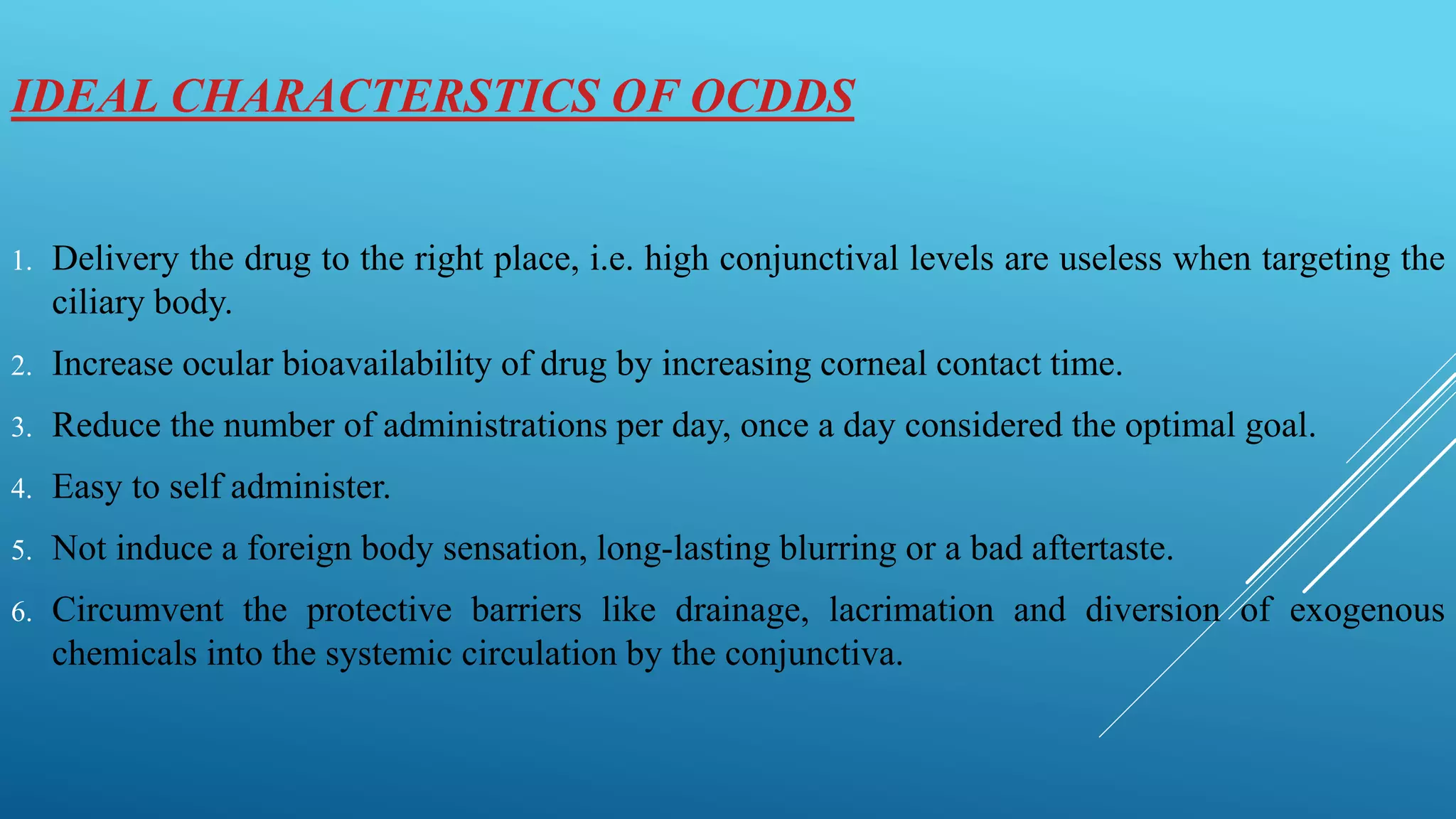
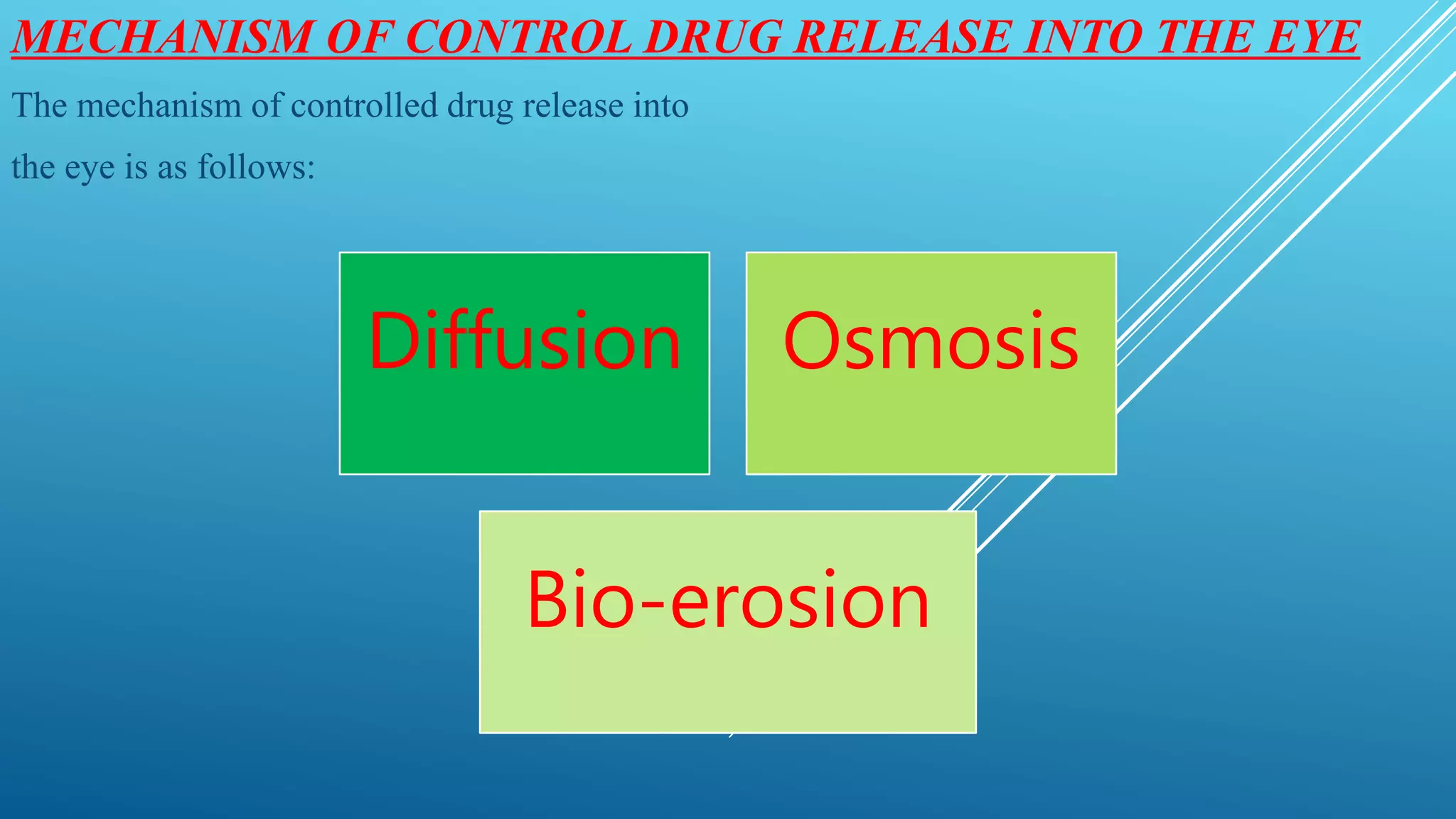
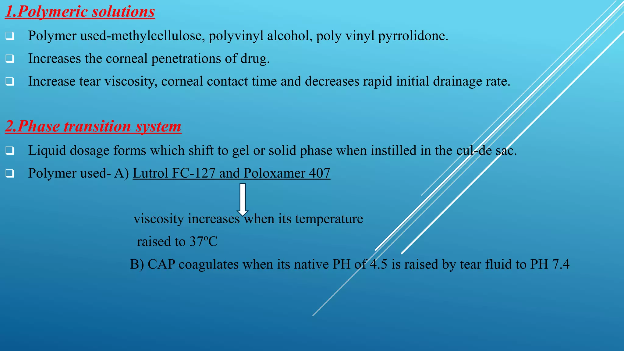
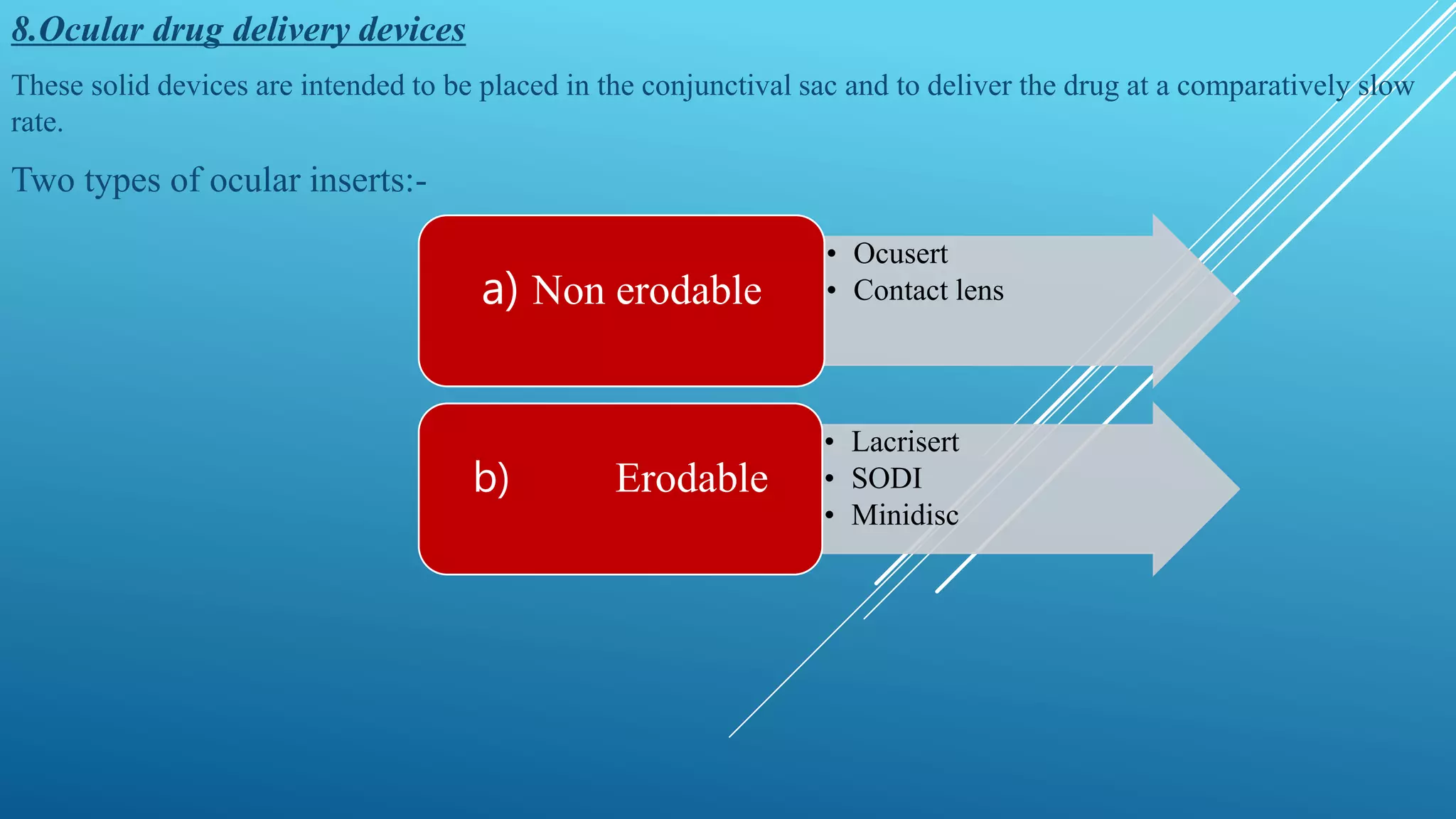
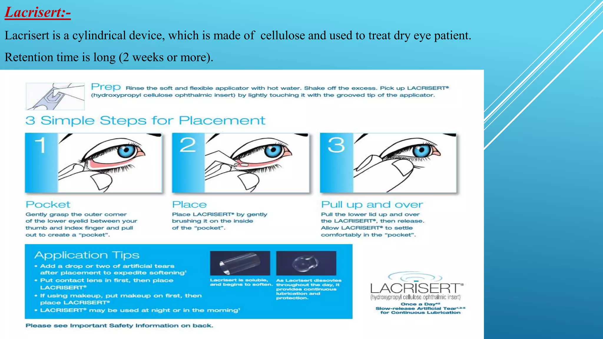
This document discusses ocular drug delivery systems (OCDDS) that aim to prolong drug release in the eye. It introduces various approaches for controlled release, including polymeric solutions, phase transition systems, mucoadhesive dosage forms, collagen shields, and ocular inserts. Specific examples are provided, such as Ocusert which releases pilocarpine at controlled rates over 4-7 days to treat glaucoma. The document outlines the ideal characteristics of OCDDS and mechanisms of controlled drug release via diffusion, osmosis and bioerosion. It also reviews factors influencing ocular drug penetration and absorption.
Call Girls In Andheri East Call 9920874524 Book Hot And Sexy Girls
Ophthalmic Drug Delivery Systems
1. PRESENTED BY:- DEBASISH SAHOO
M. PHARM 1ST YEAR
DEPARTMENT OF PHARMACEUTICS
ISF COLLEGE OF PHARMACY, MOGA, PUNJAB
2. INTRODUCTION
The ophthalmic preparations are available as sterile, buffered, isotonic solution.
Suspensions, gelled systems, ointment are also used for prolonged therapeutic action.
Prolonged drug release can be achieved using ophthalmic inserts, solid devices placed in the
eye, but the inserts must then be removed when they are no longer needed.
Ocuserts release the drug at predetermined and predictable rates thus eliminating the frequent
administration of the drug.
The elements generally include an inner layer, or core, including a therapeutic agent, and one
or more outer layers made of polymeric materials, for example substantially pure polymeric
materials.
In the area of topical ocular administration, important efforts concern the design and the
conception of new ophthalmic drug delivery systems able to prolong the residence time.
4. IDEAL CHARACTERSTICS OF OCDDS
1. Delivery the drug to the right place, i.e. high conjunctival levels are useless when targeting the
ciliary body.
2. Increase ocular bioavailability of drug by increasing corneal contact time.
3. Reduce the number of administrations per day, once a day considered the optimal goal.
4. Easy to self administer.
5. Not induce a foreign body sensation, long-lasting blurring or a bad aftertaste.
6. Circumvent the protective barriers like drainage, lacrimation and diversion of exogenous
chemicals into the systemic circulation by the conjunctiva.
5. MECHANISM OF CONTROL DRUG RELEASE INTO THE EYE
The mechanism of controlled drug release into
the eye is as follows:
Diffusion Osmosis
Bio-erosion
6. DRUG IN TEAR FLUID
OCULAR
ABSORPTION
CORNEAL
ROUTE
Primary route
Small, lipophilic
drug
CONJUNCTIVAL
AND SCLERAL
ROUTE
Large, Hydrophilic
drugs
AQUEOUS HUMOR
SYSTEMIC ABSORPTION
(~ 50-100% of dose)
Major routes:
Conjunctiva of eye
Nose
Minor routes:
Lacrimal drainage system
Pharynx
GIT
Skin at cheek lids
Aqueous humor
Inner ocular tissues
OCULAR TISSUE
ELIMINATION
Fig:-Factors and corneal barrier limitations for penetration of topically admistered drug
7. VARIOUS APPROACHES IN CR OCULAR DDS
1. Polymeric solution
2. Phase transition system
3. Mucoadhesive/ bioadhesive dosage forms
4. Collagen shields
5. Pseudolatices
6. Ocular penetration enhancers
7. Ocular iontophoresis
8. Ocular drug delivery devices
8. 1.Polymeric solutions
Polymer used-methylcellulose, polyvinyl alcohol, poly vinyl pyrrolidone.
Increases the corneal penetrations of drug.
Increase tear viscosity, corneal contact time and decreases rapid initial drainage rate.
2.Phase transition system
Liquid dosage forms which shift to gel or solid phase when instilled in the cul-de sac.
Polymer used- A) Lutrol FC-127 and Poloxamer 407
viscosity increases when its temperature
raised to 37ºC
B) CAP coagulates when its native PH of 4.5 is raised by tear fluid to PH 7.4
9. 3.Mucoadhesive/bioadhesive dosages
Polymer solution adheres to the mucin at the cornea and conjunctival surface.
interaction called mucoadhesion
increasing contact time
4.Collagen shields
Cross linking of collagen corneal shield increase ofloxacin bioavailability.
But there is some drawback.
5.Pseudolatices
Polymeric colloidal dispersion film forming agents used for topical application.
6.Ocular penetration enhancers
Enhancer like actin filament inhibitors, surfactants, bile salts, chelators and organic compound are used to
increase bioavailability.
7.Ocular iontophoresis
Direct current devices ions into cells or tissue.
10. 8.Ocular drug delivery devices
These solid devices are intended to be placed in the conjunctival sac and to deliver the drug at a comparatively slow
rate.
Two types of ocular inserts:-
• Ocusert
• Contact lensa) Non erodable
• Lacrisert
• SODI
• Minidisc
b) Erodable
11. Ocusert:-
Technology used-insoluble delicate sandwich technology.
Drug reservoir is a thin disc of pilocarpine-alginate complex
sandwiched between two transparent discs of microporous membrane
fabricated from ethylene-vinyl acetate copolymer.
Microporous membrane permit the tear fluid to penetrate into the drug
reservoir compartment to dissolve drug from the complex.
E.g-Alza-ocusert: In this pilocarpine molecules are released at constant
rate of 20 or 40µg/hr for 4 to 7 days.
Used in the management of glaucoma
Contact lense :-
Pre-soaked hydrophilic contact lenses is used for ophthalmic drug
delivery.
Therapeutic soft lenses are used to aid corneal wound healing in patient
with infections, corneal ulcers.
12.
13. Lacrisert:-
Lacrisert is a cylindrical device, which is made of cellulose and used to treat dry eye patient.
Retention time is long (2 weeks or more).
14. SODI(Soluble ocular drug insert)
It is a thin film of oval shape made from acrylamide, N-vinyl pyrrolidone and ethyl acrylate.
Wt. about 15-16 mg.
Inserted into the inferior cul-de-sac and get wets and softens in 10-15 seconds.
After 10-15 min the film turns into a viscous polymer mass, after 30-60 min it turns into polymer solutions and
delivers the drug for about 24 hrs.
Minidisc
Consists of a disc with a convex front and concave back surface in the contact with the eye ball.
Like miniature contact lenses with a diameter of 4-5mm.The minidisc is made up of silicone based prepolymer α-
omega-bis (4-methacryloxy) butyl poly-di-methyl siloxane.