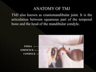
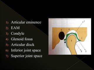
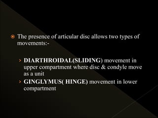
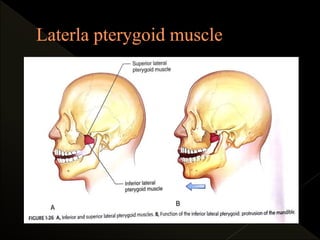
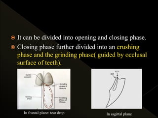
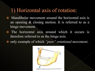
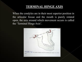
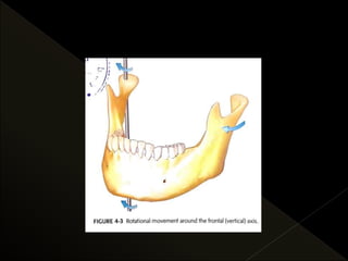
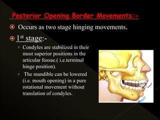
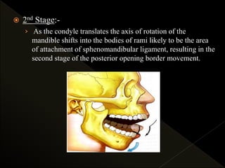
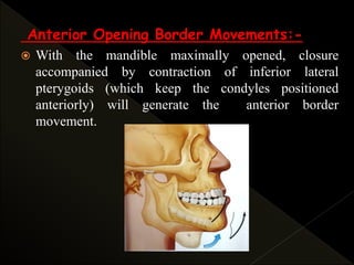
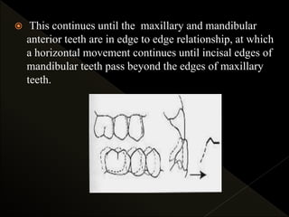
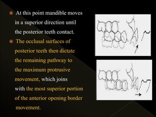
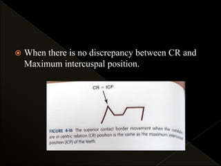
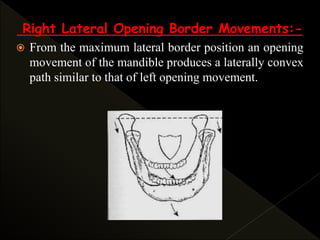
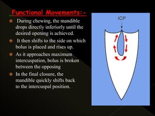
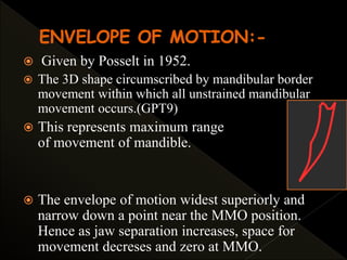
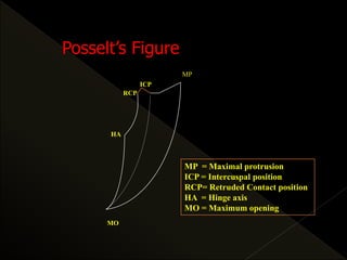
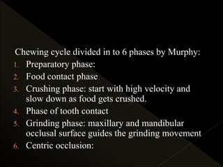
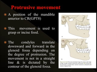
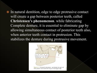
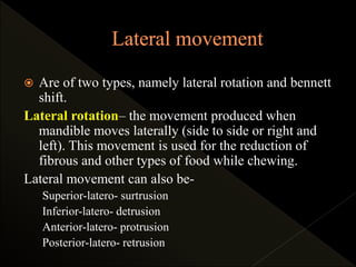
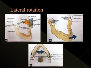
Presenter : Dr. Reena
PG Second Year
Deptt. of Prosthodontics
Guided by: Dr. Divya Dahiya
Professor,
Deptt. of Prosthodontics
 Introduction
 Definitions
 Anatomy of TMJ
 Types of mandibular movements
rotatory
translatory
 Single plane border movements
sagittal
frontal
horizontal
 Posselt’s veiw
 Determinants of mandibular movements
› Condylar guidance
› Anterior guidance
› Neuromuscular system
 Chewing stroke
 Neuromuscular regulation of mandibular motion
 Method of recording the mandibular motion
 Conclusion
 references
Masticatory system is the functional unit of the body
primarily responsible for chewing, speaking and
swallowing, tasting and breathing.
The system is made up of bones, joints, ligaments, teeth
and muscles. In addition an intricate neurologic
controlling system regulates and coordinates all these
structural components.
A sound understanding of its functional anatomy and
biomechanics is essential to the study of occlusion.
 Mandibular movement occur around the TMJ,
which is capable of making complex movements.
 The maxillomandibular relationship varies every
second during mandibular movements. There are
two basic movements:
› Functional movements
› Parafunctional movements
ANATOMY OF TMJ
FOSSA
EMINENCE
CONDYLE
TMJ also known as craniomandibular joint. It is the
articulation between squamous part of the temporal
bone and the head of the mandibular condyle.
TMJ is a bilateral bicondylar synovial
ginglioarthroidal joint. Their movements are
synchronized and act together to produce the
various mandibular movements.
 One of the most complex joints in the body.
› Ginglymoarthrodial joint.
› Compound joint.
1) Articular eminence
2) EAM
3) Condyle
4) Glenoid fossa
5) Articular disck
6) Inferior joint space
7) Superior joint space
 The presence of articular disc allows two types of
movements:-
› DIARTHROIDAL(SLIDING) movement in
upper compartment where disc & condyle move
as a unit
› GINGLYMUS( HINGE) movement in lower
compartment
 MANDIBULAR MOVEMENT (GPT-9):
any movement of the lower jaw.
 CENTRIC RELATION (GPT-8) : the maxillo-
mandibular relationship in which the condyles articulate with
the thinnest avascular portion of their respective disks with
the complex in the antero-superior position against the slopes
of articular eminences. This position is independent of tooth
contact. This position is clinically discernible when the
mandible is directed superiorly and anteriorly.
 DAWSON:
The relationship of the mandible to the maxilla when
the properly aligned condyle-disk assemblies are in
the most superior position against the eminentiae
irrespective of vertical dimension or tooth position
 The mandible moves as dictated by the movement
of its condyle in the glenoid fossa and by the
guidance of teeth. In a completely edentulous
situation, the teeth should be arranged such that they
do not interfere with the smooth, coordinated
movement of mandible during function.
 Mandibular movements occur as a complex series of
interrelated 3-dimensional rotational & transitional
activities. It is determined by the combined &
simultaneous activities of both TMJ’s.
 TMJ’s cannot function entirely independently of
each other & rarely function with identical
concurrent movements.
 To better understand the complexities mandibular
movements, it is better to isolate the movements that
occur within a single TMJ.
 Major factors that determine the mandibular
movements in general are:
oCondylar guidance/ posterior determinant
oIncisal guidance/ Anterior Determinant
oNeuromuscular factors
 Both Anterior & Posterior determinant are called as “
End- Controlling Factor.
 Defined as “mandibular guidance generated by
the condyle and articular disc traversing the
contour of glenoid fossa. It is nothing but the
path of movement taken up by the condyle in the
glenoid fossa.
 The shape of glenoid fossa which determines the
path of movement of the condyle, is called
condylar guidance.
 The slope of articular eminence determine the
angle of condylar guidance.
 This is the path travelled by the condyle in the
TMJs during various mandibular movement. It is
influenced by-
1. Inclination of Glenoid fossa
2. Tone of muscle responsible for Mandibular
movement & their Nerve control
3. Attached ligaments
4. Shape & movement of Articular disc
5. Teeth (when present)
 Fischer angle: is defined as the angle formed by
the protrusive and non-working side condylar
paths as viewed from the sagittal direction.
 It varies from 5-10º.
 This means that condylar guidance on the non
working side during laterotrusion will be 5-10
greater than the condylar guidance during
straight protrusion.
 Influence of contacting surfaces of the
mandibular and maxillary anterior teeth during
the mandibular movements.
 The fabrication of a relationship of the anterior
teeth preventing the posterior tooth contact in all
eccentric mandibular movements.
Protrusive Incisal Path
The track of the incisal edges of
the mandibular teeth from
maximum intercuspation to
edge-to-edge occlusion.
Protrusive Incisal Path Angle
The angle formed by the protrusive incisal path and the
horizontal reference plane is the protrusive incisal path
inclination. It ranges from 50 – 70 degrees and is often
5-10º steeper than the sagittal condylar guidance.
The angle formed with the horizontal
plane of occlusion and a line in the
sagittal plane between the incisal edges
of maxillary & mandibular central
incisors when the teeth are in
maximum intercuspation.
The angle formed in the sagittal plane
between the horizontal plane and the
slope of the incisal guide table.
• Opening and closing of
the mandible is simply a
rotation of the condyles in
the articular fossae.
• Anterior guidance is linked to the combination of
horizontal & vertical overlap of the anterior teeth
and can affect the occlusal surface morphology of the
posterior teeth.
• As anterior guidance is normally steeper than the
condylar guidance, the anterior teeth guide the
mandible downwards during protrusive or lateral
movement.
• during protrusive movement: produces disocclusion or
separation of the posterior teeth.
Anterior guidance can be made steeper by either increasing the vertical
overlap (overbite) ‘A-B’, or by reducing the horizontal overlap (over
jet) ‘C-A’ of the anterior teeth.
Anterior guidance can be made shallow by either decreasing the overbite
‘B-A’ or increasing the over jet ‘A-C’ of the ant. teeth.
a) The lesser the condylar guidance angle, the
shorter the cusps must be.
b) The greater the condylar guidance angle, the
longer the cusps may be.
c) The greater the horizontal overlap of the
maxillary anterior teeth, the shorter the
cusps of the posterior teeth must be.
d) The lesser the horizontal overlap the longer the
cusps of the posterior teeth may be.
e) The lesser the vertical overlap, the shorter the
cusps of the posterior teeth must be.
f) The greater the vertical overlap, the longer the
posterior cusps may be.
 The muscle of mastication are most important
determinants of mandibular movements.
 4 muscles make up a group called Muscles of Mastication
› Masseter
› Temporalis
› Medial Pterygoid
› Lateral Pterygoid
 Digastric also plays an important role in mandibular function.
Protrusion By both the lateral, medial pterygoids and superficial oblique
fibres of masseter muscle.
Retraction Posterior horizontal fibers of the temporalis and deep vertical
fibres of masseter muscle
Lateral or side to
side movements
Chewing from left side produced by right lateral pterygoid, right
medial pterygoid which push the chin to left side. Then left
temporalis ( anterior fibres), left masseter (deep fibres) chew the
food. Chewing the food from right side involves left lateral
pterygoid, left medial pterygoid, right temporalis and right
masseter.
Opening or
depression
Lateral pterygoid & the digastrics, geniohyoid and mylohyoid
muscles
Closing or
elevation
Masseter, medial pterygoid muscles of both sides & the anterior
vertical, middle oblique fibres of temporalis muscles.
Masseter muscle: Functions:- Elevation
Superficial portion: protrusion
Deep portion :stabilization against articular eminence
Temporalis muscle: It is a significant positioning muscle of the
mandible
FUNCTIONS: Elevation.
ANT PORTION: Mandible raised vertically
MIDDLE : elevation & retrusion
POSTERIOR : elevation & slight retrusion.
Medial pterygoid muscle: FUNCTION:
• Elevation and protrusion.
• Unilateral contraction –mediotrusive movement
Inferior Lateral
Pterygoid
 Simultaneous
contraction: depression
and protrusion
 Unilateral contraction:
mediotrusion &
movement to opp side.
Superior Lateral
Pterygoid
 During opening: remains
inactive.
 Becomes active only in
conjunction with elevator
muscles.
 Active during power
stroke & when teeth held
together
 To produce precise manibular movement, input
from the various sensory receptors must be
received by the CNS through the afferent fibers.
 The brainstem and cortex must assimilate and
organize this input and elicit appropriate motor
activities through efferent fibers ( generally
gamma efferent fibers).
 Within the brainstem there is a pool of neurons
that control the rhythmic muscle activities, this
pool is called as central pattern generator
(CPG)
 CPG: it is responsible for the precise timing of activity
between antagonistic muscles so that specific functions can be
carried out. For the CPG to be efficient, it must receive the
constant sensory input from masticatory system.
CPG initiates contraction of supra and
infra hyoid muscles at precise time the
elevator are to be relax.
This allows the mouth to open and
accept the food.
CPG initiates contraction of elevator
muscles while relaxing the supra &
infra hyoid muscles.
This process is repeated until the food
particles are crushed to small enough
to be swallowed easily.
 Therefore tongue, lips, teeth and PDL are
constantly feeding back information that allows
the CPG to determine the most appropriate and
efficient chewing stroke.
 Chewing stroke : each opening and closing
movement of the mandible represents a chewing
stroke. The complete chewing stroke has a pattern as
“tear-shaped”. This activity is under control of
CPG.
 It can be divided into opening and closing phase.
 Closing phase further divided into an crushing
phase and the grinding phase( guided by occlusal
surface of teeth).
In frontal plane: tear drop In sagittal plane
The movements are variable, within the borders
and influenced by:
 Consistency, bulk and type of food
 Size, number and form of teeth
 Excess or lack of saliva
 The musculature and force of chewing
 The average length of time for tooth contact
during mastication if 194ms.(Jeffrey P. okeson 7th
edition)
 Feedback mecahnism allows for alteration in
chewing stroke according to the particular food
being chewed.
 Tall cusp & deep fossae promote a predominantly
vertical chewing stroke.
 Flat/ worn teeth encourage a broader chewing
stroke.
 The neuromuscular basis and mechanism of
bruxism have been studied and is explained by
increase in tonic activity in jaw muscles.
Emotion or neuron tension, pain or discomfort,
stress, occlusal interferences are the factors that
increase muscle tonus and lead to nonfunctional
gnashing and clenching.
 Variation in environmental factor such as emotional
stress, hormonal levels, sympathetic and
parasympathetic changes in CPG , leads to the
condition like bruxism.
 Biting force applied to teeth varies from individual to
individual.
 Biting force in male is more than that of females.
 In males it ranges from 118-142 pounds (53.6-64.4 kgs) and
in females it ranges from 79-99 pounds (35.8-44.9 kgs)**
 Maximum force applied is at molar region and it was found to
be 91-198 pounds (41.3-89.8 Kgs) whereas in anterior region
29-51 pounds (13.2-23.1 Kgs).
 During chewing greatest amount of force is placed on first
molar region. With tougher food, chewing occurs
predominantly on the first molar and second premolar region.
The biting force with complete denture is only one fourth that
of subjects with natural teeth i.e. 30-35 pounds in males and
20-25 pounds in females..
** Brekhus PH. Stimulation of muscle of mastication, J Dent Res 1941;20;87-92.
I) According to Sharry:-
a) According to direction - Opening and closing movements
Protrusion and retraction
Lateral gliding movements
b) According to tooth contact - Movements with tooth contact
Movements without tooth contact
c) Limitation by joint structure - Border movements
Intra border movements
d) Functions of masticatory system - Mastication
Deglutition
Speech
Respiration
e) CNS - Innate movements – breathing & swallowing
Learned movements – speech and chewing
II) According to the type of movement occurs in
TMJ:-
a) Rotational
b) Translation
III) According to the planes of border movements:-
a) Sagittal plane border movement
b) Horizontal plane border movements
c) Frontal plane border movements
 Dorland’s Illustrated Medical Dictionary defines
rotation as “the process of turning round an axis:
movement of a body about its axis.”
 In the masticatory system, rotation occurs when
the mouth opens & closes around a fixed point or
axis within the condyles. In the TMJ rotation can
occur as movement within the inferior cavity of
the joint
 Thus, rotation is the movement between the
superior surface of the condyle & the inferior
surface of the articular disc.
 In masticatory system, rotation occurs when the
mouth opens and closes around a fixed point or
axis within the condyles.
 3 reference planes:
› Horizontal
› Frontal (vertical)
› Sagittal
 Mandibular movement around the horizontal axis is
an opening & closing motion. It is referred to as a
hinge movement.
 The horizontal axis around which it occurs is
therefore referred to as the hinge axis.
 only example of which “pure” rotational movement.
TERMINAL HINGE AXIS
When the condyles are in their most superior position in
the articular fossae and the mouth is purely rotated
open, the axis around which movement occurs is called
the ‘Terminal Hinge Axis’.
 The vertical axis runs through the condyle & the
ramus of the mandible, & the mandible rotates
around this vertical axis during lateral
movements.
 Mandibular movement around the frontal axis
occurs when one condyle moves out of the
terminal hinge position with the vertical axis of
the opposite condyle remaining in the terminal
hinge position
 This anteroposterior axis is an imaginary axis
running along the mid-sagittal plane. The
mandible shows slight rotation around this axis.
 During the movement the condyle of one side
moves downward & medially along the slope of
the entoglenoid process (medial slope of the
glenoid fossa) & the condyle of the opposite side
moves upwards & laterally.
 This type of movement is usually seen in
association with lateral movements.
 Translation occurs within the superior cavity of
the joint, between the superior surface of the
articular disc & the inferior surface of the
articular fossa.
 GPT-8:
that motion of a rigid body in which a straight
line passing through any two points always
remains parallel to its initial position. The motion
may be described as a sliding or gliding motion
 In the masticatory system, translation occurs when
the
› mandible moves forward, as in protrusion.
› The teeth, condyles, & rami all move in the same direction
& to the same degree.
› Occur in superior joint space
 During most normal movements of the mandible,
both rotation & translation occur simultaneously, i.e
while the mandible is rotating around one or more of
the axes, each of the axes is translating (i.e changing
its orientation in space).
 When the mandible moves through the outer range of
motion, reproducible and describable limits result,
which are called BORDER MOVEMENTS. The
border & functional movements of mandible
described in each reference plane.
 Border movement: It is defined as “the mandibular
movement at the limits dictated by anatomic
structures as viwed in a given plane”. Occur in 3
plane
› Horizontal plane
› Saggital plane
› Coronal plane
 Intraborder movement : occurs with in envelope of
motion.
› Functional
› Parafunctional movement
Component determined by
Posterior opening border ligaments & the morphology of TMJ’s
Anterior opening border ligaments & the morphology of TMJ’s
Superior contact border occlusal & incisal surfaces of teeth
Functional conditional responses of neuromuscular
system
 The mandibular motion is viewed in the sagittal
plane can be seen to have 4 distinct movements
components.
 A characteristic “beak tracing” is formed while
recording border movements in the sagittal plane
Posterior Opening Border Movements:-
 Occurs as two stage hinging movements.
 1st stage:-
› Condyles are stabilized in their
most superior positions in the
articular fossae.( i.e.terminal
hinge position).
› The mandible can be lowered
(i.e. mouth opening) in a pure
rotational movement without
translation of condyles.
› In CR, the mandible can be rotated around
the horizontal axis to a distance of 20-25 mm
as measured between the incisal edges of
maxillary and mandibular incisors.
› At this point of opening, the T.M. ligaments
tighten, after which continued opening
results in an anterior & inferior translation of
condyles.
 2nd Stage:-
› As the condyle translates the axis of rotation of the
mandible shifts into the bodies of rami likely to be the area
of attachment of sphenomandibular ligament, resulting in the
second stage of the posterior opening border movement.
Anterior Opening Border Movements:-
 With the mandible maximally opened, closure
accompanied by contraction of inferior lateral
pterygoids (which keep the condyles positioned
anteriorly) will generate the anterior border
movement.
 Because the maximum protrusive position is
determined in part by stylomandibular ligaments,
when closure occurs, tightening of ligaments produces
a posterior movement of the condyles.
 The posterior movement of the condyle from the
maximally open position to maximally protruded
position produces eccentricity in the anterior border
movement. Therefore, it is not a pure hinge
movement.
Superior Contact Border Movements:-
 Throughout this entire border movement tooth contact
is present.
 It depends on:-
› Amount of variation between centric relation and
maximum intercuspation.
› The steepness of the cuspal inclines of the
posterior teeth.
› Amount of vertical and horizontal overlap of
anterior teeth
› Lingual morphology of maxillary anterior teeth.
› General interarch relationships of the teeth.
 The initial tooth contact in
terminal hinge axis or centric
relation occurs between the
mesial inclines of maxillary
tooth & distal inclines of
mandibular tooth.
 When muscular force is
applied to the mandible,
a superoanterior movement
or shift will occur until the
intercuspal position is reached.
 The slide from CR to maximum intercuspation, may
have a lateral component.
Average distance is 1.25 ± 1 mm.
 In the intercuspal position, the opposing anterior teeth
usually contact.
 When the mandible is protruded,
from maximum intercuspation,
contact between the incisal
edges of the mandibular anterior
teeth & lingual inclines of
maxillary anterior teeth result
in an anteroinferior movement
of the mandible.
 This continues until the maxillary and mandibular
anterior teeth are in edge to edge relationship, at which
a horizontal movement continues until incisal edges of
mandibular teeth pass beyond the edges of maxillary
teeth.
 At this point mandible moves
in a superior direction until
the posterior teeth contact.
 The occlusal surfaces of
posterior teeth then dictate
the remaining pathway to
the maximum protrusive
movement, which joins
with the most superior portion
of the anterior opening border
movement.
 When there is no discrepancy between CR and
Maximum intercuspal position.
Functional Movements:-
 Functional movement occurs during functional activity
of the mandible. They usually take place within the
border movements & therefore, considered as free
movements.
 Most functional movements require maximum
intercuspation & therefore typically begin at & below
the intercuspal position.
 When mandible is at rest, it is found to be located
approximately 2 to 4 mm below the intercuspal
position. This is called the Clinical Rest Position.
 Postural position – Since,
clinical rest position is not a
true resting position, the
position in which mandible
is maintained is termed as
‘postural position.’
 Head position effect the
postural position.
HORIZONTAL PLANE BORDER &
FUNCTIONAL MOVEMENTS:-
 When mandibular movements are viewed in the
horizontal plane, a rhomboid-shaped/ diamond
pattern can be seen that has a functional component,
& 4 distinct movement components:-
1) Left lateral border
2) Continued left lateral border
with protrusion
3) Right lateral border
4) Continued right lateral border
with protrusion
Left Lateral Border Movements:-
 With the condyles in the centric relation position,
contraction of the right inferior lateral pterygoid move
the right condyle - anteriorly and medially.
 If left inferior pterygoid stays relaxed, with the left
condyle still in the CR & result will be left lateral
border movement.
 Left condyle- working or rotatory , Right condyle-
non-working or orbiting
Continued Left Lateral Border Movements
With Protrusion
 With the mandible in the left lateral border position,
contraction of the left inferior lateral pterygoid along
with continued contraction of right inferior lateral
pterygoid will cause the left condyle to move
anteriorly to the right.
Right Lateral Border Movements:-
 Left condyle- orbiting Right condyle- rotatory
Continued Right Lateral Border Movements
With Protrusion:-
Functional Movements:-
 As in the sagittal plane, functional movement in the horizontal
plane most often occur near the intercuspal position.
 During chewing the range of jaw movements begins some
distance from maximum intercuspal position; but as the food is
broken down into smaller particles, jaw action moves closer
and closer to intercuspal position.
FRONTAL(coronal) BORDER &
FUNCTIONAL MOVEMENTS:-
 A shield-shaped pattern can be seen that has a
functional component, & four distinct movement
components:-
1. Left lateral superior border.
2. Left lateral opening border.
3. Right lateral superior border.
4. Right lateral opening border.
Left Lateral Superior Border Movements:-
 With the mandible in maximum intercuspation, lateral
movement is made to the left.
 It depends upon morphology and interarch
relationships of maxillary and mandibular teeth.
 The maximum lateral extent of this movement is
determined by ligaments of the rotating joint.
Left Lateral Opening Border Movements:-
 From the maximum left lateral superior border position, an
opening movement of the mandible produces a laterally
convex path. As maximum opening is approached,
ligaments tighten and produce a medially directed
movement that causes a shift back in the mandibular
midline to coincide with the midline of the face.
Right Lateral Superior Border
Movements:-
 Now, the mandible is returned to maximum
intercuspation. From this position a lateral movement
is made to right that is similar to the left lateral
superior border movement.
Right Lateral Opening Border Movements:-
 From the maximum lateral border position an opening
movement of the mandible produces a laterally convex
path similar to that of left opening movement.
Functional Movements:-
 During chewing, the mandible
drops directly inferiorly until the
desired opening is achieved.
 It then shifts to the side on which
bolus is placed and rises up.
 As it approaches maximum
intercuspation, bolus is broken down
between the opposing teeth.
 In the final closure, the
mandible quickly shifts back
to the intercuspal position.
 Given by Posselt in 1952.
 The 3D shape circumscribed by mandibular border
movement within which all unstrained mandibular
movement occurs.(GPT9)
 This represents maximum range
of movement of mandible.
 The envelope of motion widest superiorly and
narrow down a point near the MMO position.
Hence as jaw separation increases, space for
movement decreses and zero at MMO.
MP
MO
ICP
RCP
HA
MP = Maximal protrusion
ICP = Intercuspal position
RCP= Retruded Contact position
HA = Hinge axis
MO = Maximum opening
Posselt’s Figure
 These movement occur with in envelope of
motion and of two types namely functional &
parafunctional.
 Functional movement include the chewing,
speech, swallowing and yawning.
 Parafunctional movements: during clenching,
bruxism and other habitual movements.
Chewing cycle divided in to 6 phases by Murphy:
1. Preparatory phase:
2. Food contact phase
3. Crushing phase: start with high velocity and
slow down as food gets crushed.
4. Phase of tooth contact
5. Grinding phase: maxillary and mandibular
occlusal surface guides the grinding movement
6. Centric occlusion:
Can be broadly classified
Hinge movement
Protrusive movement
Retrusive movement
Lateral movement
Laterotrusion / lateral rotation
Bennett movement
 A position of the mandible
anterior to CR(GPT8)
 This movement is used to
grasp or incise food.
 The condyles translate
downward and forward in the
glenoid fossa depending on
the degree of protrusion. The
movement is not in a straight
line & is dictated by the
contour of the glenoid fossa.
 In natural dentition, edge to edge protrusive contact
will create a gap between posterior teeth, called
Christensen’s phenomenon. while fabricating
Complete denture, it is essential to eliminate gap by
allowing simultaneous contact of posterior teeth also,
when anterior teeth contact in protrusion. This
stabilizes the denture during protrusive movement.
 The average path of the advancing condyle
makes an angle with the frontal plane called the
“protrusive condylar guidance/ inclination.
 Are of two types, namely lateral rotation and bennett
shift.
Lateral rotation– the movement produced when
mandible moves laterally (side to side or right and
left). This movement is used for the reduction of
fibrous and other types of food while chewing.
Lateral movement can also be-
Superior-latero- surtrusion
Inferior-latero- detrusion
Anterior-latero- protrusion
Posterior-latero- retrusion
• When the mandible moves laterally, the side to
which it moves is termed as the working side
or functional side and the other side is termed
as the non working or balancing side or non
functional side.
• The condyle on the working side is termed as
the working condyle or rotating condyle and
the condyle on the other side is termed as
orbiting condyle or nonworking condyle.
 Bennett in 1908, studied working condylar path
& called as BENNETT MOVEMENT.
 Bennett movement refers to condylar movement
on the working side &,
 Bennett Shift is the bodily side shift of the
mandible on working side in horizontal direction.
 It is defined as “ the bodily lateral movement or
lateral shift of the mandible resulting from the
movements of the condyles along the lateral inclines
along the mandibular fossae in lateral jaw
movements”.
 This lateral movement is termed as the
laterotrusion or the mandibular lateral translation
or “Bennett movement”. It has been termed
previously as Bennett shift or mandibular side
shift.
 Bennett movement is consequent to the medial
movement of orbiting condyle & is regulated by:-
› Morphology of medial wall of mandibular fossa.
› Inner horizontal part of TM ligament which attaches
to the lateral pole of rotating condyle.
 Bennett movement has 3 components:-
› Amount
› Timing
› Direction
 During lateral movement, the mandible shift
(as a whole) by 1-4mm towards the working
side. This shift is called bennett movement.
 This lateral tanslatory motion is measured
against time, which is considered as the fourth
dimension of mandibular movement.
 During laterotrusion, the non-working condyle
arcs forward and medially.
 Bennett movement is classified based on the
timing of the shift in relation to the forward
movement of the nonworking condyle.
›Immediate side shift
›Precurrent side shift
› Progressive side shift respectively.
 Immediate side shift: lateral translation occurs
before the forward movement of non-working
condyle. This shift ranges 1 to rarely 4mm (average
0.75mm) in dimension.
 Occurs in 86% of the condyle.
 Precurrent side shift: lateral translation occurs
during the first 2-3 mm forward movement of the
non-working condyle. If the major quotient of the
Bennett movement occur during the first 4mm of
anterior movement of the non-working condyle,
then its called Distributed side shift.
 Progressive side shift: lateral translation that
occurs after 2-3mm of forward movement of non
working condyle. This shift of mandible is
gradual and doesnot changes with time.
Note: Combined movement: is some patient there
may be immediate side shift for about 1mm
before lateral movement followed by progressive
side shift along with lateral movement.
 It is defined as “the angle formed by the sagittal
plane and the path of the advancing condyle
during lateral mandibular movements as viewed
in the horizontal plane”
 Graphic method record
 stereographics
 Pantographs
 Kinesiograph
 Electromyogrpahy:
 Ultrasound
 Accelerometers
 Arcus digma : by Kavo, is a high tech bite
registration system, that provide the most precise
result.
A sound understanding of mandibular movements
is essential for the Recording jaw relations,
Designing, selection and adjustment of
articulator, Developing tooth form for dental
restorations, Understanding the basic principles
of occlusion , Diagnosis and treatment of TMJ
disturbances, Preserving periodontal health.
You cannot successfully treat dysfunction
unless you understand function.”
 B.D Chaurasias; Human Anatomy; regional and
applied volume three, third edition •
 Bouchers Prosthodontic treatment for edentulous
patients 7th edition •
 Management of Temperomandibular disorders
and occlusion 7th edition ; Jeffrey p Okeson •
 Evaluation diagnosis and treatment plan of
occlusal problem 2nd edition Peter E Dawson •
Mandibular movements and determinants

Mandibular movements and determinants

  • 1.
    Presenter : Dr.Reena PG Second Year Deptt. of Prosthodontics Guided by: Dr. Divya Dahiya Professor, Deptt. of Prosthodontics
  • 2.
     Introduction  Definitions Anatomy of TMJ  Types of mandibular movements rotatory translatory  Single plane border movements sagittal frontal horizontal  Posselt’s veiw  Determinants of mandibular movements › Condylar guidance › Anterior guidance › Neuromuscular system  Chewing stroke  Neuromuscular regulation of mandibular motion  Method of recording the mandibular motion  Conclusion  references
  • 3.
    Masticatory system isthe functional unit of the body primarily responsible for chewing, speaking and swallowing, tasting and breathing. The system is made up of bones, joints, ligaments, teeth and muscles. In addition an intricate neurologic controlling system regulates and coordinates all these structural components. A sound understanding of its functional anatomy and biomechanics is essential to the study of occlusion.
  • 4.
     Mandibular movementoccur around the TMJ, which is capable of making complex movements.  The maxillomandibular relationship varies every second during mandibular movements. There are two basic movements: › Functional movements › Parafunctional movements
  • 5.
    ANATOMY OF TMJ FOSSA EMINENCE CONDYLE TMJalso known as craniomandibular joint. It is the articulation between squamous part of the temporal bone and the head of the mandibular condyle.
  • 6.
    TMJ is abilateral bicondylar synovial ginglioarthroidal joint. Their movements are synchronized and act together to produce the various mandibular movements.  One of the most complex joints in the body. › Ginglymoarthrodial joint. › Compound joint.
  • 7.
    1) Articular eminence 2)EAM 3) Condyle 4) Glenoid fossa 5) Articular disck 6) Inferior joint space 7) Superior joint space
  • 8.
     The presenceof articular disc allows two types of movements:- › DIARTHROIDAL(SLIDING) movement in upper compartment where disc & condyle move as a unit › GINGLYMUS( HINGE) movement in lower compartment
  • 9.
     MANDIBULAR MOVEMENT(GPT-9): any movement of the lower jaw.  CENTRIC RELATION (GPT-8) : the maxillo- mandibular relationship in which the condyles articulate with the thinnest avascular portion of their respective disks with the complex in the antero-superior position against the slopes of articular eminences. This position is independent of tooth contact. This position is clinically discernible when the mandible is directed superiorly and anteriorly.
  • 10.
     DAWSON: The relationshipof the mandible to the maxilla when the properly aligned condyle-disk assemblies are in the most superior position against the eminentiae irrespective of vertical dimension or tooth position
  • 11.
     The mandiblemoves as dictated by the movement of its condyle in the glenoid fossa and by the guidance of teeth. In a completely edentulous situation, the teeth should be arranged such that they do not interfere with the smooth, coordinated movement of mandible during function.
  • 12.
     Mandibular movementsoccur as a complex series of interrelated 3-dimensional rotational & transitional activities. It is determined by the combined & simultaneous activities of both TMJ’s.  TMJ’s cannot function entirely independently of each other & rarely function with identical concurrent movements.  To better understand the complexities mandibular movements, it is better to isolate the movements that occur within a single TMJ.
  • 13.
     Major factorsthat determine the mandibular movements in general are: oCondylar guidance/ posterior determinant oIncisal guidance/ Anterior Determinant oNeuromuscular factors  Both Anterior & Posterior determinant are called as “ End- Controlling Factor.
  • 14.
     Defined as“mandibular guidance generated by the condyle and articular disc traversing the contour of glenoid fossa. It is nothing but the path of movement taken up by the condyle in the glenoid fossa.  The shape of glenoid fossa which determines the path of movement of the condyle, is called condylar guidance.  The slope of articular eminence determine the angle of condylar guidance.
  • 16.
     This isthe path travelled by the condyle in the TMJs during various mandibular movement. It is influenced by- 1. Inclination of Glenoid fossa 2. Tone of muscle responsible for Mandibular movement & their Nerve control 3. Attached ligaments 4. Shape & movement of Articular disc 5. Teeth (when present)
  • 17.
     Fischer angle:is defined as the angle formed by the protrusive and non-working side condylar paths as viewed from the sagittal direction.  It varies from 5-10º.  This means that condylar guidance on the non working side during laterotrusion will be 5-10 greater than the condylar guidance during straight protrusion.
  • 18.
     Influence ofcontacting surfaces of the mandibular and maxillary anterior teeth during the mandibular movements.  The fabrication of a relationship of the anterior teeth preventing the posterior tooth contact in all eccentric mandibular movements.
  • 19.
    Protrusive Incisal Path Thetrack of the incisal edges of the mandibular teeth from maximum intercuspation to edge-to-edge occlusion.
  • 20.
    Protrusive Incisal PathAngle The angle formed by the protrusive incisal path and the horizontal reference plane is the protrusive incisal path inclination. It ranges from 50 – 70 degrees and is often 5-10º steeper than the sagittal condylar guidance.
  • 21.
    The angle formedwith the horizontal plane of occlusion and a line in the sagittal plane between the incisal edges of maxillary & mandibular central incisors when the teeth are in maximum intercuspation. The angle formed in the sagittal plane between the horizontal plane and the slope of the incisal guide table.
  • 22.
    • Opening andclosing of the mandible is simply a rotation of the condyles in the articular fossae. • Anterior guidance is linked to the combination of horizontal & vertical overlap of the anterior teeth and can affect the occlusal surface morphology of the posterior teeth.
  • 23.
    • As anteriorguidance is normally steeper than the condylar guidance, the anterior teeth guide the mandible downwards during protrusive or lateral movement. • during protrusive movement: produces disocclusion or separation of the posterior teeth.
  • 25.
    Anterior guidance canbe made steeper by either increasing the vertical overlap (overbite) ‘A-B’, or by reducing the horizontal overlap (over jet) ‘C-A’ of the anterior teeth. Anterior guidance can be made shallow by either decreasing the overbite ‘B-A’ or increasing the over jet ‘A-C’ of the ant. teeth.
  • 26.
    a) The lesserthe condylar guidance angle, the shorter the cusps must be. b) The greater the condylar guidance angle, the longer the cusps may be. c) The greater the horizontal overlap of the maxillary anterior teeth, the shorter the cusps of the posterior teeth must be.
  • 27.
    d) The lesserthe horizontal overlap the longer the cusps of the posterior teeth may be. e) The lesser the vertical overlap, the shorter the cusps of the posterior teeth must be. f) The greater the vertical overlap, the longer the posterior cusps may be.
  • 28.
     The muscleof mastication are most important determinants of mandibular movements.  4 muscles make up a group called Muscles of Mastication › Masseter › Temporalis › Medial Pterygoid › Lateral Pterygoid  Digastric also plays an important role in mandibular function.
  • 29.
    Protrusion By boththe lateral, medial pterygoids and superficial oblique fibres of masseter muscle. Retraction Posterior horizontal fibers of the temporalis and deep vertical fibres of masseter muscle Lateral or side to side movements Chewing from left side produced by right lateral pterygoid, right medial pterygoid which push the chin to left side. Then left temporalis ( anterior fibres), left masseter (deep fibres) chew the food. Chewing the food from right side involves left lateral pterygoid, left medial pterygoid, right temporalis and right masseter. Opening or depression Lateral pterygoid & the digastrics, geniohyoid and mylohyoid muscles Closing or elevation Masseter, medial pterygoid muscles of both sides & the anterior vertical, middle oblique fibres of temporalis muscles.
  • 30.
    Masseter muscle: Functions:-Elevation Superficial portion: protrusion Deep portion :stabilization against articular eminence
  • 31.
    Temporalis muscle: Itis a significant positioning muscle of the mandible FUNCTIONS: Elevation. ANT PORTION: Mandible raised vertically MIDDLE : elevation & retrusion POSTERIOR : elevation & slight retrusion.
  • 32.
    Medial pterygoid muscle:FUNCTION: • Elevation and protrusion. • Unilateral contraction –mediotrusive movement
  • 34.
    Inferior Lateral Pterygoid  Simultaneous contraction:depression and protrusion  Unilateral contraction: mediotrusion & movement to opp side. Superior Lateral Pterygoid  During opening: remains inactive.  Becomes active only in conjunction with elevator muscles.  Active during power stroke & when teeth held together
  • 35.
     To produceprecise manibular movement, input from the various sensory receptors must be received by the CNS through the afferent fibers.  The brainstem and cortex must assimilate and organize this input and elicit appropriate motor activities through efferent fibers ( generally gamma efferent fibers).  Within the brainstem there is a pool of neurons that control the rhythmic muscle activities, this pool is called as central pattern generator (CPG)
  • 36.
     CPG: itis responsible for the precise timing of activity between antagonistic muscles so that specific functions can be carried out. For the CPG to be efficient, it must receive the constant sensory input from masticatory system. CPG initiates contraction of supra and infra hyoid muscles at precise time the elevator are to be relax. This allows the mouth to open and accept the food. CPG initiates contraction of elevator muscles while relaxing the supra & infra hyoid muscles. This process is repeated until the food particles are crushed to small enough to be swallowed easily.
  • 37.
     Therefore tongue,lips, teeth and PDL are constantly feeding back information that allows the CPG to determine the most appropriate and efficient chewing stroke.  Chewing stroke : each opening and closing movement of the mandible represents a chewing stroke. The complete chewing stroke has a pattern as “tear-shaped”. This activity is under control of CPG.
  • 38.
     It canbe divided into opening and closing phase.  Closing phase further divided into an crushing phase and the grinding phase( guided by occlusal surface of teeth). In frontal plane: tear drop In sagittal plane
  • 39.
    The movements arevariable, within the borders and influenced by:  Consistency, bulk and type of food  Size, number and form of teeth  Excess or lack of saliva  The musculature and force of chewing  The average length of time for tooth contact during mastication if 194ms.(Jeffrey P. okeson 7th edition)
  • 40.
     Feedback mecahnismallows for alteration in chewing stroke according to the particular food being chewed.  Tall cusp & deep fossae promote a predominantly vertical chewing stroke.  Flat/ worn teeth encourage a broader chewing stroke.
  • 42.
     The neuromuscularbasis and mechanism of bruxism have been studied and is explained by increase in tonic activity in jaw muscles. Emotion or neuron tension, pain or discomfort, stress, occlusal interferences are the factors that increase muscle tonus and lead to nonfunctional gnashing and clenching.  Variation in environmental factor such as emotional stress, hormonal levels, sympathetic and parasympathetic changes in CPG , leads to the condition like bruxism.
  • 43.
     Biting forceapplied to teeth varies from individual to individual.  Biting force in male is more than that of females.  In males it ranges from 118-142 pounds (53.6-64.4 kgs) and in females it ranges from 79-99 pounds (35.8-44.9 kgs)**  Maximum force applied is at molar region and it was found to be 91-198 pounds (41.3-89.8 Kgs) whereas in anterior region 29-51 pounds (13.2-23.1 Kgs).  During chewing greatest amount of force is placed on first molar region. With tougher food, chewing occurs predominantly on the first molar and second premolar region. The biting force with complete denture is only one fourth that of subjects with natural teeth i.e. 30-35 pounds in males and 20-25 pounds in females.. ** Brekhus PH. Stimulation of muscle of mastication, J Dent Res 1941;20;87-92.
  • 44.
    I) According toSharry:- a) According to direction - Opening and closing movements Protrusion and retraction Lateral gliding movements b) According to tooth contact - Movements with tooth contact Movements without tooth contact c) Limitation by joint structure - Border movements Intra border movements d) Functions of masticatory system - Mastication Deglutition Speech Respiration e) CNS - Innate movements – breathing & swallowing Learned movements – speech and chewing
  • 45.
    II) According tothe type of movement occurs in TMJ:- a) Rotational b) Translation III) According to the planes of border movements:- a) Sagittal plane border movement b) Horizontal plane border movements c) Frontal plane border movements
  • 46.
     Dorland’s IllustratedMedical Dictionary defines rotation as “the process of turning round an axis: movement of a body about its axis.”  In the masticatory system, rotation occurs when the mouth opens & closes around a fixed point or axis within the condyles. In the TMJ rotation can occur as movement within the inferior cavity of the joint  Thus, rotation is the movement between the superior surface of the condyle & the inferior surface of the articular disc.
  • 48.
     In masticatorysystem, rotation occurs when the mouth opens and closes around a fixed point or axis within the condyles.  3 reference planes: › Horizontal › Frontal (vertical) › Sagittal
  • 49.
     Mandibular movementaround the horizontal axis is an opening & closing motion. It is referred to as a hinge movement.  The horizontal axis around which it occurs is therefore referred to as the hinge axis.  only example of which “pure” rotational movement.
  • 50.
    TERMINAL HINGE AXIS Whenthe condyles are in their most superior position in the articular fossae and the mouth is purely rotated open, the axis around which movement occurs is called the ‘Terminal Hinge Axis’.
  • 51.
     The verticalaxis runs through the condyle & the ramus of the mandible, & the mandible rotates around this vertical axis during lateral movements.  Mandibular movement around the frontal axis occurs when one condyle moves out of the terminal hinge position with the vertical axis of the opposite condyle remaining in the terminal hinge position
  • 53.
     This anteroposterioraxis is an imaginary axis running along the mid-sagittal plane. The mandible shows slight rotation around this axis.  During the movement the condyle of one side moves downward & medially along the slope of the entoglenoid process (medial slope of the glenoid fossa) & the condyle of the opposite side moves upwards & laterally.  This type of movement is usually seen in association with lateral movements.
  • 55.
     Translation occurswithin the superior cavity of the joint, between the superior surface of the articular disc & the inferior surface of the articular fossa.  GPT-8: that motion of a rigid body in which a straight line passing through any two points always remains parallel to its initial position. The motion may be described as a sliding or gliding motion
  • 56.
     In themasticatory system, translation occurs when the › mandible moves forward, as in protrusion. › The teeth, condyles, & rami all move in the same direction & to the same degree. › Occur in superior joint space  During most normal movements of the mandible, both rotation & translation occur simultaneously, i.e while the mandible is rotating around one or more of the axes, each of the axes is translating (i.e changing its orientation in space).
  • 57.
     When themandible moves through the outer range of motion, reproducible and describable limits result, which are called BORDER MOVEMENTS. The border & functional movements of mandible described in each reference plane.
  • 58.
     Border movement:It is defined as “the mandibular movement at the limits dictated by anatomic structures as viwed in a given plane”. Occur in 3 plane › Horizontal plane › Saggital plane › Coronal plane  Intraborder movement : occurs with in envelope of motion. › Functional › Parafunctional movement
  • 59.
    Component determined by Posterioropening border ligaments & the morphology of TMJ’s Anterior opening border ligaments & the morphology of TMJ’s Superior contact border occlusal & incisal surfaces of teeth Functional conditional responses of neuromuscular system  The mandibular motion is viewed in the sagittal plane can be seen to have 4 distinct movements components.
  • 60.
     A characteristic“beak tracing” is formed while recording border movements in the sagittal plane
  • 61.
    Posterior Opening BorderMovements:-  Occurs as two stage hinging movements.  1st stage:- › Condyles are stabilized in their most superior positions in the articular fossae.( i.e.terminal hinge position). › The mandible can be lowered (i.e. mouth opening) in a pure rotational movement without translation of condyles.
  • 62.
    › In CR,the mandible can be rotated around the horizontal axis to a distance of 20-25 mm as measured between the incisal edges of maxillary and mandibular incisors. › At this point of opening, the T.M. ligaments tighten, after which continued opening results in an anterior & inferior translation of condyles.
  • 63.
     2nd Stage:- ›As the condyle translates the axis of rotation of the mandible shifts into the bodies of rami likely to be the area of attachment of sphenomandibular ligament, resulting in the second stage of the posterior opening border movement.
  • 64.
    Anterior Opening BorderMovements:-  With the mandible maximally opened, closure accompanied by contraction of inferior lateral pterygoids (which keep the condyles positioned anteriorly) will generate the anterior border movement.
  • 65.
     Because themaximum protrusive position is determined in part by stylomandibular ligaments, when closure occurs, tightening of ligaments produces a posterior movement of the condyles.  The posterior movement of the condyle from the maximally open position to maximally protruded position produces eccentricity in the anterior border movement. Therefore, it is not a pure hinge movement.
  • 66.
    Superior Contact BorderMovements:-  Throughout this entire border movement tooth contact is present.  It depends on:- › Amount of variation between centric relation and maximum intercuspation. › The steepness of the cuspal inclines of the posterior teeth. › Amount of vertical and horizontal overlap of anterior teeth › Lingual morphology of maxillary anterior teeth. › General interarch relationships of the teeth.
  • 67.
     The initialtooth contact in terminal hinge axis or centric relation occurs between the mesial inclines of maxillary tooth & distal inclines of mandibular tooth.  When muscular force is applied to the mandible, a superoanterior movement or shift will occur until the intercuspal position is reached.
  • 68.
     The slidefrom CR to maximum intercuspation, may have a lateral component. Average distance is 1.25 ± 1 mm.  In the intercuspal position, the opposing anterior teeth usually contact.  When the mandible is protruded, from maximum intercuspation, contact between the incisal edges of the mandibular anterior teeth & lingual inclines of maxillary anterior teeth result in an anteroinferior movement of the mandible.
  • 69.
     This continuesuntil the maxillary and mandibular anterior teeth are in edge to edge relationship, at which a horizontal movement continues until incisal edges of mandibular teeth pass beyond the edges of maxillary teeth.
  • 70.
     At thispoint mandible moves in a superior direction until the posterior teeth contact.  The occlusal surfaces of posterior teeth then dictate the remaining pathway to the maximum protrusive movement, which joins with the most superior portion of the anterior opening border movement.
  • 71.
     When thereis no discrepancy between CR and Maximum intercuspal position.
  • 72.
    Functional Movements:-  Functionalmovement occurs during functional activity of the mandible. They usually take place within the border movements & therefore, considered as free movements.  Most functional movements require maximum intercuspation & therefore typically begin at & below the intercuspal position.
  • 73.
     When mandibleis at rest, it is found to be located approximately 2 to 4 mm below the intercuspal position. This is called the Clinical Rest Position.  Postural position – Since, clinical rest position is not a true resting position, the position in which mandible is maintained is termed as ‘postural position.’  Head position effect the postural position.
  • 78.
    HORIZONTAL PLANE BORDER& FUNCTIONAL MOVEMENTS:-  When mandibular movements are viewed in the horizontal plane, a rhomboid-shaped/ diamond pattern can be seen that has a functional component, & 4 distinct movement components:- 1) Left lateral border 2) Continued left lateral border with protrusion 3) Right lateral border 4) Continued right lateral border with protrusion
  • 79.
    Left Lateral BorderMovements:-  With the condyles in the centric relation position, contraction of the right inferior lateral pterygoid move the right condyle - anteriorly and medially.  If left inferior pterygoid stays relaxed, with the left condyle still in the CR & result will be left lateral border movement.  Left condyle- working or rotatory , Right condyle- non-working or orbiting
  • 80.
    Continued Left LateralBorder Movements With Protrusion  With the mandible in the left lateral border position, contraction of the left inferior lateral pterygoid along with continued contraction of right inferior lateral pterygoid will cause the left condyle to move anteriorly to the right.
  • 81.
    Right Lateral BorderMovements:-  Left condyle- orbiting Right condyle- rotatory
  • 82.
    Continued Right LateralBorder Movements With Protrusion:-
  • 84.
    Functional Movements:-  Asin the sagittal plane, functional movement in the horizontal plane most often occur near the intercuspal position.  During chewing the range of jaw movements begins some distance from maximum intercuspal position; but as the food is broken down into smaller particles, jaw action moves closer and closer to intercuspal position.
  • 85.
    FRONTAL(coronal) BORDER & FUNCTIONALMOVEMENTS:-  A shield-shaped pattern can be seen that has a functional component, & four distinct movement components:- 1. Left lateral superior border. 2. Left lateral opening border. 3. Right lateral superior border. 4. Right lateral opening border.
  • 86.
    Left Lateral SuperiorBorder Movements:-  With the mandible in maximum intercuspation, lateral movement is made to the left.  It depends upon morphology and interarch relationships of maxillary and mandibular teeth.  The maximum lateral extent of this movement is determined by ligaments of the rotating joint.
  • 87.
    Left Lateral OpeningBorder Movements:-  From the maximum left lateral superior border position, an opening movement of the mandible produces a laterally convex path. As maximum opening is approached, ligaments tighten and produce a medially directed movement that causes a shift back in the mandibular midline to coincide with the midline of the face.
  • 88.
    Right Lateral SuperiorBorder Movements:-  Now, the mandible is returned to maximum intercuspation. From this position a lateral movement is made to right that is similar to the left lateral superior border movement.
  • 89.
    Right Lateral OpeningBorder Movements:-  From the maximum lateral border position an opening movement of the mandible produces a laterally convex path similar to that of left opening movement.
  • 90.
    Functional Movements:-  Duringchewing, the mandible drops directly inferiorly until the desired opening is achieved.  It then shifts to the side on which bolus is placed and rises up.  As it approaches maximum intercuspation, bolus is broken down between the opposing teeth.  In the final closure, the mandible quickly shifts back to the intercuspal position.
  • 92.
     Given byPosselt in 1952.  The 3D shape circumscribed by mandibular border movement within which all unstrained mandibular movement occurs.(GPT9)  This represents maximum range of movement of mandible.  The envelope of motion widest superiorly and narrow down a point near the MMO position. Hence as jaw separation increases, space for movement decreses and zero at MMO.
  • 93.
    MP MO ICP RCP HA MP = Maximalprotrusion ICP = Intercuspal position RCP= Retruded Contact position HA = Hinge axis MO = Maximum opening Posselt’s Figure
  • 94.
     These movementoccur with in envelope of motion and of two types namely functional & parafunctional.  Functional movement include the chewing, speech, swallowing and yawning.  Parafunctional movements: during clenching, bruxism and other habitual movements.
  • 95.
    Chewing cycle dividedin to 6 phases by Murphy: 1. Preparatory phase: 2. Food contact phase 3. Crushing phase: start with high velocity and slow down as food gets crushed. 4. Phase of tooth contact 5. Grinding phase: maxillary and mandibular occlusal surface guides the grinding movement 6. Centric occlusion:
  • 96.
    Can be broadlyclassified Hinge movement Protrusive movement Retrusive movement Lateral movement Laterotrusion / lateral rotation Bennett movement
  • 97.
     A positionof the mandible anterior to CR(GPT8)  This movement is used to grasp or incise food.  The condyles translate downward and forward in the glenoid fossa depending on the degree of protrusion. The movement is not in a straight line & is dictated by the contour of the glenoid fossa.
  • 98.
     In naturaldentition, edge to edge protrusive contact will create a gap between posterior teeth, called Christensen’s phenomenon. while fabricating Complete denture, it is essential to eliminate gap by allowing simultaneous contact of posterior teeth also, when anterior teeth contact in protrusion. This stabilizes the denture during protrusive movement.
  • 99.
     The averagepath of the advancing condyle makes an angle with the frontal plane called the “protrusive condylar guidance/ inclination.
  • 100.
     Are oftwo types, namely lateral rotation and bennett shift. Lateral rotation– the movement produced when mandible moves laterally (side to side or right and left). This movement is used for the reduction of fibrous and other types of food while chewing. Lateral movement can also be- Superior-latero- surtrusion Inferior-latero- detrusion Anterior-latero- protrusion Posterior-latero- retrusion
  • 102.
    • When themandible moves laterally, the side to which it moves is termed as the working side or functional side and the other side is termed as the non working or balancing side or non functional side. • The condyle on the working side is termed as the working condyle or rotating condyle and the condyle on the other side is termed as orbiting condyle or nonworking condyle.
  • 103.
     Bennett in1908, studied working condylar path & called as BENNETT MOVEMENT.  Bennett movement refers to condylar movement on the working side &,  Bennett Shift is the bodily side shift of the mandible on working side in horizontal direction.
  • 104.
     It isdefined as “ the bodily lateral movement or lateral shift of the mandible resulting from the movements of the condyles along the lateral inclines along the mandibular fossae in lateral jaw movements”.  This lateral movement is termed as the laterotrusion or the mandibular lateral translation or “Bennett movement”. It has been termed previously as Bennett shift or mandibular side shift.
  • 105.
     Bennett movementis consequent to the medial movement of orbiting condyle & is regulated by:- › Morphology of medial wall of mandibular fossa. › Inner horizontal part of TM ligament which attaches to the lateral pole of rotating condyle.  Bennett movement has 3 components:- › Amount › Timing › Direction
  • 106.
     During lateralmovement, the mandible shift (as a whole) by 1-4mm towards the working side. This shift is called bennett movement.  This lateral tanslatory motion is measured against time, which is considered as the fourth dimension of mandibular movement.  During laterotrusion, the non-working condyle arcs forward and medially.
  • 107.
     Bennett movementis classified based on the timing of the shift in relation to the forward movement of the nonworking condyle. ›Immediate side shift ›Precurrent side shift › Progressive side shift respectively.
  • 108.
     Immediate sideshift: lateral translation occurs before the forward movement of non-working condyle. This shift ranges 1 to rarely 4mm (average 0.75mm) in dimension.  Occurs in 86% of the condyle.
  • 109.
     Precurrent sideshift: lateral translation occurs during the first 2-3 mm forward movement of the non-working condyle. If the major quotient of the Bennett movement occur during the first 4mm of anterior movement of the non-working condyle, then its called Distributed side shift.
  • 110.
     Progressive sideshift: lateral translation that occurs after 2-3mm of forward movement of non working condyle. This shift of mandible is gradual and doesnot changes with time.
  • 111.
    Note: Combined movement:is some patient there may be immediate side shift for about 1mm before lateral movement followed by progressive side shift along with lateral movement.
  • 112.
     It isdefined as “the angle formed by the sagittal plane and the path of the advancing condyle during lateral mandibular movements as viewed in the horizontal plane”
  • 113.
     Graphic methodrecord  stereographics  Pantographs  Kinesiograph  Electromyogrpahy:  Ultrasound  Accelerometers  Arcus digma : by Kavo, is a high tech bite registration system, that provide the most precise result.
  • 114.
    A sound understandingof mandibular movements is essential for the Recording jaw relations, Designing, selection and adjustment of articulator, Developing tooth form for dental restorations, Understanding the basic principles of occlusion , Diagnosis and treatment of TMJ disturbances, Preserving periodontal health. You cannot successfully treat dysfunction unless you understand function.”
  • 115.
     B.D Chaurasias;Human Anatomy; regional and applied volume three, third edition •  Bouchers Prosthodontic treatment for edentulous patients 7th edition •  Management of Temperomandibular disorders and occlusion 7th edition ; Jeffrey p Okeson •  Evaluation diagnosis and treatment plan of occlusal problem 2nd edition Peter E Dawson •