The liver is located in the upper right portion of the abdomen under the ribs. It has four lobes and performs many important functions like producing bile, filtering blood, and synthesizing proteins and clotting factors. The liver receives blood from both the hepatic artery and portal vein. It drains into the hepatic vein which empties into the inferior vena cava. The liver's main cell type is hepatocytes which are arranged in lobules and acini centered around portal triads and central veins to efficiently filter blood and perform metabolic functions.
This is not a substitute for Books. Let it just help you understand some concepts in liver anatomy.
Continuation of this work will depend on your feedback. Stay Blessed.
This is not a substitute for Books. Let it just help you understand some concepts in liver anatomy.
Continuation of this work will depend on your feedback. Stay Blessed.
Actions-Finance propose la formation Gestion de crise
Cette formation en finance permet notamment de:
•Faire face à l’effet de surprise provoqué par la crise.
•Savoir prendre du recul et faire de la crise une opportunité.
•Comprendre les mécanismes émotionnels liés à la crise pour mieux se gérer et mieux gérer les autres.
Pour plus de renseignements sur la formation Gestion de crise, N’hésitez pas à nous contacter par téléphone au + 33 (0)1 47 20 37 30, ou par email sur contact@actions-finance.com
Ricerca sull'uso dei social media da parte delle aziende di moda italiane condotta dall'Istituto di Comunicazione IULM. Primo settore esaminato: moda. Risultati della prima fase: ricerca desk.
Connaître la réforme de Bâle II - Bâle III dans son ensemble , Maîtriser les différentes approches introduites par la réforme de Bâle II , Savoir appréhender les impacts de la réforme
Bâle III et ses implications
Objectifs et enjeux
Historique des objectifs et de la mise en place de Bâle II
Les raisons de l’évolution Bâle III
3 piliers de Bâle II, vers Bâle III…
Pilier 1 : les exigences de solvabilité et de liquidité face aux risques
Evolution des ratios réglementaires
Zoom sur les différents ratios
Calendrier de mise en place
Pilier 2 : la procédure de surveillance et le contrôle des risques
Exigences et implications pour les banques
Pilier 3 : la discipline de marché et la transparence
Renforcement des Fonds propres
Contrôle des risques (crédit, opérationnels)
Renforcement du reporting réglementaire (SURFI)
Impacts de la réforme
Sur l’organisation interne
Sur les opérations clientèle et les stratégies de la banque
Sur les marchés financiers
Le projet Solvency 2 pour les Compagnies d’Assurance
Après Bâle III, les travaux en cours du Comité de Bâle
Voir notre formation Réforme de Bâle II et ses implications, vers Bale III:
http://formation.actions-finance.com/reforme-de-bale-ii-et-ses-implications-vers-bale-iii/
Plus d'infos sur:
http://www.actions-finance.com/
twitter.com/Actions_finance
Pour tout renseignement, contactez nous au
+ 33 (0)1 47 20 37 30
Liver is the largest internal organ of the body weighing about 1500g in adults. It occupies the right hypochondrium and extends into the epigastrium and left hypochondrium .
Consists of liver, biliary tree, gall bladder
Liver is the largest gland in the body
Multiple functions
Disease of the liver and biliary tree influences drug actions
Every anesthetic drugs are metabolized by the liver
So liver is very much important for anesthetists
To define the hepatobiliary system
To outline the embryological development and congenital anomalies of the hepatobiliary system.
To describe the gross anatomy and histology of the hepatobiliary system.
To outline the clinical anomalies associated with the hepatobiliary system
Composed of the liver and the bile ducts.
Mainly concerned with formation, transport, concentration and secretion of bile.
Bile is produced by the liver and transported by the bile ducts into the small intestines
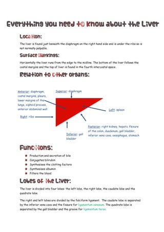
1. The liver is found just beneath the diaphragm on the right hand side and is under the ribs so is
not normally palpable.
Horizontally the liver runs from the edge to the midline. The bottom of the liver follows the
costal margins and the top of liver is found in the fourth intercostal space.
Production and secretion of bile
Conjugates bilirubin
Synthesises the clotting factors
Synthesises albumin
Filters the blood
The liver is divided into four lobes: the left lobe, the right lobe, the caudate lobe and the
quadrate lobe.
The right and left lobes are divided by the falciform ligament. The caudate lobe is separated
by the inferior vena cava and the fissure for ligamentum venosum. The quadrate lobe is
separated by the gall bladder and the groove for ligamentum teres.
Posterior: right kidney, hepatic flexure
of the colon, duodenum, gall bladder,
inferior vena cava, oesophagus, stomach
Superior: diaphragm
Left: spleen
Inferior: gall
bladder
Right: ribs
Anterior: diaphragm,
costal margins, pleura,
lower margins of the
lungs, xiphoid process,
anterior abdominal wall
2. The caudate and quadrate lobes are anatomically part of the right lobe but functionally part of
the left lobe of the liver.
The liver has its own hilum known as the porta hepatis. It is found on the posterorinferior
surface between the caudate and quadrate lobes.
It contains:
Right hepatic duct
Left hepatic duct
Right branch of the hepatic artery
Left branch of the hepatic artery
Portal vein
Nerve fibres
Lymph nodes
The lesser omentum is attached to porta hepatis.
The liver is covered by peritoneum. All of the liver is covered by peritoneum except for the
bare area of the liver.
The liver is supplied by both the portal vein (80%) and the hepatic artery (20%). The hepatic
artery brings oxygen rich blood. The portal vein brings blood rich in the products of digestion.
The liver is drained by the hepatic vein which drains into the inferior vena cava.
Lymph produced by the liver passes into lymph node in the porta hepatis and then into the celiac
lymph nodes. The bare area of the liver drains into posterior mediastinal lymph nodes.
The liver is supplied by the hepatic branch of the anterior vagal nerve.
remnant of ductus venosum which allows blood to bypass the liver in a foetus
: remnant of the left umbilical vein
3. The liver is made up mainly of hepatocytes. They have large central prominent nuclei, abundant
mitochondria, prominent endoplasmic reticulum, active golgi apparatus and numerous
peroxisomes (features of metabolically active secreting cells). Hepatocytes are surrounded by
sinusoids.
Hepatocytes have three different types of surfaces:
Sinusoidal – permits exchange of material with the blood
Canalicular – for excretion of bile
Intercellular
The Space of Disse is the space between the sinusoidal epithelium and hepatocytes. It contains:
Kupffer cells- phagocytic cells
Ito cells- synthesise collagen
Plasma
The basement membrane of the liver is made from reticulin- type III collagen.
There are two ways of dividing up liver cells: lobules and acini. A lobule is the area drained by
one central vein. Acini are based on blood supply and are clinically more useful.
Lobules:
Portal triad
Bile duct
Portal vein
Portal artery
Central vein
Sinusoid: capillaries with fenestrated epitheilium
Caniculi: thin tubes which collect bile and drain into bile ducts
4. A portal triad consists of:
A portal artery
A portal vein
A bile duct
They are held together by loose fibrous connective tissue
Central veins drain blood into the hepatic veins.
Acini:
Zone 2
Zone 3: furthest away from the blood supply
Zone 1: closest to the blood supply