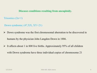
ANEUPLOIDY
MDE 605
PRESENTED BY
NJILLA STEEV-BECCA NJISSIC
(SC17P276)
MDE 605. Njilla steeve 15/5/2019
OUTLINE
 Introduction
 Causes of aneuploidy
 Disease conditions
 Conclusion.
MDE 605. Njilla steeve 25/5/2019
INTRODUCTION
 It is a category of chromosomal aberration in which the chromosome number
is abnormal.
 It involves changes in chromosome number by additions or deletions of less
than a whole chromosome set .
 Organism with this conditions are called aneuploids.
MDE 605. Njilla steeve 35/5/2019
 Aneuploid nomenclature (see table 1) is based on the number of copies of
the specific chromosome in the aneuploid state. For autosomes in diploid
organisms, the aneuploid (2n +1) is trisomic, (2n -1) is monosomic, and
(2n- 2) is nullisomic. In haploids, ( n+1) is disomic.
 Special notation is used to describe sex chromosome aneuploids because it
must deal with the two different chromosomes. The notation merely lists
the copies of each sex chromosome, such as XXY, XXY, XXX or XO (O
stands fors absence of a chromosome).
MDE 605. Njilla steeve 45/5/2019
Table 1: Partial List of Terms to Describe Aneuploidy, Using Drosophila as
an Example (Eight Chromosomes: X, X, 2, 2, 3, 3, 4, 4)
MDE 605. Njilla steeve 55/5/2019
Causes of Aneuploidy
 The primary cause of aneuploidy is nondisjunction during mitosis or
meiosis.
 Nondisjunction is a process by which homologous chromosomes or
chromatids fail to segregate normaly, that is all the chromosomes move to
one end of the pole and none to the other.
MDE 605. Njilla steeve 65/5/2019
Figure 1:The origin of aneuploid
gametes by nondisjunction at the first
or second meiotic division
MESSAGE: Crossovers are
needed to maintain the intact
tetrad until anaphase I. If
crossing-over fails for some
reason, first-division
nondisjunction occurs.
MDE 605. Njilla steeve 75/5/2019
Diseases conditions resulting from aneuploidy.
Trisomics (2n+1)
Down syndrome; (47,XX, XY+21)
 Down syndrome was the first chromosomal aberration to be discovered in
humans by the physician John Langdon Down in 1886.
 It affects about 1 in 800 live births. Approximately 95% of all children
with Down syndrome have three individual copies of chromosome 21
MDE 605. Njilla steeve 85/5/2019
 Most affected individuals are mildly to moderately mentally retarded,
have congenital heart defects, and a high (1/100) risk of acute
leukemia. They are usually short, have a broad, short skull, and
possess hyper flexible joints, mental retardation, females may
reproduce but not males. Life expectancy is 17 years, however, 8%
make it past 4o years.
MDE 605. Njilla steeve 95/5/2019
 About 95% of infected individuals have an extra copy of chromosome 21
caused by nondisjunction of chromosome 21 in a parent who is
chromosomally normal. In this sporadic type of Down syndrome, there is no
family history of aneuploidy.
 An interesting aspect of this syndrome is its increased incidence among
children born to older mothers, a fact known more than 25 years before the
discovery of its genetic cause
MDE 605. Njilla steeve 105/5/2019
 Recently, molecular genetic techniques identified the origins of the
three chromosome 21 copies in a large sample of individuals with
Down syndrome. As expected, 95% of the extra copies of
chromosome 21 were of maternal origin. Of these cases,
approximately 70% resulted from errors in meiosis I and 25% were
errors in meiosis II. Less than 1% of the Down syndrome children
resulted from nondisjuntion in meiosis II in the father.
MDE 605. Njilla steeve 115/5/2019
 The remaining 5% of Down syndrome individuals that contain three
free copies of chromosome 21 were produced by mitotic nondisjunction
in the zygote or embryo. In these cases, the extra copy of chromosome
21 originated equally from either parent and there was no increased
incidence with older parents.
 About 5% of individuals with Down syndrome do not have trisomy 21,
but rather have a translocation of the long arm of chromosome 21 to
usually chromosome 14 or 15. This type of Down syndrome is termed
familial down syndrom), based on the pattern of inheritance from
generation to generationMDE 605. Njilla steeve 125/5/2019
Figurer 2: characteristic of down syndrome Figure 3: Karyotype of a male with down syndromeMDE 605. Njilla steeve 135/5/2019
Trisomy 18 ( Edwards syndrome);
 Edwards syndrome affects 1 in 10 thousand live births. The karyotype for this
syndrome is 47, XX or XY,+18. Most affected fetuses fail to survive to birth.
Those that do survive are usually female, with 80–90% mortality by 2 years
of age.
MDE 605. Njilla steeve 145/5/2019
 An affected infant is usually born with major heart defects and a displaced
liver. In addition, the infant usually has a small nose and mouth, a
receding lower jaw, abnormal ears, and a lack of distal flexion creases on
the fingers. The distal joints have limited motion, and the fingers display a
characteristic posturing in which the little and index fingers overlap the
middle two. The syndrome is usually accompanied by severe mental
retardation.
MDE 605. Njilla steeve 155/5/2019
Trisomy 13 (Patau syndrome);
 Patau syndrome, karyotype 47,XX or XY,+13, affects 1 in 20 thousand
live births. A very high percentage of fetuses with trisomy 13 fail to
survive until birth.
 Individuals who are born with trisomy 13 often have small or missing
eyes, although in rare cases they exhibit a single fused eye structure in
the center of the face. Additional features include cleft palate, cleft lip,
congenital heart defects, polydactyly, and severe mental retardation.
Mortality is very high in the first year of life.
MDE 605. Njilla steeve 165/5/2019
XXY (Klinefelter syndrome);
 The incidence of Klinefelter syndrome (47,XXY) is about 1 in 1000 live births.
 The phenotypic expressions include midly impaired IQ, tall stature,
underdeveloped testes, and infertility. Some problems with behavior and
speech development are associated with this syndrome.
 If discovered early, testosterone injections during adolescence can help to
induce the secondary sexual characteristics (pubic and facial hair) that are often
reduced in Klinefelter individuals.
MDE 605. Njilla steeve 175/5/2019
Figure 4: Characteristic of Klinefelter syndromeMDE 605. Njilla steeve 185/5/2019
XYY Karyotype;
 Approximately 1 in 1000 live male births is an individual with a 47,XYY
karyotype.
 There is some indication that XYY men may have some speech and reading
problems, which could lead to lower test scores and lowered intelligence test
results. Criminal tendencies could therefore be attributed to lower intelligence,
rather than to the extra Y chromosome. But this idea is highly speculative. Males
with XYY are usually fertile.
MDE 605. Njilla steeve 195/5/2019
Tripple X chromosomes;
 A triple-X female (47,XXX) appears in about 1 in 1000 female live births.
Fertility can be normal, but these individuals are usually mildly mentally
retarded. Delayed growth, as well as congenital malformations, are also
sometimes present.
MDE 605. Njilla steeve 205/5/2019
Figure 5: XYY karyotype Figure 6: Karyotype of a tripple-X female.
MDE 605. Njilla steeve 215/5/2019
MONOSOMICS (2n -1)
X0 (Turner Syndrome);
 Turner syndrome occurs in about 1 in 2000 live female births, with
approximately 99% of Turner syndrome fetuses failing to survive until
birth. The karyotype designation is 45,X .
 Individuals with Turner syndrome usually exhibit normal intelligence
but underdeveloped ovaries, infertility, abnormal jaws, webbed necks,
and shieldlike chests. They are often short in stature.
MDE 605. Njilla steeve 225/5/2019
Figure 7: Characteristic of turner syndrome
MDE 605. Njilla steeve 235/5/2019
Conclusion
Aneuploidy refers to abnormal number of chromosomes. It is
primarily caused by nondisjunction in mitosis or meiosis but more
predorminant in meiosis. Aneuploidogens like colchicine, tobacco
smoke, benzene can also cause aneuploidy. The resulting disease
conditions are dentrimental to affected individuals and most aneuploid
states in human fetus lead to dead. Aneuploidy is important in the
determination of tumorigenic cells like colorectal cancer
MDE 605. Njilla steeve 245/5/2019
References
 Griffiths AJ, Miller JH, Suzuki DT. 2000. An introduction to genetic analysis.
8th edition. pp 490- 495, chapter 15.
 Sens S. 2000. “ Aneuploidy and cancer”. Current opinion in oncology. 12(1):
82- 88.
 Templado C, Uroz L, Estop A . 2013. “New insights on the origin and
relevance of aneuploidy in human spermatozoa”. Molecular human
reproduction. 19 (10):634-643.
 Yang AH, Kaushual D, Rehen SK. 2003. Chromosome segregation defects
contribute to aneuploidy in normal progenitor cells. The journal of
neuroscience. 23(32). MDE 605. Njilla steeve 255/5/2019
Thanks for listening
MDE 605. Njilla steeve 265/5/2019

Njilla steeve . aneuploidy presentation

  • 1.
    ANEUPLOIDY MDE 605 PRESENTED BY NJILLASTEEV-BECCA NJISSIC (SC17P276) MDE 605. Njilla steeve 15/5/2019
  • 2.
    OUTLINE  Introduction  Causesof aneuploidy  Disease conditions  Conclusion. MDE 605. Njilla steeve 25/5/2019
  • 3.
    INTRODUCTION  It isa category of chromosomal aberration in which the chromosome number is abnormal.  It involves changes in chromosome number by additions or deletions of less than a whole chromosome set .  Organism with this conditions are called aneuploids. MDE 605. Njilla steeve 35/5/2019
  • 4.
     Aneuploid nomenclature(see table 1) is based on the number of copies of the specific chromosome in the aneuploid state. For autosomes in diploid organisms, the aneuploid (2n +1) is trisomic, (2n -1) is monosomic, and (2n- 2) is nullisomic. In haploids, ( n+1) is disomic.  Special notation is used to describe sex chromosome aneuploids because it must deal with the two different chromosomes. The notation merely lists the copies of each sex chromosome, such as XXY, XXY, XXX or XO (O stands fors absence of a chromosome). MDE 605. Njilla steeve 45/5/2019
  • 5.
    Table 1: PartialList of Terms to Describe Aneuploidy, Using Drosophila as an Example (Eight Chromosomes: X, X, 2, 2, 3, 3, 4, 4) MDE 605. Njilla steeve 55/5/2019
  • 6.
    Causes of Aneuploidy The primary cause of aneuploidy is nondisjunction during mitosis or meiosis.  Nondisjunction is a process by which homologous chromosomes or chromatids fail to segregate normaly, that is all the chromosomes move to one end of the pole and none to the other. MDE 605. Njilla steeve 65/5/2019
  • 7.
    Figure 1:The originof aneuploid gametes by nondisjunction at the first or second meiotic division MESSAGE: Crossovers are needed to maintain the intact tetrad until anaphase I. If crossing-over fails for some reason, first-division nondisjunction occurs. MDE 605. Njilla steeve 75/5/2019
  • 8.
    Diseases conditions resultingfrom aneuploidy. Trisomics (2n+1) Down syndrome; (47,XX, XY+21)  Down syndrome was the first chromosomal aberration to be discovered in humans by the physician John Langdon Down in 1886.  It affects about 1 in 800 live births. Approximately 95% of all children with Down syndrome have three individual copies of chromosome 21 MDE 605. Njilla steeve 85/5/2019
  • 9.
     Most affectedindividuals are mildly to moderately mentally retarded, have congenital heart defects, and a high (1/100) risk of acute leukemia. They are usually short, have a broad, short skull, and possess hyper flexible joints, mental retardation, females may reproduce but not males. Life expectancy is 17 years, however, 8% make it past 4o years. MDE 605. Njilla steeve 95/5/2019
  • 10.
     About 95%of infected individuals have an extra copy of chromosome 21 caused by nondisjunction of chromosome 21 in a parent who is chromosomally normal. In this sporadic type of Down syndrome, there is no family history of aneuploidy.  An interesting aspect of this syndrome is its increased incidence among children born to older mothers, a fact known more than 25 years before the discovery of its genetic cause MDE 605. Njilla steeve 105/5/2019
  • 11.
     Recently, moleculargenetic techniques identified the origins of the three chromosome 21 copies in a large sample of individuals with Down syndrome. As expected, 95% of the extra copies of chromosome 21 were of maternal origin. Of these cases, approximately 70% resulted from errors in meiosis I and 25% were errors in meiosis II. Less than 1% of the Down syndrome children resulted from nondisjuntion in meiosis II in the father. MDE 605. Njilla steeve 115/5/2019
  • 12.
     The remaining5% of Down syndrome individuals that contain three free copies of chromosome 21 were produced by mitotic nondisjunction in the zygote or embryo. In these cases, the extra copy of chromosome 21 originated equally from either parent and there was no increased incidence with older parents.  About 5% of individuals with Down syndrome do not have trisomy 21, but rather have a translocation of the long arm of chromosome 21 to usually chromosome 14 or 15. This type of Down syndrome is termed familial down syndrom), based on the pattern of inheritance from generation to generationMDE 605. Njilla steeve 125/5/2019
  • 13.
    Figurer 2: characteristicof down syndrome Figure 3: Karyotype of a male with down syndromeMDE 605. Njilla steeve 135/5/2019
  • 14.
    Trisomy 18 (Edwards syndrome);  Edwards syndrome affects 1 in 10 thousand live births. The karyotype for this syndrome is 47, XX or XY,+18. Most affected fetuses fail to survive to birth. Those that do survive are usually female, with 80–90% mortality by 2 years of age. MDE 605. Njilla steeve 145/5/2019
  • 15.
     An affectedinfant is usually born with major heart defects and a displaced liver. In addition, the infant usually has a small nose and mouth, a receding lower jaw, abnormal ears, and a lack of distal flexion creases on the fingers. The distal joints have limited motion, and the fingers display a characteristic posturing in which the little and index fingers overlap the middle two. The syndrome is usually accompanied by severe mental retardation. MDE 605. Njilla steeve 155/5/2019
  • 16.
    Trisomy 13 (Patausyndrome);  Patau syndrome, karyotype 47,XX or XY,+13, affects 1 in 20 thousand live births. A very high percentage of fetuses with trisomy 13 fail to survive until birth.  Individuals who are born with trisomy 13 often have small or missing eyes, although in rare cases they exhibit a single fused eye structure in the center of the face. Additional features include cleft palate, cleft lip, congenital heart defects, polydactyly, and severe mental retardation. Mortality is very high in the first year of life. MDE 605. Njilla steeve 165/5/2019
  • 17.
    XXY (Klinefelter syndrome); The incidence of Klinefelter syndrome (47,XXY) is about 1 in 1000 live births.  The phenotypic expressions include midly impaired IQ, tall stature, underdeveloped testes, and infertility. Some problems with behavior and speech development are associated with this syndrome.  If discovered early, testosterone injections during adolescence can help to induce the secondary sexual characteristics (pubic and facial hair) that are often reduced in Klinefelter individuals. MDE 605. Njilla steeve 175/5/2019
  • 18.
    Figure 4: Characteristicof Klinefelter syndromeMDE 605. Njilla steeve 185/5/2019
  • 19.
    XYY Karyotype;  Approximately1 in 1000 live male births is an individual with a 47,XYY karyotype.  There is some indication that XYY men may have some speech and reading problems, which could lead to lower test scores and lowered intelligence test results. Criminal tendencies could therefore be attributed to lower intelligence, rather than to the extra Y chromosome. But this idea is highly speculative. Males with XYY are usually fertile. MDE 605. Njilla steeve 195/5/2019
  • 20.
    Tripple X chromosomes; A triple-X female (47,XXX) appears in about 1 in 1000 female live births. Fertility can be normal, but these individuals are usually mildly mentally retarded. Delayed growth, as well as congenital malformations, are also sometimes present. MDE 605. Njilla steeve 205/5/2019
  • 21.
    Figure 5: XYYkaryotype Figure 6: Karyotype of a tripple-X female. MDE 605. Njilla steeve 215/5/2019
  • 22.
    MONOSOMICS (2n -1) X0(Turner Syndrome);  Turner syndrome occurs in about 1 in 2000 live female births, with approximately 99% of Turner syndrome fetuses failing to survive until birth. The karyotype designation is 45,X .  Individuals with Turner syndrome usually exhibit normal intelligence but underdeveloped ovaries, infertility, abnormal jaws, webbed necks, and shieldlike chests. They are often short in stature. MDE 605. Njilla steeve 225/5/2019
  • 23.
    Figure 7: Characteristicof turner syndrome MDE 605. Njilla steeve 235/5/2019
  • 24.
    Conclusion Aneuploidy refers toabnormal number of chromosomes. It is primarily caused by nondisjunction in mitosis or meiosis but more predorminant in meiosis. Aneuploidogens like colchicine, tobacco smoke, benzene can also cause aneuploidy. The resulting disease conditions are dentrimental to affected individuals and most aneuploid states in human fetus lead to dead. Aneuploidy is important in the determination of tumorigenic cells like colorectal cancer MDE 605. Njilla steeve 245/5/2019
  • 25.
    References  Griffiths AJ,Miller JH, Suzuki DT. 2000. An introduction to genetic analysis. 8th edition. pp 490- 495, chapter 15.  Sens S. 2000. “ Aneuploidy and cancer”. Current opinion in oncology. 12(1): 82- 88.  Templado C, Uroz L, Estop A . 2013. “New insights on the origin and relevance of aneuploidy in human spermatozoa”. Molecular human reproduction. 19 (10):634-643.  Yang AH, Kaushual D, Rehen SK. 2003. Chromosome segregation defects contribute to aneuploidy in normal progenitor cells. The journal of neuroscience. 23(32). MDE 605. Njilla steeve 255/5/2019
  • 26.
    Thanks for listening MDE605. Njilla steeve 265/5/2019

Editor's Notes

  • #4 Usually involves changes in a single chromosome or a small number of chromosomes.
  • #5 2n-2 is nullisomic and 2 represents the loss of two non homologous chromosomes.
  • #10 Account for symptoms
  • #11 Sporadic down syndrome Explain age related susceptibility
  • #12 Why do down syndrome easily results from the mother
  • #13 Read on translocation
  • #17 Other autosomal trisomics are known but are extremely rare. These include trisomy 8 (47,XX or XY,+8) and cat’s eye syndrome, a trisomy of an unidentified, small acrocentric chromosome (47,XX or XY,[+acrocentric]).
  • #18 Diagnosis is usually by buccal (cheek tissue) smear to ascertain the presence of a Barr body, which in a male would indicate an XXY karyotype.
  • #20 We avoid the term syndrome here because XYY men have no clearly defined series of attributes, other than often being taller than normal Meioses show normal pairing of the X with one of the Y’s; the other Y does not pair and is not transmitted to gametes. Therefore the gametes contain either X or Y, never YY or XY.
  • #23 The severe consequences of missing one entire chromosome allow only three different human chromosomes to be monosomic (X, 21, or 22) and still yield a viable individual.
  • #25 Flourescence insitu hybridization, quantitative PCR of short tandem repeats, quantitative flourescence PCR, Quantitative PCR doseage.