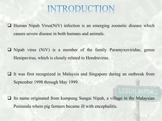
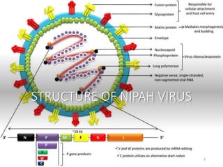
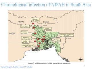
By
Arman Firoz
PhD Scholar
VIT University
1
 Human Nipah Virus(NiV) infection is an emerging zoonotic disease which
causes severe disease in both humans and animals.
 Nipah virus (NiV) is a member of the family Paramyxoviridae, genus
Henipavirus, which is closely related to Hendravirus.
 It was first recognized in Malaysia and Singapore during an outbreak from
September 1998 through May 1999.
 Its name originated from kampung Sungai Nipah, a village in the Malaysian
Peninsula where pig farmers became ill with encephalitis.
2
STRUCTURE OF NIPAH VIRUS
3
Though Nipah virus has caused only a few outbreaks, it infects a wide range of
animals and causes severe disease and death in people, making it a public health
concern.
4
Laura T Mazzola 1, Cassandra Kelly-Cirino 1
5
Gurjeet Singh1 , Raksha , Anant D. Urhekar.
Graph.2. Representation of Nipah spread across south Asia
• The recent outbreak in Kerala is
thought to have been caused by dead
bats found in a well of a family's
home in the village of Changaroth.
• The infection reportedly spread
among family members and was
passed on to others who had been in
contact with the family.
The Nipah virus has already claimed 10 lives in the Indian state of Kerala, including a 31-year-
old nurse who was treating the infected. 6
Table.1 Mortality and Morbidity due to Nipah infection in South East Asia
Bangladesh
During the collection of date palm
sap, fruit bats drink from the sap and
contaminate the sap with Nipah virus
through saliva, urine, or feces. People
drinking the date palm sap become
infected with Nipah virus and transmit
the virus to close contacts.
Malaysia
Pteropid fruit bats are the natural reservoir
of Nipah virus. Bats roosting in fruit trees
on pig farms transmitted the virus to pigs.
Pigs transmitted Nipah virus to people in
close contact with them.
7
 The pathologic findings in the brain
of Nipah encephalitis cases showed
evidence of necrotizing vasculitis.
 The main pathology appeared to be
widespread ischemia and infarction
caused by vasculitis-induced
thrombosis, although direct neuronal
invasion may also play a major role
in the pathogenesis of the
encephalitis.
 Alveolar hemorrhage, pulmonary edema and aspiration pneumonia were often
encountered in the lungs.
 These may lead to pneumonia and acute respiratory distress syndrome (ARDS)
ultimately.
8
 Acute onset of
cough with
shortness of
breath
 Severe Headache
 Acute onset of
altered mental
status
 High Grade Fever
 Muscle pain
 Convulsion
 Diarrhoea
9
DIAGNOSIS, TREATMENT AND
PREVENTIVE MEASURES
10
The samples should be collected as early as possible (preferably
within 4-5 days on onset of illness.
The samples may be as follows:
 Throat swab to be collected in viral transport medium
 Urine approx 10 ml in universal sterile container
 Blood in plain vial (at least 5ml)
 CSF (at least 1 ml) in a sterile container
 Samples should be safely packed in triple container packing and should be
transported under cold chain (2-8°C) to the testing laboratory with prior
intimation.
 Before dispatching the sample, disinfect the outer surface of container using
1:100 dilution of bleach or 5% Lysol solution.
11
Serology
RT-PCR
ELISA
Virus Isolation
Neutralization test
Immunohistochemistry
12
Yet No Specific treatment for NIPAH
 Treatment is limited to supportive care. May require intensive care monitoring
 Mechanical ventilation for air way protection for neurological deterioration.
 Proper barrier nursing techniques are important in preventing hospital-acquired
infections
13
Antiviral Species Efficacy
Ribavirin
Human (Malaysia
outbreak)
36 % reduction in mortality
Syrian hamster No beneficial effect
Chloroquine
Syrian hamster No beneficial effect
Ferret No beneficial effect
Neutralizing
antibodies
Syrian hamster
100 % survival with pretreatment;
partial protection with posttreatment
m102.4 antibody Ferret
100 % survival when treated 24 h after
inoculation
poly(I)-poly(C 12 U) Syrian hamster
80 % survival when treated 2 h after
inoculation and 9 additional days
VIKI-PEG4-chol Syrian hamster
40 % survival when treated 2 days after
inoculation
Table.2 The efficacy of antiviral treatments tested in Nipah virus animal models
14
15
Table.3 The efficacy of vaccine candidates tested in Nipah virus animal disease models
Respiratory Hygiene
Hand hygiene Injection safety facial protection
Gloves and gown
wearing
Environmental cleaning
16
Avoid intake of fruits bitten by animals
Proper handling of specimen
Strong Disease Surveillance
17
 Mazzola, L. T., & Kelly-Cirino, C. (2019). Diagnostics for Nipah virus: a zoonotic
pathogen endemic to Southeast Asia. BMJ global health, 4(Suppl 2).
 Gurjeet Singh1 , Raksha , Anant D. Urhekar. Nipah: A killer virus,
Int.J.Adv.Microbiol.Health.Res.2018; 2(2):40-55
 Mohd Nor MN, Gan CH, Ong BL. Nipah virus infection of pigs in peninsular
Malaysia. Rev Sci Tech. 2000;19:160–5.
 Chua KB, Bellini WJ, Rota PA, Harcourt BH, Tamin A, et al. Nipah virus: a recently
emergent deadly paramyxovirus. Science. 2000;288:1432–5.
 Halpin K, Hyatt AD, Fogarty R, Middleton D, Bingham J, et al. Pteropid bats are
confirmed as the reservoir hosts of henipaviruses: a comprehensive experimental study
of virus transmission. Am J Trop Med Hyg. 2011;85:946–51.
18
19

Nipah virus ppt

  • 1.
  • 2.
     Human NipahVirus(NiV) infection is an emerging zoonotic disease which causes severe disease in both humans and animals.  Nipah virus (NiV) is a member of the family Paramyxoviridae, genus Henipavirus, which is closely related to Hendravirus.  It was first recognized in Malaysia and Singapore during an outbreak from September 1998 through May 1999.  Its name originated from kampung Sungai Nipah, a village in the Malaysian Peninsula where pig farmers became ill with encephalitis. 2
  • 3.
  • 4.
    Though Nipah virushas caused only a few outbreaks, it infects a wide range of animals and causes severe disease and death in people, making it a public health concern. 4 Laura T Mazzola 1, Cassandra Kelly-Cirino 1
  • 5.
    5 Gurjeet Singh1 ,Raksha , Anant D. Urhekar. Graph.2. Representation of Nipah spread across south Asia
  • 6.
    • The recentoutbreak in Kerala is thought to have been caused by dead bats found in a well of a family's home in the village of Changaroth. • The infection reportedly spread among family members and was passed on to others who had been in contact with the family. The Nipah virus has already claimed 10 lives in the Indian state of Kerala, including a 31-year- old nurse who was treating the infected. 6 Table.1 Mortality and Morbidity due to Nipah infection in South East Asia
  • 7.
    Bangladesh During the collectionof date palm sap, fruit bats drink from the sap and contaminate the sap with Nipah virus through saliva, urine, or feces. People drinking the date palm sap become infected with Nipah virus and transmit the virus to close contacts. Malaysia Pteropid fruit bats are the natural reservoir of Nipah virus. Bats roosting in fruit trees on pig farms transmitted the virus to pigs. Pigs transmitted Nipah virus to people in close contact with them. 7
  • 8.
     The pathologicfindings in the brain of Nipah encephalitis cases showed evidence of necrotizing vasculitis.  The main pathology appeared to be widespread ischemia and infarction caused by vasculitis-induced thrombosis, although direct neuronal invasion may also play a major role in the pathogenesis of the encephalitis.  Alveolar hemorrhage, pulmonary edema and aspiration pneumonia were often encountered in the lungs.  These may lead to pneumonia and acute respiratory distress syndrome (ARDS) ultimately. 8
  • 9.
     Acute onsetof cough with shortness of breath  Severe Headache  Acute onset of altered mental status  High Grade Fever  Muscle pain  Convulsion  Diarrhoea 9
  • 10.
  • 11.
    The samples shouldbe collected as early as possible (preferably within 4-5 days on onset of illness. The samples may be as follows:  Throat swab to be collected in viral transport medium  Urine approx 10 ml in universal sterile container  Blood in plain vial (at least 5ml)  CSF (at least 1 ml) in a sterile container  Samples should be safely packed in triple container packing and should be transported under cold chain (2-8°C) to the testing laboratory with prior intimation.  Before dispatching the sample, disinfect the outer surface of container using 1:100 dilution of bleach or 5% Lysol solution. 11
  • 12.
  • 13.
    Yet No Specifictreatment for NIPAH  Treatment is limited to supportive care. May require intensive care monitoring  Mechanical ventilation for air way protection for neurological deterioration.  Proper barrier nursing techniques are important in preventing hospital-acquired infections 13
  • 14.
    Antiviral Species Efficacy Ribavirin Human(Malaysia outbreak) 36 % reduction in mortality Syrian hamster No beneficial effect Chloroquine Syrian hamster No beneficial effect Ferret No beneficial effect Neutralizing antibodies Syrian hamster 100 % survival with pretreatment; partial protection with posttreatment m102.4 antibody Ferret 100 % survival when treated 24 h after inoculation poly(I)-poly(C 12 U) Syrian hamster 80 % survival when treated 2 h after inoculation and 9 additional days VIKI-PEG4-chol Syrian hamster 40 % survival when treated 2 days after inoculation Table.2 The efficacy of antiviral treatments tested in Nipah virus animal models 14
  • 15.
    15 Table.3 The efficacyof vaccine candidates tested in Nipah virus animal disease models
  • 16.
    Respiratory Hygiene Hand hygieneInjection safety facial protection Gloves and gown wearing Environmental cleaning 16
  • 17.
    Avoid intake offruits bitten by animals Proper handling of specimen Strong Disease Surveillance 17
  • 18.
     Mazzola, L.T., & Kelly-Cirino, C. (2019). Diagnostics for Nipah virus: a zoonotic pathogen endemic to Southeast Asia. BMJ global health, 4(Suppl 2).  Gurjeet Singh1 , Raksha , Anant D. Urhekar. Nipah: A killer virus, Int.J.Adv.Microbiol.Health.Res.2018; 2(2):40-55  Mohd Nor MN, Gan CH, Ong BL. Nipah virus infection of pigs in peninsular Malaysia. Rev Sci Tech. 2000;19:160–5.  Chua KB, Bellini WJ, Rota PA, Harcourt BH, Tamin A, et al. Nipah virus: a recently emergent deadly paramyxovirus. Science. 2000;288:1432–5.  Halpin K, Hyatt AD, Fogarty R, Middleton D, Bingham J, et al. Pteropid bats are confirmed as the reservoir hosts of henipaviruses: a comprehensive experimental study of virus transmission. Am J Trop Med Hyg. 2011;85:946–51. 18
  • 19.

Editor's Notes

  • #3 Extras - In the 1999 outbreak, Nipah virus caused a relatively mild disease in pigs, but nearly 300 human cases with over 100 deaths were reported. In order to stop the outbreak, more than a million pigs were euthanized, causing tremendous trade loss for Malaysia. Since this outbreak, no subsequent cases (in neither swine nor human) have been reported in either Malaysia or Singapore.
  • #4 SINCE this contain single stranded rna this is an RNA VIRUS This is an enveloped virus
  • #6 Red spots are the area which is affected by nipah virus
  • #7 Which shows the number of death around “ “ with the fatality rate ranging from 30 – 100%. Recently, in 2018 May kerala was struck with Nipah incidence and the source was found to be the dead bat in a well which spreaded the disease across families.
  • #8 • In malaysian outbreaks human infections occurred through direct contact with respiratory secretions and urine from infected pigs. Occupational contacts in abattoir workers, pork sellers • In Bangladesh Nipah virus infection in human resulted from consumption of raw date palm sap contaminated by Bat saliva and human to human transmission. • In philippines, there was evidence of horse to human and human to human transmission.
  • #9  Viremia- entering of virus in blood stream Vasculitis- inflammation Infarction – tissue death due to inadequate blood supply Infection is mainly associated with encaphilitis(inflammation of brain) After exposure it has an incubation period of 5 to 14 days, illness present with 3-14 days of fever and headache followed by drowsiness, disorientation and mental confusion.
  • #13 Laboratory diagnosis of a patient with a clinical history of NiV can be made during the acute and convalescent phases of the disease by using a combination of tests. Virus isolation attempts and real time polymerase chain reaction (RT-PCR) from throat and nasal swabs, cerebrospinal fluid, urine, and blood should be performed in the early stages of disease. Antibody detection by ELISA (IgG and IgM) can be used later. In fatal cases, immunohistochemistry on tissues collected during autopsy may be the only way to confirm a diagnosis.
  • #14 There is no specific treatment for Nipah Virus. Supportive care is the general treatment for this disease. • Treatment is limited to supportive care. May require intensive care monitoring • Mechanical ventilation for air way protection should be initiated with onset of neurological deterioration. • Because Nipah virus encephalitis can be transmitted person-to-person, standard infection control practices and proper barrier nursing techniques are important in preventing hospital-acquired infections (nosocomial transmission).
  • #15 Due to the severity of Nipah virus outbreaks and their sporadic nature, antiviral treatment options would be very valuable. Currently, the only treatment available to Nipah virus patients is supportive care in hospital settings. Attempts to develop antivirals against Nipah virus are under way. An overview of antiviral treatments tested in one or more of the Nipah virus animal models is given in Table.
  • #16 Since 2012, an equine Hendra virus vaccine is available in Australia. This vaccine, Equivac HeV, consisting of soluble glycoprotein G, aims to protect horses from acquiring Hendra virus. Since humans are largely exposed to Hendra virus through infected horses, the vaccine at the same time aims to prevent transmission of Hendra virus to humans. Since there is no intermediate reservoir involved in the transmission of Nipah virus to humans in the Bangladeshi outbreaks, a similar vaccination strategy cannot be adopted there. Moreover, Nipah virus outbreaks in Bangladesh are small and sporadic, thereby making it unlikely that largescale vaccination campaigns will ever be used in the human population. However, a Nipah virus vaccine may be useful to employ a ring vaccination strategy during Nipah virus outbreaks. For such a strategy to be successful, fast-acting vaccines need to be developed. An overview of vaccine candidates tested in one or more of the Nipah virus animal models is presented in this table.