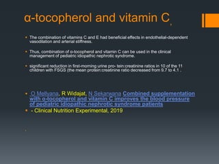
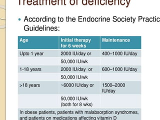
Nephrotic syndrom and
vitamins
Ahmad Ali
Nikibakhsh Pediatric
Nephrologist
Urmia University of
Medical Sciences
Abnormal vitamin D metabolism in
idiopathic NS is multi-factorial
 losses of both vitamin D binding protein and 25(OH)D in the
urine
 The urinary losses of vitamin D binding protein may be
secondary to proteinuria.
 Deficiency in 25(OH)D may lead to hypocalcemia,
hyperparathyroidism, and diminished bone mineral
density/content.
 Vitamin D deficiency has also been associated with multiple
systemic effects including elevated blood pressure ,metabolic
syndrome, cardiovascular disease ,anemia and impaired
immune system regulation .
 Roos BA. Urinary and plasma vitamin D3 metabolites in the nephrotic syndrome. Metab
Bone Dis Relat Res. 1982;4:7–15.
25(OH)D levels25(OH)D levels
 There has been an increasing amount of data suggesting
that 25(OH)D levels may not completely normalize when
children go into remission
 Data suggest that abnormalities in 25(OH)D levels persist
and occur at a greater frequency than would be expected in
the general population.
 Banerjee S, Basu S, Sengupta J. Vitamin D in nephrotic
syndrome remission: a case–control study. Pediatr Nephrol.
2013;28
Study by
Nikibakhsh.Ahmadali,
Mahmoodzadeh.Hashem,Valizade
h.Mohammad
Effect of supplementary
use of Vitamin D on
serum level of vitamin D3
in patients with nephrotic
syndrome receiving
steroid therapy
Study by
Nikibakhsh.Ahmadali,
Mahmoodzadeh.Hashem,Valizade
h.Mohammad
 Out of 30 children 60% were male and 40% were female with
mean age of 6.91 + 3.34 years.
 Before intervention 70% of patients had sever deficiency,
26.7% mild to moderate deficiency.
 Mean level of 25(OH) D was 8.277±0.84 ng/ml rising to
14.364±1.14 ng/ml after two months that was statistically
significant (P=0.001).
Study by
Nikibakhsh.Ahmadali,
Mahmoodzadeh.Hashem,Valizade
h.Mohammad
Severe deficiency;
 The serum level of 25-hydroxy vitamin-D3 less than 10 ng/ml
Mild to moderate ;
 Patients with the levels between 10 and 20 ng/ml
Insufficient
 those with the levels between 20 and 30 ng/ml
Normal ;
 serum levels of more than 30 ng/ml
 Before intervention none of the patients had normal serum level
of 25-(OH)-D and after one month, only one patient gained
normal level which was not statistically significant (P=0.500).
 vitamin D levels at three times (beginning, one month, and two
month after intervention). According to the results of this table, it
can be seen that the mean serum level of vitamin D before
intervention was 8.27 ± 0.84 ng/ml, with increasing to 14.36±
1.14 and finally reaching 27.78 ±2.80 after two month of
treatment.
 After two months of intervention, 12 patients escaped deficiency
but still insufficient followed by 8 people with deficiency and 10
reached normal values which was statistically significant
(P=0.002)
Time Baseline After 1month
Treatment
After2Month Treatment P value
Severe Deficiency 21 (70) 5 (16.7) 1 (3.3) 0.001
Mild-Moderate Deficiency 8 (26.7) 21 (70) 7 (23.3)
Insufficient 1 (3.3) 3 (10) 12 (40)
Normal --- 1 (3.3) 10 (33.3)
Total 30 (100) 30 (100) 30 (100)
Study by
Nikibakhsh.Ahmadali,
Mahmoodzadeh.Hashem,Valizade
h.Mohammad
We suggest that children may benefit
from routine measurement of their
serum vitamin D levels at the time of
diagnosis and later on fallow-up visits
so an individual strategy for treatment
with vitamin D can be given.
 A daily dose of systemic steroids as low as 5 mg per day has
been shown to contribute to osteoporosis in adults .
 This is of concern in pediatric patients following a new
diagnosis of NS where steroid treatment protocols exceed
the dose at which adult osteoporosis risk increases with
repeated exposures to steroids during subsequent relapses
 Leonard MB, Zemel BS. Current concepts in pediatric bone
disease. Pediatr Clin N Am. 2002;49:143–173.
 Data suggest that, all children with incident NS had
25(OH)D deficiency at diagnosis and the majority
continued to have a deficiency at 2–4 months.
 Furthermore, supplemental vitamin D decreased the
odds of 25(OH)D deficiency, suggesting the need for
future interventional studies addressing vitamin D
supplementation in incident NS to evaluate optimal
vitamin D dosing and long-term impact in these
children.
 Selewski DT Vitamin D in incident nephrotic syndrome: a
Midwest Pediatric Nephrology Consortium study. Pediatric
Nephrology (Berlin, Germany), 22 Oct 2015, 31(3):465-472
 Short-term, high-dose glucocorticoid therapy decreases
the BMC of the lumbar spine in steroid-naïve children
with NS. Vitamin D and calcium co-administration not
only prevents this decline, but also enhances BMC of
the lumbar spine.
 The 1000 IU/day dose is marginally more effective than
400 IU/day and it is likely than an even larger dose is
required.
 Further research is required to assess the efficacy and
safety of vitamin D doses higher than 1000 IU/day.
 S Choudhary, Calcium and vitamin D for
osteoprotection in children with new-onset
nephrotic syndrome treated with steroids Pediatric
Nephrology, 2014,
 Vitamin D plus calcium therapy at the current doses(vitamin
D 40 0 IU plus calcium 1 g daily) reduces but does not
completely prevent bone loss, with no additional adverse
effects
 S Choudhary, Calcium and vitamin D for osteoprotection
in children with new-onset nephrotic syndrome treated
with steroids Pediatric Nephrology, 2014
 Several studies indicate a relationship between
hypovitaminosis D, survival, vascular calcification and
inflammation.
 In addition to its central role in the regulation of bone mineral
metabolism, vitamin D also contributes to other systems,
including the immune, cardiovascular and endocrine
systems.
 Vitamin D analogs reduces proteinuria, in particular through
suppression of the renin-angiotensin-aldosterone system
(RAAS) and exerts anti-inflammatory and immunomodulatory
effects.
 A Passantino, C Aloisi, V Cernaro, D Santoro New insights
on the role of vitamin D in the progression of renal
damage- Kidney and Blood …, 2013
 …,
 Vitamin D deficiency contribute to an inappropriately activated
RAAS, as a mechanism for progression of chronic kidney
disease (CKD) and/or cardiovascular disease.
 Vitamin D may interact with B and T lymphocytes and influence
the phenotype and function of the antigen presenting cells and
dendritic cells, promoting properties that favor the induction of
tolerogenic T regulators rather than T effectory.
 A Passantino, C Aloisi, V Cernaro, D Santoro New insights on
the role of vitamin D in the progression of renal damage-
Kidney and Blood …, 2013
 Interstitial fibrosis may be prevented through vitamin D
supplementation.
 It turns out that calcitriol effectively blocks myofibroblast
activation from interstitial fibroblasts, as evidenced by
suppression of TGF-β1-mediated α-SMA expression.
 A Passantino, C Aloisi, V Cernaro, D Santoro New insights
on the role of vitamin D in the progression of renal
damage- Kidney and Blood …, 2013
 previous studies confirmed that vitamin D receptors reduced
proteinuria, lessened the degree of glomerular sclerosis, and
exerted protective effects against kidney diseases by
inhibiting the renin angiotensin system, and having an anti-
inflammatory and anti-fibrotic effect.
 Supplementation with 1,25-vitamin D3or1,25-vitamin
D2prevented podocyte effacement or reversed glomerular
and tubulointerstitial damage in1,25-vitamin D3edeficient
animals, thereby preserving and restoring renal function,
respectively.
 Ramon Sonneveld 1,25-Vitamin D3 Deficiency Induces
Albuminuria Am J Pathol 2016 Apr;
vitamin B6
 Patients with the nephrotic syndrome have multiple risk factors for
thrombosis.
 Some reports indicate that they frequently have low circulating levels of
vitamin B6, which associate with a heightened risk for venous and
arterial thrombosis.
 GM Podda, F Lussana, G Moroni ,Abnormalities of homocysteine
and B vitamins in the nephrotic syndrome
 - Thrombosis research, 2007
 .
α-tocopherol and vitamin C,
 The combination of vitamins C and E had beneficial effects in endothelial-dependent
vasodilation and arterial stiffness.
 Thus, combination of α-tocopherol and vitamin C can be used in the clinical
management of pediatric idiopathic nephrotic syndrome.
 significant reduction in first-morning urine pro- tein:creatinine ratios in 10 of the 11
children with FSGS (the mean protein:creatinine ratio decreased from 9.7 to 4.1 .
 O Mellyana, R Widajat, N Sekarwana Combined supplementation
with α-tocopherol and vitamin C improves the blood pressure
of pediatric idiopathic nephrotic syndrome patients
 - Clinical Nutrition Experimental, 2019

Thanks
Nephrotic syndrom and vitamins

Nephrotic syndrom and vitamins

  • 1.
    Nephrotic syndrom and vitamins AhmadAli Nikibakhsh Pediatric Nephrologist Urmia University of Medical Sciences
  • 2.
    Abnormal vitamin Dmetabolism in idiopathic NS is multi-factorial  losses of both vitamin D binding protein and 25(OH)D in the urine  The urinary losses of vitamin D binding protein may be secondary to proteinuria.  Deficiency in 25(OH)D may lead to hypocalcemia, hyperparathyroidism, and diminished bone mineral density/content.  Vitamin D deficiency has also been associated with multiple systemic effects including elevated blood pressure ,metabolic syndrome, cardiovascular disease ,anemia and impaired immune system regulation .  Roos BA. Urinary and plasma vitamin D3 metabolites in the nephrotic syndrome. Metab Bone Dis Relat Res. 1982;4:7–15.
  • 3.
    25(OH)D levels25(OH)D levels There has been an increasing amount of data suggesting that 25(OH)D levels may not completely normalize when children go into remission  Data suggest that abnormalities in 25(OH)D levels persist and occur at a greater frequency than would be expected in the general population.  Banerjee S, Basu S, Sengupta J. Vitamin D in nephrotic syndrome remission: a case–control study. Pediatr Nephrol. 2013;28
  • 4.
    Study by Nikibakhsh.Ahmadali, Mahmoodzadeh.Hashem,Valizade h.Mohammad Effect ofsupplementary use of Vitamin D on serum level of vitamin D3 in patients with nephrotic syndrome receiving steroid therapy
  • 5.
    Study by Nikibakhsh.Ahmadali, Mahmoodzadeh.Hashem,Valizade h.Mohammad  Outof 30 children 60% were male and 40% were female with mean age of 6.91 + 3.34 years.  Before intervention 70% of patients had sever deficiency, 26.7% mild to moderate deficiency.  Mean level of 25(OH) D was 8.277±0.84 ng/ml rising to 14.364±1.14 ng/ml after two months that was statistically significant (P=0.001).
  • 6.
    Study by Nikibakhsh.Ahmadali, Mahmoodzadeh.Hashem,Valizade h.Mohammad Severe deficiency; The serum level of 25-hydroxy vitamin-D3 less than 10 ng/ml Mild to moderate ;  Patients with the levels between 10 and 20 ng/ml Insufficient  those with the levels between 20 and 30 ng/ml Normal ;  serum levels of more than 30 ng/ml
  • 7.
     Before interventionnone of the patients had normal serum level of 25-(OH)-D and after one month, only one patient gained normal level which was not statistically significant (P=0.500).  vitamin D levels at three times (beginning, one month, and two month after intervention). According to the results of this table, it can be seen that the mean serum level of vitamin D before intervention was 8.27 ± 0.84 ng/ml, with increasing to 14.36± 1.14 and finally reaching 27.78 ±2.80 after two month of treatment.  After two months of intervention, 12 patients escaped deficiency but still insufficient followed by 8 people with deficiency and 10 reached normal values which was statistically significant (P=0.002)
  • 8.
    Time Baseline After1month Treatment After2Month Treatment P value Severe Deficiency 21 (70) 5 (16.7) 1 (3.3) 0.001 Mild-Moderate Deficiency 8 (26.7) 21 (70) 7 (23.3) Insufficient 1 (3.3) 3 (10) 12 (40) Normal --- 1 (3.3) 10 (33.3) Total 30 (100) 30 (100) 30 (100)
  • 9.
    Study by Nikibakhsh.Ahmadali, Mahmoodzadeh.Hashem,Valizade h.Mohammad We suggestthat children may benefit from routine measurement of their serum vitamin D levels at the time of diagnosis and later on fallow-up visits so an individual strategy for treatment with vitamin D can be given.
  • 10.
     A dailydose of systemic steroids as low as 5 mg per day has been shown to contribute to osteoporosis in adults .  This is of concern in pediatric patients following a new diagnosis of NS where steroid treatment protocols exceed the dose at which adult osteoporosis risk increases with repeated exposures to steroids during subsequent relapses  Leonard MB, Zemel BS. Current concepts in pediatric bone disease. Pediatr Clin N Am. 2002;49:143–173.
  • 11.
     Data suggestthat, all children with incident NS had 25(OH)D deficiency at diagnosis and the majority continued to have a deficiency at 2–4 months.  Furthermore, supplemental vitamin D decreased the odds of 25(OH)D deficiency, suggesting the need for future interventional studies addressing vitamin D supplementation in incident NS to evaluate optimal vitamin D dosing and long-term impact in these children.  Selewski DT Vitamin D in incident nephrotic syndrome: a Midwest Pediatric Nephrology Consortium study. Pediatric Nephrology (Berlin, Germany), 22 Oct 2015, 31(3):465-472
  • 12.
     Short-term, high-doseglucocorticoid therapy decreases the BMC of the lumbar spine in steroid-naïve children with NS. Vitamin D and calcium co-administration not only prevents this decline, but also enhances BMC of the lumbar spine.  The 1000 IU/day dose is marginally more effective than 400 IU/day and it is likely than an even larger dose is required.  Further research is required to assess the efficacy and safety of vitamin D doses higher than 1000 IU/day.  S Choudhary, Calcium and vitamin D for osteoprotection in children with new-onset nephrotic syndrome treated with steroids Pediatric Nephrology, 2014,
  • 13.
     Vitamin Dplus calcium therapy at the current doses(vitamin D 40 0 IU plus calcium 1 g daily) reduces but does not completely prevent bone loss, with no additional adverse effects  S Choudhary, Calcium and vitamin D for osteoprotection in children with new-onset nephrotic syndrome treated with steroids Pediatric Nephrology, 2014
  • 14.
     Several studiesindicate a relationship between hypovitaminosis D, survival, vascular calcification and inflammation.  In addition to its central role in the regulation of bone mineral metabolism, vitamin D also contributes to other systems, including the immune, cardiovascular and endocrine systems.  Vitamin D analogs reduces proteinuria, in particular through suppression of the renin-angiotensin-aldosterone system (RAAS) and exerts anti-inflammatory and immunomodulatory effects.  A Passantino, C Aloisi, V Cernaro, D Santoro New insights on the role of vitamin D in the progression of renal damage- Kidney and Blood …, 2013  …,
  • 15.
     Vitamin Ddeficiency contribute to an inappropriately activated RAAS, as a mechanism for progression of chronic kidney disease (CKD) and/or cardiovascular disease.  Vitamin D may interact with B and T lymphocytes and influence the phenotype and function of the antigen presenting cells and dendritic cells, promoting properties that favor the induction of tolerogenic T regulators rather than T effectory.  A Passantino, C Aloisi, V Cernaro, D Santoro New insights on the role of vitamin D in the progression of renal damage- Kidney and Blood …, 2013
  • 16.
     Interstitial fibrosismay be prevented through vitamin D supplementation.  It turns out that calcitriol effectively blocks myofibroblast activation from interstitial fibroblasts, as evidenced by suppression of TGF-β1-mediated α-SMA expression.  A Passantino, C Aloisi, V Cernaro, D Santoro New insights on the role of vitamin D in the progression of renal damage- Kidney and Blood …, 2013
  • 17.
     previous studiesconfirmed that vitamin D receptors reduced proteinuria, lessened the degree of glomerular sclerosis, and exerted protective effects against kidney diseases by inhibiting the renin angiotensin system, and having an anti- inflammatory and anti-fibrotic effect.  Supplementation with 1,25-vitamin D3or1,25-vitamin D2prevented podocyte effacement or reversed glomerular and tubulointerstitial damage in1,25-vitamin D3edeficient animals, thereby preserving and restoring renal function, respectively.  Ramon Sonneveld 1,25-Vitamin D3 Deficiency Induces Albuminuria Am J Pathol 2016 Apr;
  • 18.
    vitamin B6  Patientswith the nephrotic syndrome have multiple risk factors for thrombosis.  Some reports indicate that they frequently have low circulating levels of vitamin B6, which associate with a heightened risk for venous and arterial thrombosis.  GM Podda, F Lussana, G Moroni ,Abnormalities of homocysteine and B vitamins in the nephrotic syndrome  - Thrombosis research, 2007  .
  • 19.
    α-tocopherol and vitaminC,  The combination of vitamins C and E had beneficial effects in endothelial-dependent vasodilation and arterial stiffness.  Thus, combination of α-tocopherol and vitamin C can be used in the clinical management of pediatric idiopathic nephrotic syndrome.  significant reduction in first-morning urine pro- tein:creatinine ratios in 10 of the 11 children with FSGS (the mean protein:creatinine ratio decreased from 9.7 to 4.1 .  O Mellyana, R Widajat, N Sekarwana Combined supplementation with α-tocopherol and vitamin C improves the blood pressure of pediatric idiopathic nephrotic syndrome patients  - Clinical Nutrition Experimental, 2019 
  • 21.