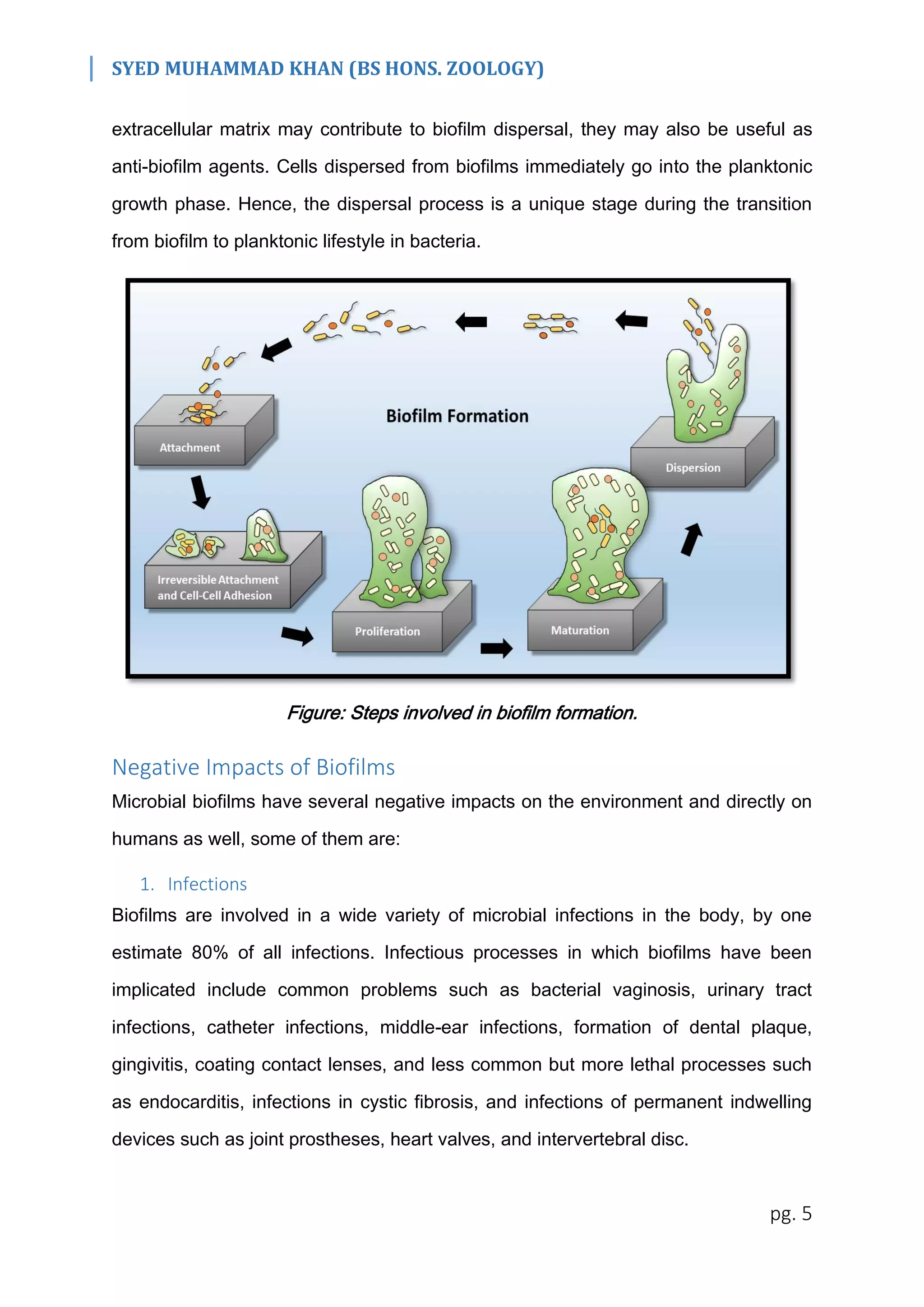
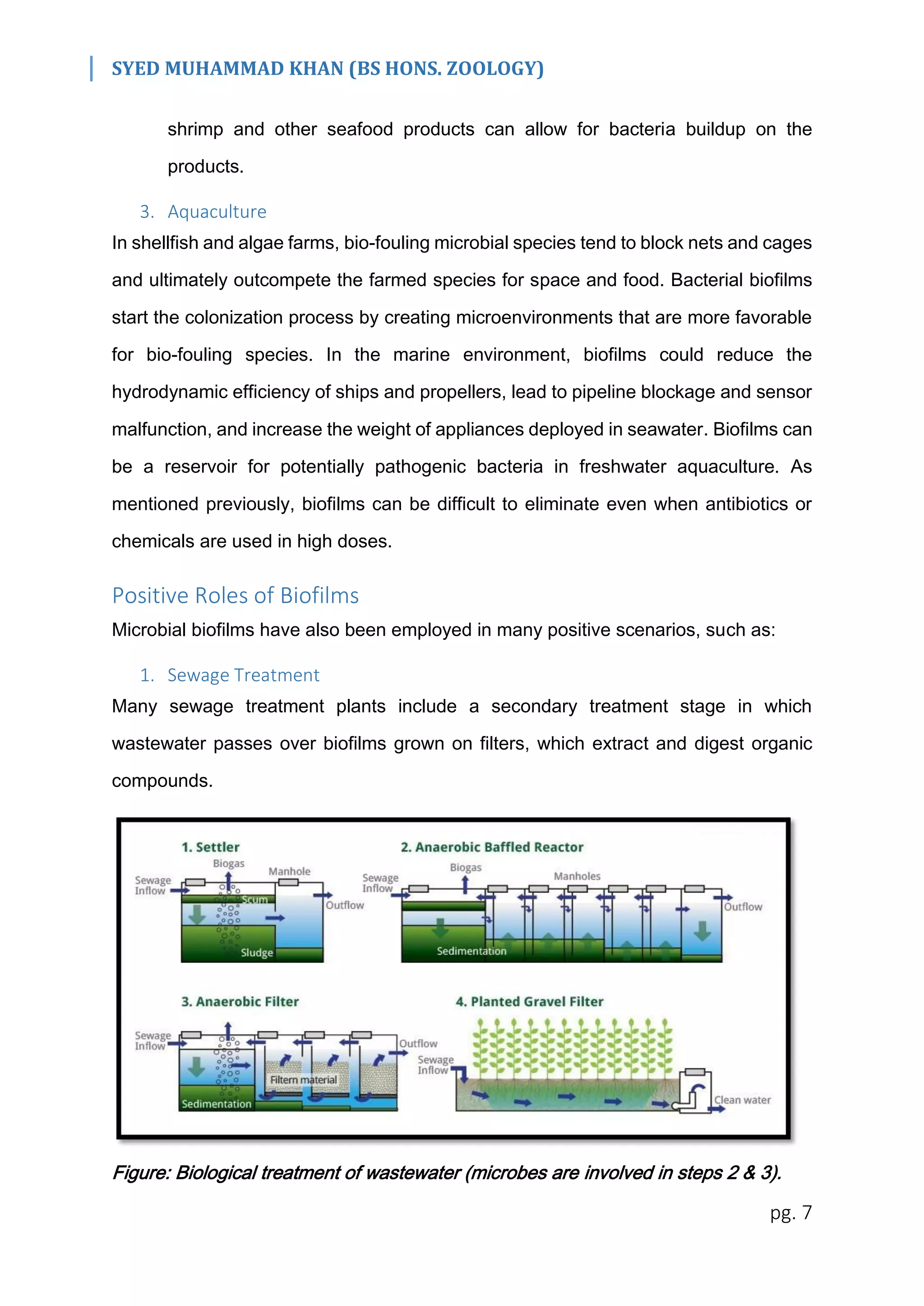
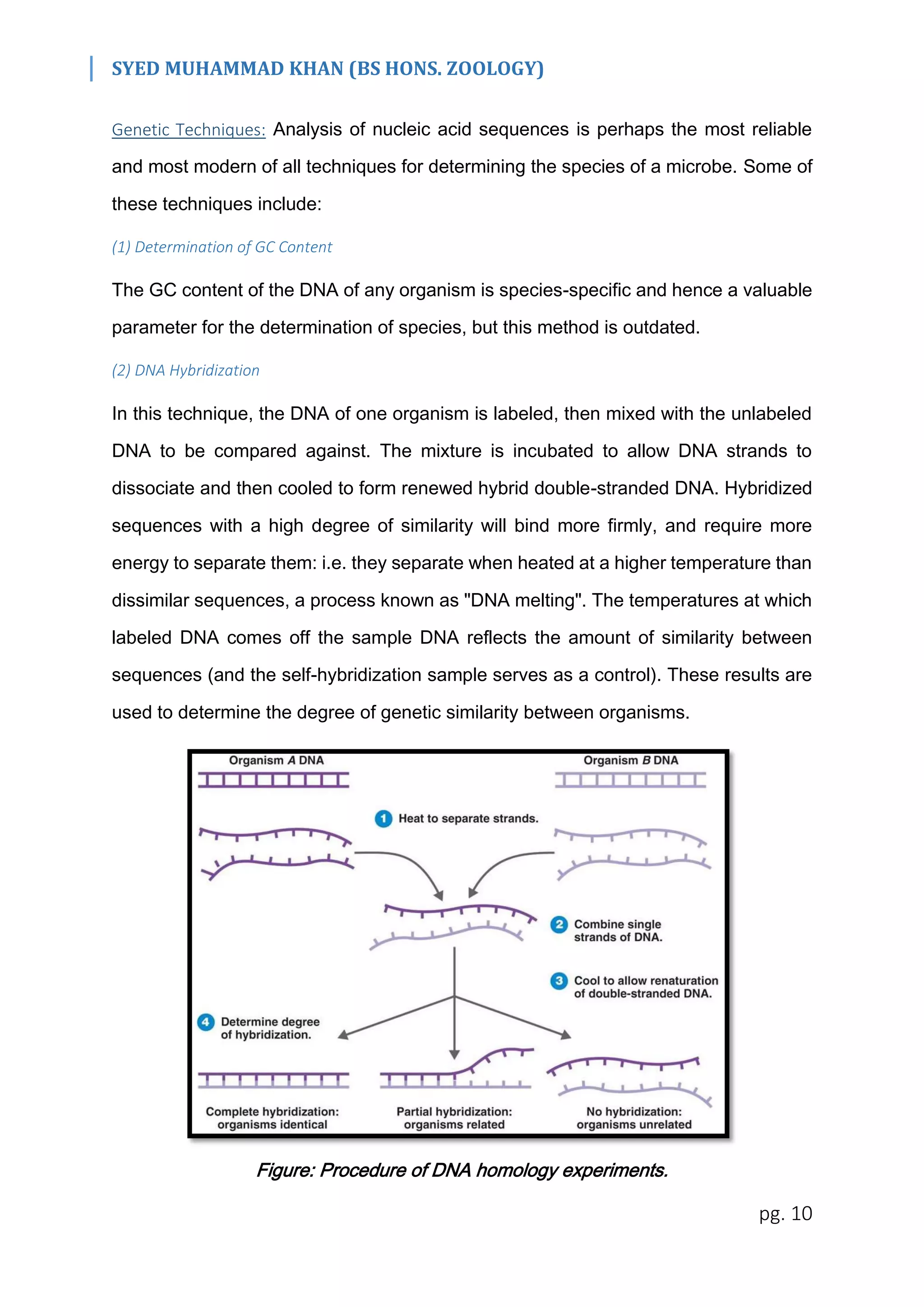
This document discusses microbial communities and biofilms. It begins by explaining that microbes thrive in diverse ecosystems under a range of conditions. Microbial communities are heterogeneous mixtures that interact. Biofilms provide advantages like nutrient sharing and protection. The document then discusses techniques to analyze microbial communities, including genetic methods. It covers positive and negative impacts of biofilms in areas like infections, food production, and wastewater treatment. Stress can impact microbial diversity by selecting certain organisms. Modern techniques allow direct analysis of constituent populations in communities.
Microbial interactions are ubiquitous, diverse, critically important in the function of any biological community.
The most common cooperative interactions seen in microbial systems are mutually beneficial. The interactions between the two populations are classified according to whether both populations and one of them benefit from the associations, or one or both populations are negatively affected.
•Introduction of bioremediation: Bioremediation refers to the process of using microorganisms to remove the environmental pollutants i.e. toxic wastes found in soil, water, air etc.
•In situ bioremediation:
It involves a direct approach for the microbial
degradation of xenobiotics at the sites of pollution
(soil, ground water).
•Types of in situ bioremediation:
Natural attenuation.
Engineered in situ bioremediation.
- Bioventing, biosparging, bioslurping,
phytoremediation.
•Ex situ bioremediation:
Waste or toxic pollutants can be collected from the polluted sites and bioremediation can be carried out at a designated place or site.
• Types of ex situ bioremediation
Land farming, windrow, biopiles, bioreactors.
•Microorganisms use in bioremediation:
A number of naturally occurring marine microbes
such as Pseudomonas sp. is capable of degrading oil and other hydrocarbons.
•Factors affecting bioremediation:
Nutrient availability, moisture content, pH, temperature, contaminant availability.
•References:
Satyanarayana U. Biotechnology. BOOKS AND ALLIED (P) Ltd.
Sharma P.D. Environmental Microbiology. RASTOGI PUBLICATIONS.
Gupta P.K. Biotechnology and Genomics. RASTOGI PUBLICATIONS.
Dubey R.C. A Textbook of Biotechnology. S Chand And Company Ltd.
Dubey R.C. A Textbook of Microbiology. S Chand And Company Ltd.
Willey/Sherwood/Woolverton. Prescott’s Microbiology. McGRAW-HILL INTERNATIONAL EDITION.
www.sciencedirect.com/bioremediation.
“Bioleaching" or "bio-oxidation" employs the use of naturally occurring bacteria, harmless to both humans and the environment, to extract of metals from their ores.
Conversion of insoluble metal sulfides into water-soluble metal sulfates.
It is mainly used to recover certain metals from sulfide ores. This is much cleaner than the traditional leaching.
Introduction to biofilm
Examples of biofilm
Form of biofilm
Discovery of biofilm
Properties of biofilm
Composition of biofilm
Formation of biofilm
Bacterial biofilm
Impact of biofilm
Problem caused by biofilm
Uses of biofilm
Antibiotic Tolerance/Resistance Of Bacterial Biofilms
Antibiofilm approach
Control strategies of Biofilm
Microorganisms, those minuscule entities that elude the naked eye, take centre stage in Class 8 Science Chapter 2, titled "Microorganisms: Friend and Foe." This chapter delves into the intricate world of these tiny beings, exploring their dual nature as both friends and foes, with profound implications for our environment, health, and daily life.
BIOFILMS_which cause the our theeth coatingummeed2024
it's ppt on biofims, which is a cause of our mouth, in this ppt we described about how that can cause, also what the reason we got biofilms, what pracuosan we have to take and how to take care for not happening it.
Microorganisms cause virtually all pathoses of the pulp and periapical tissues.
Once bacterial invasion of pulp tissues has taken place, both non-specific inflammation and specific immunologic response of the host have a profound effect on the progress of the disease.
Endodontic infection develops in root canals devoid of host defenses,
pulp necrosis (as a sequel to caries, trauma, periodontal disease,or iatrogenic operative procedures)
or pulp removal for treatment.
Biofilm-induced oral diseases.
ROUTES OF ROOT CANAL INFECTION
Caries
• Trauma-induced fractures
• Cracks
• Restorative procedures
• Scaling and root planing
• Attrition
• Abrasion
• Gaps in the cementoenamel junction
at the cervical root surface
• Dentinal tubules
• Direct pulp exposure
• Periodontal disease
• Anachoresis
Mechanisms of Microbial Pathogenicity and Virulence Factors
Pathogenicity : The ability of a microorganism to cause disease.
Virulence: Degree of pathogenicity of a microorganism.
Some microorganisms routinely cause disease in a given host and are called primary pathogens.
Other microorganisms cause disease only when host defenses are impaired and are called opportunistic pathogens by changing the balance of the host–bacteria relationship.
Bacterial strategies that contribute to pathogenicity include the ability to coaggregate and form biofilms.
In the pathogenesis of primary apical periodontitis
Bacteria in caries lesions form authentic biofilms adhered to dentin.
Diffusion of bacterial products through dentinal tubules induces pulpal inflammation
After pulp exposure, the exposed pulp tissue is in direct contact with bacteria and their products
and responds with severe inflammation. Some tissue invasion by bacteria may also occur.
Bacteria in the battlefront have to survive the attack from the host defenses and at the same time acquire nutrients to keep themselves alive.
In this bacteria–pulp clash, the latter invariably is “defeated” and becomes necrotic, so bacteria move forward and “occupy the territory”—that is, they colonize the necrotic tissue.
These events advance through tissue compartments, coalesce, and move toward the apical part of the canal until virtually the entire root canal is necrotic and infected.
At this stage, involved bacteria can be regarded as the early root canal colonizers or pioneer species (play an important role in the initiation of the apical periodontitis disease process, modify the environment, making it conducive to the establishment of other bacterial groups)
Slide 1: Title Slide
Extrachromosomal Inheritance
Slide 2: Introduction to Extrachromosomal Inheritance
Definition: Extrachromosomal inheritance refers to the transmission of genetic material that is not found within the nucleus.
Key Components: Involves genes located in mitochondria, chloroplasts, and plasmids.
Slide 3: Mitochondrial Inheritance
Mitochondria: Organelles responsible for energy production.
Mitochondrial DNA (mtDNA): Circular DNA molecule found in mitochondria.
Inheritance Pattern: Maternally inherited, meaning it is passed from mothers to all their offspring.
Diseases: Examples include Leber’s hereditary optic neuropathy (LHON) and mitochondrial myopathy.
Slide 4: Chloroplast Inheritance
Chloroplasts: Organelles responsible for photosynthesis in plants.
Chloroplast DNA (cpDNA): Circular DNA molecule found in chloroplasts.
Inheritance Pattern: Often maternally inherited in most plants, but can vary in some species.
Examples: Variegation in plants, where leaf color patterns are determined by chloroplast DNA.
Slide 5: Plasmid Inheritance
Plasmids: Small, circular DNA molecules found in bacteria and some eukaryotes.
Features: Can carry antibiotic resistance genes and can be transferred between cells through processes like conjugation.
Significance: Important in biotechnology for gene cloning and genetic engineering.
Slide 6: Mechanisms of Extrachromosomal Inheritance
Non-Mendelian Patterns: Do not follow Mendel’s laws of inheritance.
Cytoplasmic Segregation: During cell division, organelles like mitochondria and chloroplasts are randomly distributed to daughter cells.
Heteroplasmy: Presence of more than one type of organellar genome within a cell, leading to variation in expression.
Slide 7: Examples of Extrachromosomal Inheritance
Four O’clock Plant (Mirabilis jalapa): Shows variegated leaves due to different cpDNA in leaf cells.
Petite Mutants in Yeast: Result from mutations in mitochondrial DNA affecting respiration.
Slide 8: Importance of Extrachromosomal Inheritance
Evolution: Provides insight into the evolution of eukaryotic cells.
Medicine: Understanding mitochondrial inheritance helps in diagnosing and treating mitochondrial diseases.
Agriculture: Chloroplast inheritance can be used in plant breeding and genetic modification.
Slide 9: Recent Research and Advances
Gene Editing: Techniques like CRISPR-Cas9 are being used to edit mitochondrial and chloroplast DNA.
Therapies: Development of mitochondrial replacement therapy (MRT) for preventing mitochondrial diseases.
Slide 10: Conclusion
Summary: Extrachromosomal inheritance involves the transmission of genetic material outside the nucleus and plays a crucial role in genetics, medicine, and biotechnology.
Future Directions: Continued research and technological advancements hold promise for new treatments and applications.
Slide 11: Questions and Discussion
Invite Audience: Open the floor for any questions or further discussion on the topic.
Introduction:
RNA interference (RNAi) or Post-Transcriptional Gene Silencing (PTGS) is an important biological process for modulating eukaryotic gene expression.
It is highly conserved process of posttranscriptional gene silencing by which double stranded RNA (dsRNA) causes sequence-specific degradation of mRNA sequences.
dsRNA-induced gene silencing (RNAi) is reported in a wide range of eukaryotes ranging from worms, insects, mammals and plants.
This process mediates resistance to both endogenous parasitic and exogenous pathogenic nucleic acids, and regulates the expression of protein-coding genes.
What are small ncRNAs?
micro RNA (miRNA)
short interfering RNA (siRNA)
Properties of small non-coding RNA:
Involved in silencing mRNA transcripts.
Called “small” because they are usually only about 21-24 nucleotides long.
Synthesized by first cutting up longer precursor sequences (like the 61nt one that Lee discovered).
Silence an mRNA by base pairing with some sequence on the mRNA.
Discovery of siRNA?
The first small RNA:
In 1993 Rosalind Lee (Victor Ambros lab) was studying a non- coding gene in C. elegans, lin-4, that was involved in silencing of another gene, lin-14, at the appropriate time in the
development of the worm C. elegans.
Two small transcripts of lin-4 (22nt and 61nt) were found to be complementary to a sequence in the 3' UTR of lin-14.
Because lin-4 encoded no protein, she deduced that it must be these transcripts that are causing the silencing by RNA-RNA interactions.
Types of RNAi ( non coding RNA)
MiRNA
Length (23-25 nt)
Trans acting
Binds with target MRNA in mismatch
Translation inhibition
Si RNA
Length 21 nt.
Cis acting
Bind with target Mrna in perfect complementary sequence
Piwi-RNA
Length ; 25 to 36 nt.
Expressed in Germ Cells
Regulates trnasposomes activity
MECHANISM OF RNAI:
First the double-stranded RNA teams up with a protein complex named Dicer, which cuts the long RNA into short pieces.
Then another protein complex called RISC (RNA-induced silencing complex) discards one of the two RNA strands.
The RISC-docked, single-stranded RNA then pairs with the homologous mRNA and destroys it.
THE RISC COMPLEX:
RISC is large(>500kD) RNA multi- protein Binding complex which triggers MRNA degradation in response to MRNA
Unwinding of double stranded Si RNA by ATP independent Helicase
Active component of RISC is Ago proteins( ENDONUCLEASE) which cleave target MRNA.
DICER: endonuclease (RNase Family III)
Argonaute: Central Component of the RNA-Induced Silencing Complex (RISC)
One strand of the dsRNA produced by Dicer is retained in the RISC complex in association with Argonaute
ARGONAUTE PROTEIN :
1.PAZ(PIWI/Argonaute/ Zwille)- Recognition of target MRNA
2.PIWI (p-element induced wimpy Testis)- breaks Phosphodiester bond of mRNA.)RNAse H activity.
MiRNA:
The Double-stranded RNAs are naturally produced in eukaryotic cells during development, and they have a key role in regulating gene expression .
Nutraceutical market, scope and growth: Herbal drug technologyLokesh Patil
As consumer awareness of health and wellness rises, the nutraceutical market—which includes goods like functional meals, drinks, and dietary supplements that provide health advantages beyond basic nutrition—is growing significantly. As healthcare expenses rise, the population ages, and people want natural and preventative health solutions more and more, this industry is increasing quickly. Further driving market expansion are product formulation innovations and the use of cutting-edge technology for customized nutrition. With its worldwide reach, the nutraceutical industry is expected to keep growing and provide significant chances for research and investment in a number of categories, including vitamins, minerals, probiotics, and herbal supplements.
Multi-source connectivity as the driver of solar wind variability in the heli...Sérgio Sacani
The ambient solar wind that flls the heliosphere originates from multiple
sources in the solar corona and is highly structured. It is often described
as high-speed, relatively homogeneous, plasma streams from coronal
holes and slow-speed, highly variable, streams whose source regions are
under debate. A key goal of ESA/NASA’s Solar Orbiter mission is to identify
solar wind sources and understand what drives the complexity seen in the
heliosphere. By combining magnetic feld modelling and spectroscopic
techniques with high-resolution observations and measurements, we show
that the solar wind variability detected in situ by Solar Orbiter in March
2022 is driven by spatio-temporal changes in the magnetic connectivity to
multiple sources in the solar atmosphere. The magnetic feld footpoints
connected to the spacecraft moved from the boundaries of a coronal hole
to one active region (12961) and then across to another region (12957). This
is refected in the in situ measurements, which show the transition from fast
to highly Alfvénic then to slow solar wind that is disrupted by the arrival of
a coronal mass ejection. Our results describe solar wind variability at 0.5 au
but are applicable to near-Earth observatories.
Deep Behavioral Phenotyping in Systems Neuroscience for Functional Atlasing a...Ana Luísa Pinho
Functional Magnetic Resonance Imaging (fMRI) provides means to characterize brain activations in response to behavior. However, cognitive neuroscience has been limited to group-level effects referring to the performance of specific tasks. To obtain the functional profile of elementary cognitive mechanisms, the combination of brain responses to many tasks is required. Yet, to date, both structural atlases and parcellation-based activations do not fully account for cognitive function and still present several limitations. Further, they do not adapt overall to individual characteristics. In this talk, I will give an account of deep-behavioral phenotyping strategies, namely data-driven methods in large task-fMRI datasets, to optimize functional brain-data collection and improve inference of effects-of-interest related to mental processes. Key to this approach is the employment of fast multi-functional paradigms rich on features that can be well parametrized and, consequently, facilitate the creation of psycho-physiological constructs to be modelled with imaging data. Particular emphasis will be given to music stimuli when studying high-order cognitive mechanisms, due to their ecological nature and quality to enable complex behavior compounded by discrete entities. I will also discuss how deep-behavioral phenotyping and individualized models applied to neuroimaging data can better account for the subject-specific organization of domain-general cognitive systems in the human brain. Finally, the accumulation of functional brain signatures brings the possibility to clarify relationships among tasks and create a univocal link between brain systems and mental functions through: (1) the development of ontologies proposing an organization of cognitive processes; and (2) brain-network taxonomies describing functional specialization. To this end, tools to improve commensurability in cognitive science are necessary, such as public repositories, ontology-based platforms and automated meta-analysis tools. I will thus discuss some brain-atlasing resources currently under development, and their applicability in cognitive as well as clinical neuroscience.
(May 29th, 2024) Advancements in Intravital Microscopy- Insights for Preclini...Scintica Instrumentation
Intravital microscopy (IVM) is a powerful tool utilized to study cellular behavior over time and space in vivo. Much of our understanding of cell biology has been accomplished using various in vitro and ex vivo methods; however, these studies do not necessarily reflect the natural dynamics of biological processes. Unlike traditional cell culture or fixed tissue imaging, IVM allows for the ultra-fast high-resolution imaging of cellular processes over time and space and were studied in its natural environment. Real-time visualization of biological processes in the context of an intact organism helps maintain physiological relevance and provide insights into the progression of disease, response to treatments or developmental processes.
In this webinar we give an overview of advanced applications of the IVM system in preclinical research. IVIM technology is a provider of all-in-one intravital microscopy systems and solutions optimized for in vivo imaging of live animal models at sub-micron resolution. The system’s unique features and user-friendly software enables researchers to probe fast dynamic biological processes such as immune cell tracking, cell-cell interaction as well as vascularization and tumor metastasis with exceptional detail. This webinar will also give an overview of IVM being utilized in drug development, offering a view into the intricate interaction between drugs/nanoparticles and tissues in vivo and allows for the evaluation of therapeutic intervention in a variety of tissues and organs. This interdisciplinary collaboration continues to drive the advancements of novel therapeutic strategies.
Cancer cell metabolism: special Reference to Lactate PathwayAADYARAJPANDEY1
Normal Cell Metabolism:
Cellular respiration describes the series of steps that cells use to break down sugar and other chemicals to get the energy we need to function.
Energy is stored in the bonds of glucose and when glucose is broken down, much of that energy is released.
Cell utilize energy in the form of ATP.
The first step of respiration is called glycolysis. In a series of steps, glycolysis breaks glucose into two smaller molecules - a chemical called pyruvate. A small amount of ATP is formed during this process.
Most healthy cells continue the breakdown in a second process, called the Kreb's cycle. The Kreb's cycle allows cells to “burn” the pyruvates made in glycolysis to get more ATP.
The last step in the breakdown of glucose is called oxidative phosphorylation (Ox-Phos).
It takes place in specialized cell structures called mitochondria. This process produces a large amount of ATP. Importantly, cells need oxygen to complete oxidative phosphorylation.
If a cell completes only glycolysis, only 2 molecules of ATP are made per glucose. However, if the cell completes the entire respiration process (glycolysis - Kreb's - oxidative phosphorylation), about 36 molecules of ATP are created, giving it much more energy to use.
IN CANCER CELL:
Unlike healthy cells that "burn" the entire molecule of sugar to capture a large amount of energy as ATP, cancer cells are wasteful.
Cancer cells only partially break down sugar molecules. They overuse the first step of respiration, glycolysis. They frequently do not complete the second step, oxidative phosphorylation.
This results in only 2 molecules of ATP per each glucose molecule instead of the 36 or so ATPs healthy cells gain. As a result, cancer cells need to use a lot more sugar molecules to get enough energy to survive.
introduction to WARBERG PHENOMENA:
WARBURG EFFECT Usually, cancer cells are highly glycolytic (glucose addiction) and take up more glucose than do normal cells from outside.
Otto Heinrich Warburg (; 8 October 1883 – 1 August 1970) In 1931 was awarded the Nobel Prize in Physiology for his "discovery of the nature and mode of action of the respiratory enzyme.
WARNBURG EFFECT : cancer cells under aerobic (well-oxygenated) conditions to metabolize glucose to lactate (aerobic glycolysis) is known as the Warburg effect. Warburg made the observation that tumor slices consume glucose and secrete lactate at a higher rate than normal tissues.
Seminar of U.V. Spectroscopy by SAMIR PANDASAMIR PANDA
Spectroscopy is a branch of science dealing the study of interaction of electromagnetic radiation with matter.
Ultraviolet-visible spectroscopy refers to absorption spectroscopy or reflect spectroscopy in the UV-VIS spectral region.
Ultraviolet-visible spectroscopy is an analytical method that can measure the amount of light received by the analyte.
Earliest Galaxies in the JADES Origins Field: Luminosity Function and Cosmic ...Sérgio Sacani
We characterize the earliest galaxy population in the JADES Origins Field (JOF), the deepest
imaging field observed with JWST. We make use of the ancillary Hubble optical images (5 filters
spanning 0.4−0.9µm) and novel JWST images with 14 filters spanning 0.8−5µm, including 7 mediumband filters, and reaching total exposure times of up to 46 hours per filter. We combine all our data
at > 2.3µm to construct an ultradeep image, reaching as deep as ≈ 31.4 AB mag in the stack and
30.3-31.0 AB mag (5σ, r = 0.1” circular aperture) in individual filters. We measure photometric
redshifts and use robust selection criteria to identify a sample of eight galaxy candidates at redshifts
z = 11.5 − 15. These objects show compact half-light radii of R1/2 ∼ 50 − 200pc, stellar masses of
M⋆ ∼ 107−108M⊙, and star-formation rates of SFR ∼ 0.1−1 M⊙ yr−1
. Our search finds no candidates
at 15 < z < 20, placing upper limits at these redshifts. We develop a forward modeling approach to
infer the properties of the evolving luminosity function without binning in redshift or luminosity that
marginalizes over the photometric redshift uncertainty of our candidate galaxies and incorporates the
impact of non-detections. We find a z = 12 luminosity function in good agreement with prior results,
and that the luminosity function normalization and UV luminosity density decline by a factor of ∼ 2.5
from z = 12 to z = 14. We discuss the possible implications of our results in the context of theoretical
models for evolution of the dark matter halo mass function.
Richard's entangled aventures in wonderlandRichard Gill
Since the loophole-free Bell experiments of 2020 and the Nobel prizes in physics of 2022, critics of Bell's work have retreated to the fortress of super-determinism. Now, super-determinism is a derogatory word - it just means "determinism". Palmer, Hance and Hossenfelder argue that quantum mechanics and determinism are not incompatible, using a sophisticated mathematical construction based on a subtle thinning of allowed states and measurements in quantum mechanics, such that what is left appears to make Bell's argument fail, without altering the empirical predictions of quantum mechanics. I think however that it is a smoke screen, and the slogan "lost in math" comes to my mind. I will discuss some other recent disproofs of Bell's theorem using the language of causality based on causal graphs. Causal thinking is also central to law and justice. I will mention surprising connections to my work on serial killer nurse cases, in particular the Dutch case of Lucia de Berk and the current UK case of Lucy Letby.
1. SYED MUHAMMAD KHAN (BS HONS. ZOOLOGY)
pg. 1
Nature of Microbial Communities
All natural ecosystems host a diverse array of microorganisms. Some of these
microbes tend to thrive under moderate living conditions (i.e. moderate temperature,
tolerable pH, ample nutrient supply, etc.) while there are others, known as
extremophiles, which “love” extreme conditions such as high acidity or alkalinity,
extremely high or low temperatures, high pressures, etc.
These organisms flourish under conditions that are normally fatal to most other
organisms. Hence it is without doubt that every inch of the planet Earth is inhabited by
microorganism communities, from the highest mountain summits to the abyssal depths
of the oceans, from the freezing cold of the Polar Regions to the boiling hot water
springs, from simple freshwater bodies to hypersaline environments.
But these microbial communities do not live apart from one another, as previously
thought, modern research indicates that these populations intermingle in the
environment to produce a heterogeneous mixture.
Spatial Organization
The distribution of microbes in the environment is mostly affected by certain
physiological gradients, i.e. temperature, pH, nutrient availability, chemicals released
by other microbes, etc. These gradients may be vertical or horizontal or both at once,
and hence affect the distribution likewise.
The result is a heterogeneous mixture of microbial species, with each constituent
population being present in the range most favorable for it. But they are in no way
isolated, since many microbes depend upon one another due to symbiotic
relationships, their populations tend to overlap.
In fact in small-sized "micro" environments, even microbes with conflicting interests
may be pinned close together. There is a very high degree of organization of microbial
communities, which appears to integrate the growth conditions and the species
2. SYED MUHAMMAD KHAN (BS HONS. ZOOLOGY)
pg. 2
composition in such a way that resources available in the environment are used
effectively (at an optimum rate).
Biofilms
Biofilm is an aggregate of microorganisms in which cells that are frequently embedded
within a self-produced matrix of extracellular polymeric substances adhere to each
other and/or to a surface.
Extracellular Polymeric Substances
The cells within the biofilm produce the extracellular polymeric substances (EPS)
components, which are typically a polymeric conglomeration of extracellular
polysaccharides, proteins, lipids, DNA, and mostly water. This matrix makes up around
50 – 95% of the dry weight of any biofilm.
Figure: Structure of a microbial biofilm.
Advantages of Biofilms to Microbes
Spatial organization of microbes in biofilms and similar situations grants them many of
the advantages of multicellular life such as co-metabolism, cross-feeding, increased
3. SYED MUHAMMAD KHAN (BS HONS. ZOOLOGY)
pg. 3
resilience to stress (i.e. electromagnetic radiations such as UV, heat shocks, pH
fluctuations, inhibitory substances, and most importantly dehydration – owing to the
large amount of water in the matrix). Biofilms are not just bacterial slime layers but
biological systems; the bacteria organize themselves into a coordinated functional
community. Biofilms can attach to a surface such as a tooth, rock, or surface, and may
include a single species or a diverse group of microorganisms. The biofilm bacteria
can share nutrients and are sheltered from harmful factors in the environment, such
as desiccation, antibiotics, and a host body's immune system.
Figure: A summary of how members of a microbial community in a biofilm (analogous
to a city) receive several advantages.
Effect of environment on biofilms
Biofilms may form on living or non-living surfaces and can be prevalent in natural,
industrial, and hospital settings. The microbial cells growing in a biofilm are
physiologically distinct from planktonic cells of the same organism (planktonic cells are
single-cells that may float or swim in a liquid medium). The thickness of the biofilm
varies, depending upon the environmental conditions, i.e. biofilms in nutrient-deficient
environments tend to be only a few micrometers thick whereas soil crumbs, microbial
mats, sewage flocs, and biofilms in nutrient-rich environment are comparatively much
4. SYED MUHAMMAD KHAN (BS HONS. ZOOLOGY)
pg. 4
thicker. Microbes form a biofilm in response to various factors, which may include
cellular recognition of specific or non-specific attachment sites on a surface, nutritional
cues, or in some cases, by exposure of planktonic cells to sub-inhibitory
concentrations of antibiotics.
Formation of a Biofilm
Biofilms are the product of a microbial developmental process. The process of biofilm
formation consists of the following stages:
1. Initial Attachment
The formation of a biofilm begins with the attachment of free-floating microorganisms
to a surface. The first colonist bacteria of a biofilm may simply adhere to the surface
due to physical factors such as van der Waal’s forces, hydrophobic effects, etc.
2. Irreversible Attachment
If the colonists are not immediately separated from the surface, they can anchor
themselves more permanently using cell adhesion structures such as pili. Some
bacteria species are not able to attach to a surface on their success due to their limited
motility but are instead able to anchor themselves to the matrix or directly to other,
earlier bacterial colonists. Non-motile bacteria cannot recognize surfaces or aggregate
together as easily as motile bacteria.
3. Maturation
Once the colonization has begun, the biofilm grows by a combination of cell division
and recruitment. Polysaccharide matrices typically enclose bacterial biofilms. In
addition to the polysaccharides, these matrices may also contain material from the
surrounding environment, including but not limited to minerals, soil particles, and blood
components, such as erythrocytes and fibrin.
4. Dispersion
The final stage of biofilm formation is known as dispersion and is the stage in which
the biofilm is established and may only change in shape and size. Dispersal enables
biofilms to spread and colonize new surfaces. Enzymes that degrade the biofilm
5. SYED MUHAMMAD KHAN (BS HONS. ZOOLOGY)
pg. 5
extracellular matrix may contribute to biofilm dispersal, they may also be useful as
anti-biofilm agents. Cells dispersed from biofilms immediately go into the planktonic
growth phase. Hence, the dispersal process is a unique stage during the transition
from biofilm to planktonic lifestyle in bacteria.
Figure: Steps involved in biofilm formation.
Negative Impacts of Biofilms
Microbial biofilms have several negative impacts on the environment and directly on
humans as well, some of them are:
1. Infections
Biofilms are involved in a wide variety of microbial infections in the body, by one
estimate 80% of all infections. Infectious processes in which biofilms have been
implicated include common problems such as bacterial vaginosis, urinary tract
infections, catheter infections, middle-ear infections, formation of dental plaque,
gingivitis, coating contact lenses, and less common but more lethal processes such
as endocarditis, infections in cystic fibrosis, and infections of permanent indwelling
devices such as joint prostheses, heart valves, and intervertebral disc.
6. SYED MUHAMMAD KHAN (BS HONS. ZOOLOGY)
pg. 6
Infections associated with the biofilm growth usually are challenging to eradicate. This
is mostly because mature biofilms display tolerance towards antibiotics and the
immune response. Microbial infections can develop on all medical devices and tissue
engineering constructs. 60 to 70% of nosocomial or hospital-acquired infections are
associated with the implantation of a biomedical device.
2. Food Industry
Biofilms have become problematic in several food industries due to the ability to form
on plants and during industrial processes. Along with economic problems, biofilm
formation on food poses a health risk to consumers due to the ability to make the food
more resistant to disinfectants. During the washing process, biofilms resist sanitization
and allow bacteria to spread across the product. This problem is also found in ready-
to-eat foods because the foods go through limited cleaning procedures before
consumption.
Dairy Products: Due to the perishability of dairy products and limitations in
cleaning procedures, resulting in the buildup of bacteria, dairy is susceptible to
biofilm formation and contamination. The bacteria can spoil the products more
readily and contaminated products pose a health risk to consumers.
Poultry (Salmonella Infestation): One species of bacteria that can be found in
various industries and is a major cause of foodborne disease is Salmonella spp.
Large amounts of Salmonella contamination can be found in the poultry
processing industry as about 50% of Salmonella strains can produce biofilms
on poultry farms. Salmonella increases the risk of foodborne illnesses when the
poultry products are not cleaned and cooked correctly.
Seafood (Salmonella Infestation): Salmonella is also found in the seafood industry
where biofilms form from seafood borne pathogens on the seafood itself as well
as in water. Shrimp products are commonly affected by Salmonella because of
unhygienic processing and handling techniques. The preparation practices of
7. SYED MUHAMMAD KHAN (BS HONS. ZOOLOGY)
pg. 7
shrimp and other seafood products can allow for bacteria buildup on the
products.
3. Aquaculture
In shellfish and algae farms, bio-fouling microbial species tend to block nets and cages
and ultimately outcompete the farmed species for space and food. Bacterial biofilms
start the colonization process by creating microenvironments that are more favorable
for bio-fouling species. In the marine environment, biofilms could reduce the
hydrodynamic efficiency of ships and propellers, lead to pipeline blockage and sensor
malfunction, and increase the weight of appliances deployed in seawater. Biofilms can
be a reservoir for potentially pathogenic bacteria in freshwater aquaculture. As
mentioned previously, biofilms can be difficult to eliminate even when antibiotics or
chemicals are used in high doses.
Positive Roles of Biofilms
Microbial biofilms have also been employed in many positive scenarios, such as:
1. Sewage Treatment
Many sewage treatment plants include a secondary treatment stage in which
wastewater passes over biofilms grown on filters, which extract and digest organic
compounds.
Figure: Biological treatment of wastewater (microbes are involved in steps 2 & 3).
8. SYED MUHAMMAD KHAN (BS HONS. ZOOLOGY)
pg. 8
In such biofilms, bacteria are mainly responsible for the removal of organic matter,
while protozoa and rotifers are mainly responsible for the removal of suspended solids,
including pathogens and other microorganisms. Slow sand filters rely on biofilm
development in the same way to filter surface water from lake, spring, or river sources
for drinking purposes.
2. Removal of Petroleum from Marine Waters
Biofilms can help eliminate petroleum oil from contaminated oceans or marine
systems. The oil is eliminated by the hydrocarbon-degrading activities of microbial
communities.
3. Microbial Fuel Cells
Biofilms are used in microbial fuel cells to generate electricity from a variety of starting
materials, including complex organic waste and renewable biomass.
4. Bioleaching
Biofilms are also relevant for the improvement of metal dissolution in the bioleaching
industry. Bioleaching is the extraction of metals from their ores through the use of living
organisms. This is much cleaner than the traditional heap leaching using cyanide.
Bioleaching is one of several applications within biohydrometallurgy and several
methods are used to recover copper, zinc, lead, arsenic, antimony, nickel,
molybdenum, gold, silver, and cobalt.
Effect of Stress on Microbial Communities
Stress is an abiotic factor or a set of factors that limit the production of biomass. Stress
factors such as temperature variations, pH fluctuations, changes in nutrient availability
and water supply have a direct effect on the growth of microbes and the diversity of
microbial communities on a whole (as many species might be wiped out altogether
from an area because of their inability to resist such changes).
The degree of effectiveness of any stress factor is determined by three parameters:
(1) nature of the factor, (2) extent of damage done by the factor, and (3) rapidity of
application.
9. SYED MUHAMMAD KHAN (BS HONS. ZOOLOGY)
pg. 9
If the stress factor is applied slowly, the microbes gain an opportunity to adapt (within
certain limits) to that factor, this may lead to the formation of a much more resilient
population than before. Stress may also provide a selective advantage to some
microbes, at the expense of others, for instance, extremophiles such as thermophiles
will be able to resist heat shocks, while all of their competitors (mesophiles) will be
wiped out. Alternatively, endospore-forming bacteria will be able to repopulate an
environment, after it has been cleared of all viable cells, owing to the germination of
the endospores. In many natural environments, cyanobacteria are among the first
organisms to recolonize barren areas, hence making them pioneers in ecological
succession.
Determination of the Structure & Diversity of Constituent Populations
The constituent populations of a microbial community are hard to determine via
conventional methods such as culturing them on nutrient media. These media fail to
emulate the conditions of the natural environment for the microbes, for instance, they
are mostly rich in nutrients, and hence only the copiotrophs from the sample will grow
properly whereas the oligotrophs will be inhibited simply by the high concentration of
nutrients. Similarly, no single medium is suitable for growing all microbial species.
1. Modern Techniques
Modern techniques have allowed to take a direct peek into the microbial communities
and hence determine the constituent populations, these techniques include:
Microscopy: Direct microscopic analysis, owing to the modern advances in
microscopes and microscopic techniques have allowed the diagnosis of many
microbial species based on their morphology (distinct features, such as trichomes and
akinetes of cyanobacteria).
Analysis of Cellular Constituents: Various cellular constituents such as types of fatty acids
are distinct for each species and if they are detected in a culture then the presence of
that particular species (or at least that genus) can be confirmed.
10. SYED MUHAMMAD KHAN (BS HONS. ZOOLOGY)
pg. 10
Genetic Techniques: Analysis of nucleic acid sequences is perhaps the most reliable
and most modern of all techniques for determining the species of a microbe. Some of
these techniques include:
(1) Determination of GC Content
The GC content of the DNA of any organism is species-specific and hence a valuable
parameter for the determination of species, but this method is outdated.
(2) DNA Hybridization
In this technique, the DNA of one organism is labeled, then mixed with the unlabeled
DNA to be compared against. The mixture is incubated to allow DNA strands to
dissociate and then cooled to form renewed hybrid double-stranded DNA. Hybridized
sequences with a high degree of similarity will bind more firmly, and require more
energy to separate them: i.e. they separate when heated at a higher temperature than
dissimilar sequences, a process known as "DNA melting". The temperatures at which
labeled DNA comes off the sample DNA reflects the amount of similarity between
sequences (and the self-hybridization sample serves as a control). These results are
used to determine the degree of genetic similarity between organisms.
Figure: Procedure of DNA homology experiments.
11. SYED MUHAMMAD KHAN (BS HONS. ZOOLOGY)
pg. 11
(3) PCR
Polymerase chain reaction has allowed us to amplify (not merely replicate, amplify to
millions of copies) a DNA sample so that it can be analyzed via gel electrophoresis
later on and be compared with the known sequence of a known species to judge the
degree of similarity.
(4) Hybridization Probes
Within the field of microbial ecology, oligonucleotide probes are used to determine the
presence of microbial species, genera, or microorganisms classified on a more broad
level, such as bacteria, archaea, and eukaryotes via fluorescence in situ hybridization
(FISH) – Fluorescence in situ hybridization (FISH) is a molecular cytogenetic
technique that uses fluorescent probes that bind to only those parts of a nucleic acid
sequence with a high degree of sequence complementarity.
(5) 16S & 23S rRNA sequencing
The 16S rRNA gene is used for phylogenetic studies as it is highly conserved between
different species of bacteria and archaea. It is suggested that the 16S rRNA gene can
be used as a reliable molecular clock because 16S rRNA sequences from distantly
related bacterial lineages are shown to have similar functionalities. In some instances,
differentiation between species may be problematic when using 16S rRNA sequences
due to similarity. In such instances, 23S rRNA may be a better alternative. The global
standard library of rRNA sequences is constantly becoming larger and continuously
being updated.
Some Constituent Populations of Biofilms
Biofilms are formed by both prokaryotes and eukaryotes, modern techniques have
allowed us to identify many of these constituent populations:
1. Prokaryotic Biofilms
Many different bacteria form biofilms, including gram-positive (e.g. Bacillus spp.,
Listeria monocytogenes, Staphylococcus spp., and lactic acid bacteria, including
12. SYED MUHAMMAD KHAN (BS HONS. ZOOLOGY)
pg. 12
Lactobacillus plantarum and Lactococcus lactis) and gram-negative species (e.g.
Escherichia coli, or Pseudomonas aeruginosa). Cyanobacteria also form biofilms in
aquatic environments.
Biofilms are also formed by bacteria that colonize plants, i.e. Pseudomonas putida,
Pseudomonas fluorescens, and related pseudomonads which are common plant-
associated bacteria found on leaves, roots, and in the soil, and the majority of their
natural isolates form biofilms. Several nitrogen-fixing symbionts of legumes such as
Rhizobium leguminosarum and Sinorhizobium meliloti form biofilms on legume roots
and other inert surfaces. Many bacterial species which cause diseases in animals such
as Streptococcus pneumoniae (causes community-acquired pneumonia and
meningitis in children and the elderly, and sepsis in HIV-infected persons),
Pseudomonas aeroginosa (causes chronic wounds, chronic otitis media, chronic
prostatitis, and chronic lung infections in cystic fibrosis patients), etc. also form
biofilms.
2. Eukaryotic Biofilms
Along with bacteria, biofilms are often initiated and produced by eukaryotic microbes.
The biofilms produced by eukaryotes are usually occupied by bacteria and other
eukaryotes alike, however the surface is cultivated and EPS is secreted initially by
eukaryotes. Both fungi and microalgae are known to form biofilms in such a way.
Biofilms of fungal origin are important aspects of human infection and fungal
pathogenicity, as the fungal infection is more resistant to antifungals.
In the environment, fungal biofilms are an area of ongoing research. One key area of
research is fungal biofilms on plants. For example, in the soil, plant-associated fungi
including mycorrhiza have been shown to decompose organic matter and protect
plants from bacterial pathogens.
Biofilms in aquatic environments are often founded by diatoms. The exact purpose of
these biofilms is unknown, however, there is evidence that the EPS produced by
diatoms helps them resist both cold and salinity stresses. These eukaryotes interact
13. SYED MUHAMMAD KHAN (BS HONS. ZOOLOGY)
pg. 13
with a diverse range of other organisms within a region known as the phycosphere
(phycosphere is a microscale mucus region that is rich in organic matter and surrounds
a phytoplankton cell), but importantly are the bacteria associated with diatoms, as it
has been shown that although diatoms excrete EPS, they only do so when interacting
with certain bacterial species.