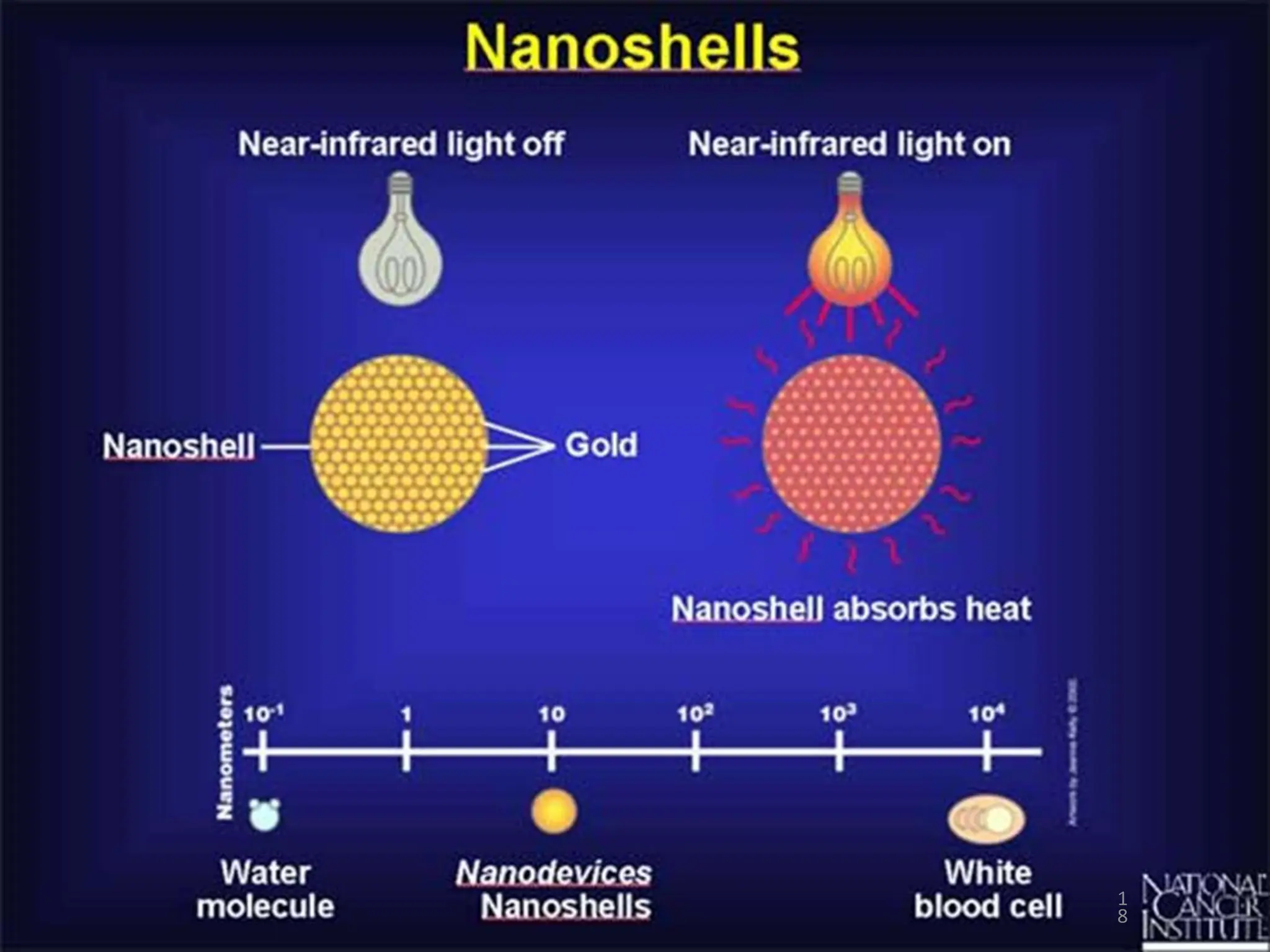
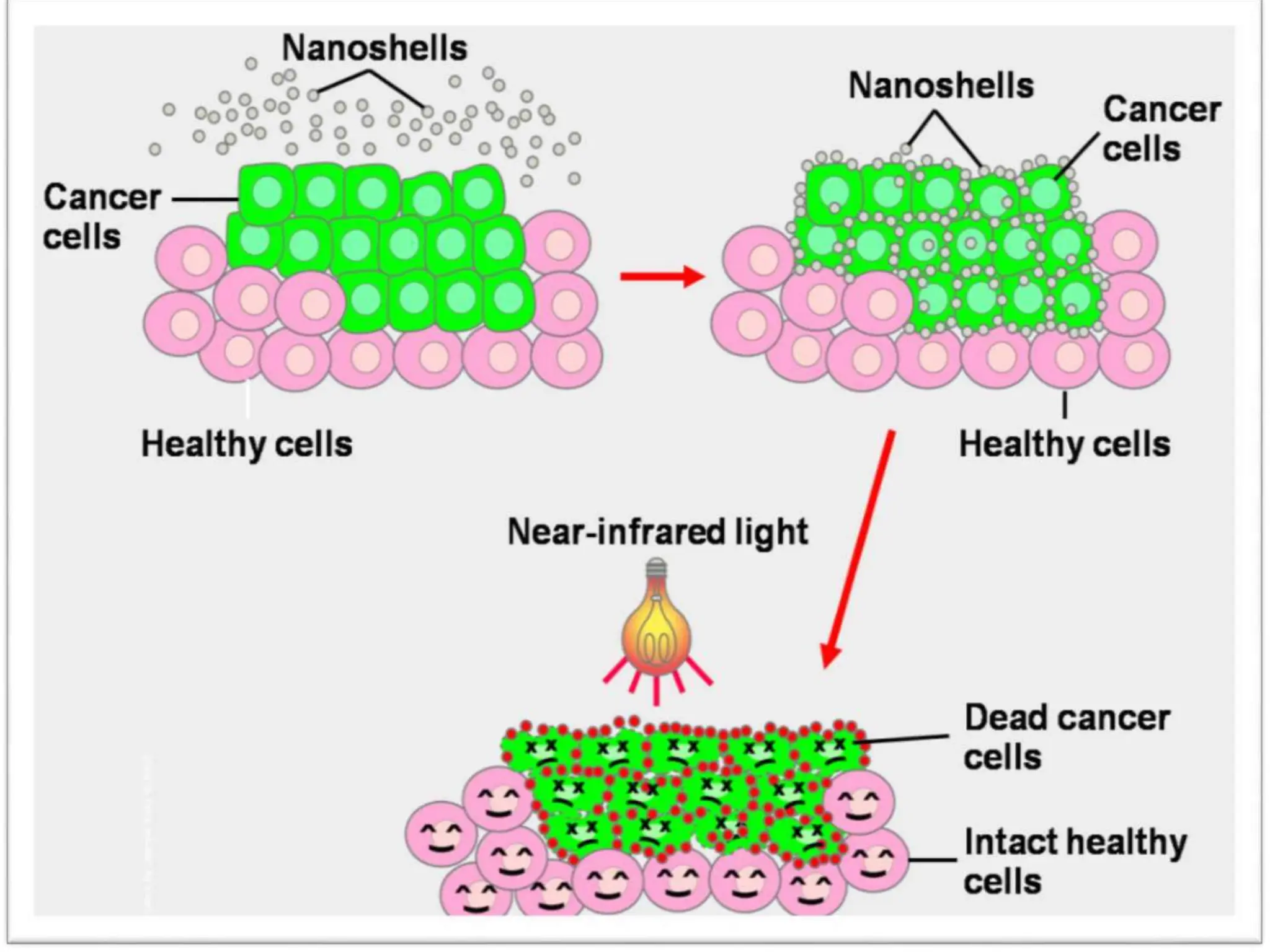
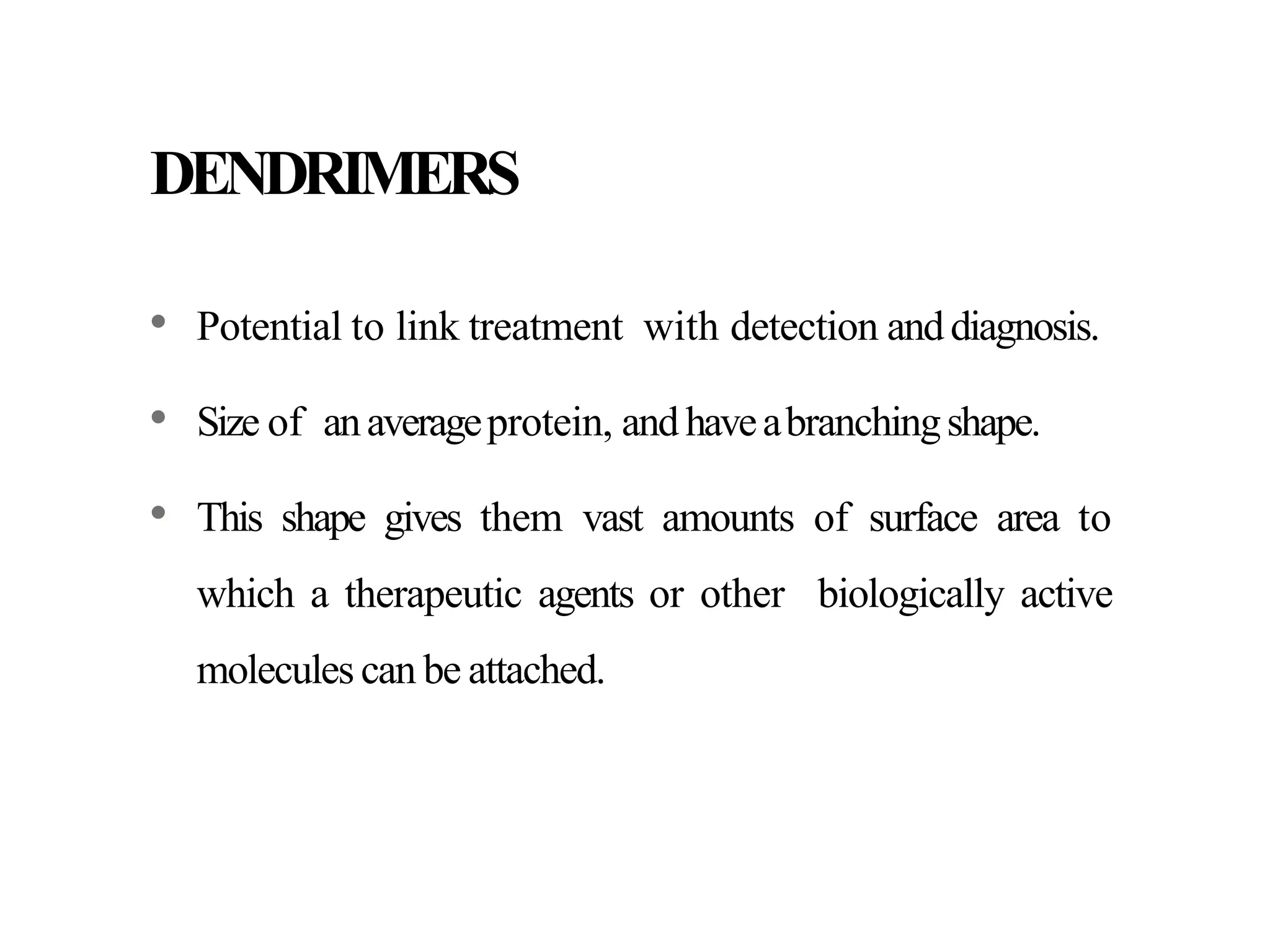
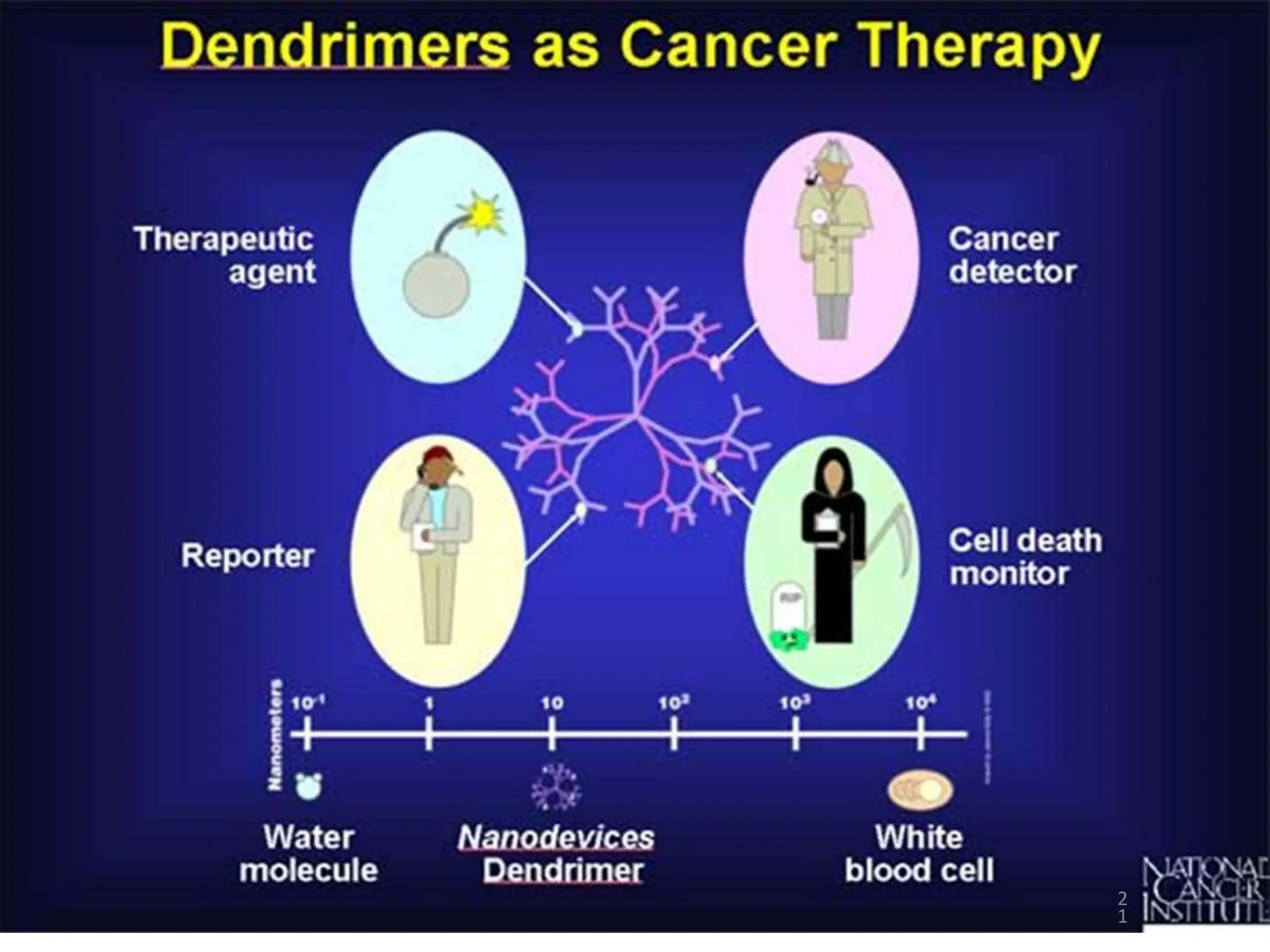
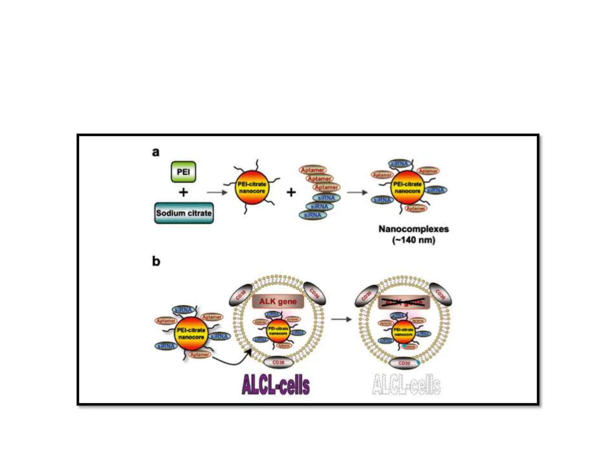
Nanotechnology
and it's applications in
haematological
malignancies
Presented by
Sonali Dixit
Moderator
Dr. Meera Sikka
What is Nanotechnology?
Definition
• Nanotechnology is the study of
manipulating matter on an
atomic scale.
• Nanotechnology refers to the
constructing and engineering of
the functional systems at very
micro level or we can say at
atomic level.
• A Nanometer is one billionth of
a meter, roughly the width of
three or four atoms. The average
human hair is about 25,000
nanometers wide.
Nanotechnology is defined by the
National Nanotechnology Initiative
as
“The understanding and control of
matter at dimensions between
approximately 1 and 100 nm, where
unique phenomena enable novel
applications”
History
• The first ever concept was presented in 1959
by the famous professor of physics Dr.
Richard P.Feynman.
• Invention of the scanning tunneling
microscope in 1981 and the discovery of
fullerene(C60) in 1985 lead to the emergence
of nanotechnology.
• The term “Nano-technology" had been
coined by Norio Taniguchi in 1974
Nanomaterials formedicaldiagnosis
 Nanomaterials exhibit higher chemical reactivity, increased mechanical strength, faster
electrical and magneticresponsesowingto its highsurfaceto unit volume ratio.
 Nanoparticles can attach to biomolecules, allowing detection of disease biomarkers in
a lab sample at a very early stage.
 Because of their small size,nanomaterials canreadily interact with biomoleculesand
gaining access to so many areas of the human body by passing through intracellular
spaces.
 Medical application of nanotechnology has ability to enable early detection,
prevention, treatment and follow up of many life-threatening diseases including
cancer, cardiovascular disease, diabetes, Alzheimer’s and AIDS as well as infectious
diseases.
Application of nanotechnology in Medicine
Diagnostic
Imaging
Early detection of cancer and infection
Automation
Therapeutic
Delivering medication to the exact location
Killing of bacteria, viruses and cancer cells
Repair of damage tissue
Oxygen transport
Augmentation of immune system
 There is variety of methods to synthesize NPs such as physical,
chemical andbiologicalsynthesis.
 Thecommon waysto produce nanomaterialsare:
Topdown Bottom up
Variousnanodevicesusedin diagnostics
 Cantilevers
 Nanopores
 Nanotubes
 Quantum dots
 Nanoshells
 Dendrimers
 MagneticNanoparticles
 Gold/Silver nanoparticles etc.
Cantilevers
• Tiny levers, which are anchored at one end, can be engineered to bind to molecules
that represent someof the changesassociatedwithcancer.
• Bind to altered DNAsequencesor proteins that are present in certain types of
cancers.
• When these molecules bind to the cantilevers, surface tension changes, causing the
cantileverstobend.
• By monitoring the bendingof the cantilevers,Presence of specific molecule can
be detected.
• Effective when cancer-associated molecules are present even in very low
concentrations making cantilevers a potential tool for early detecting
malignancies in early stage.
1
1
NANOPORES
• Nanopores, tiny holes that allow DNAto pass through one strand at a
time, will makeDNAsequencing more efficient.
• As DNApasses through a nanopore, one can monitor the shape and
electricalpropertiesof each base,or letter, on thestrand.
• Passage of DNA through a nanopore can be used to decipher the
encoded information, including errors in the code known to be
associatedwithcancer.
NANOTUBES-MARKINGMUTATIONS
• Identify DNAchangesassociatedwith cancer.
• Carbon rods about half the diameter of a molecule of DNAthat not only can
detect the presence of altered genes, but they may help to pinpoint the exact
location ofthose changes.
• DNA for nanotube analysis, can be prepared by attaching a bulky molecule to
regionsof the DNA that are associatedwith cancer.
• Tags can be designed to seek out specific mutations in the DNA and bind to
them.
NANOTUBES-MAPPINGMUTATIONS
• Once the mutation has been tagged, a nanotube tip can be used
resembling the needle on arecord player to trace the physical shape
of DNAandpinpoint the mutated regions.
• The nanotube creates a map showing the shape of the DNAmolecule,
including the tagsidentifying important mutations.
• Sincethe location of mutations can influence the effects they have
on acell, these techniques willbe important in predictingdisease.
1
6
NANOSHELLS
• Nanoshellsaremini beadscoatedwithgold.
• Optically active and have emission/absorption properties that
range from UVto the infrared.
• The absorption of light by the nanoshells creates an intense heat
that islethal to cells.
1
8
DENDRIMERS
• Potential to link treatment with detection anddiagnosis.
• Size of anaverageprotein, andhaveabranchingshape.
• This shape gives them vast amounts of surface area to
which a therapeutic agents or other biologically active
molecules can be attached.
2
1
QUANTUM DOTS (QD):
• Tiny crystals thatglow when they are stimulated by UV light.
• The wavelength, or color, of the light dependson thesize of the crystal.
• Latex beads filled with these crystals can be designed to bind to specific
DNAsequences.
• By combining different sized quantum dots within a single bead, probes
can be createdthat releasedistinct colors and intensities of light.
• When the crystals are stimulated by UVlight, each bead emits light
that serves as a sort of spectral bar code, identifying a particular
region of DNA.
2
3
Gold Nanoparticles
• Nanometer scale particles, various size and shapes.
• Chemical inertness, non toxicity, ease of functionalization
• Scattered visible light: contrast agent
• To specifically target tumor cells the gold particles are conjugated
with an antibody.
• Nanothermameter: Ca cells appear to have a more elevated
temperature than normal cells, a local temperature mapping can be
used to determine the spread of tumor.
Silver Nanoparticles
• Can be used as biosensor and numerous assays where the silver
nanoparticles materials can be used as biological tags for quantitative
detection.
MAGNETICNANOP
ARTICLES (MNP):
• MNPs are employed in multiple disciplines such as biosensors,
magnetic resonanceimaging and nanoelectronics etc.
• Commonly consist of magnetic element such as iron, nickel, and their
derivatives. Versatile diagnostic tool as it is manipulated using external
magnetic field.
• Super-paramagnetic iron oxide nanoparticles (SPION) are a versatile agent
for early diagnosisof cancer, atherosclerosis and
other diseases.
 Circulating tumor Cells (CTCs) are a hallmark of invasive behavior of cancer,
responsible for the development of metastasis. Their detection and analysis have
significant impactsin cancerbiology andclinicalpractice.
 Nanotechnologyshowsstrong promisesfor C
T
C enrichment anddetection owning to the
uniquestructuralandfunctional properties of nanoscale materials.
Nanobiosensors
• Nanosensors used for detection of chemical and biological materials.
• Viral Nanobiosensors:
• Herpes simplex virus and adenovirus can be used to trigger the
assembly of magnetic nanobeads.
• It is possible to detect as few as 5 viral particles in a 10 ml serum
sample.
• More sensitive than ELISA and is an improvement over PCR based
detection because it is cheaper and faster and has fewer artifacts.
Nanotechnology in Haematological
malignancies
Application of Nanotechnology in Acute
leukemia
• Aptamer-conjugated nanoparticles for selective detection of
leukemic cells.
• As antibodies used in FCM may not detect all the molecular events
associated with the development of malignancy.
• Study of cell surface proteins with nanotechnology may improve
treatment strategies.
• Creating probes to detect surface proteins can be used to classify tumors
depending on the molecular features of these cells rather than their
tissues of origin
• Aptamers are synthetic nucleic acid ligands that can be produced against
many targets including proteins, drugs and amino acids.
• Compared with antibodies, aptamers have several advantages. higher affinity
and are easily synthesized with limited toxicity.
• In addition, aptamers can also fold into three-dimensional conformations
with unique ability to bind biomolecular ligands.
• Aptamer-conjugated nanoparticles are useful for quick recognition of acute
leukemic cells.
Enhanced leukemic cell detection using a novel magnetic needle
and Nanoparticles
• Traditional methods for detection of MRD include FCM, polymerase chain
reaction (PCR) as well as immunoglobulin (Ig) and T-cell receptor (TCR) gene
rearrangements.
• These methods have a detection limit of 0.001% leukemic cells therefore
decreasing dependence on morphological examination for detection of MRD.
• In order to improve the sensitivity of detection of leukemic cells in the bone
marrow, a new device was developed that uses antibodies coupled to
superparamagnetic iron oxide nanoparticles (SPION) that were directed against
the acute leukemia antigen CD34 linked with a magnetic needle biopsy.
• CD34-conjugated nanoparticles bound highly to CD34 expressing cell lines.
• In addition, the magnetic needle allow the recognition of cells from cell lines
and patient leukemic cells diluted into normal blood at low concentrations.
• Magnetic needle improved the percentage of blasts visible by light
microscopy by 10-fold indicating that using this needle may improve the
detection levels for MRD.
Modifications of all-trans retinoic acid for treatment of acute
promyelocytic leukemia.
• APML cells are induced by all-trans retinoic acid (ATRA) to differentiate
into mature myeloid cells and ATRA may induce complete remission in APL
patients.
• ATRA resistance may develop due to lower plasma concentration of the
drug leading to recurrence of the disease.
• A new nanoparticles, based on creating a complex between low molecular
weight water soluble chitosan (LMWSC) and ATRA.
• LMWSC has many advantages, in addition to being water soluble,
including ease of modification and potential use as a gene or drug
carrier.
• ATRA was released from the nanoparticles for 10 days and had superior
effects on CT-26 colon carcinoma cells on day 1, but the same
cytotoxicity on day 2.
• These results provide proof-of-concept of using LMWSC nanoparticles
in the drug delivery field.
Nanotechnology for reversal of multidrug resistance (MDR) in
leukemia
• A major problem that represents an obstacle to the success of
treatment of leukemia is the development of MDR, which is
responsible for ~90% of cancer treatment failure.
• Need for the development of a new approach combining conventional
methods with new strategies to increase the delivery and
concentration of drugs in target tissues.
• One of the common mechanisms is the development of efflux pumps, such
as P-glycoprotein and MDR-associated protein (MRP).
• An approach that may be used to overcome MDR is utilizing antibody drug
conjugates (ADCs) by linking an antibody (or an antibody fragment) to a
cytotoxic drug to improve the anticancer effect of antibodies and decrease
the toxicity of the conjugated drugs.
• Fe3O4-MNPs (magnetic nanoparticles) loaded with adriamycin and
tetrandrine can counter act the effects of MDR by its polymerization.
• FE3O4 MNP + CT+ Hyperthermia= Apoptosis of Cancer cells
• Another promising approach to overcome MDR is the use of
nanodiamonds (NDs).
• Advantages of NDs: being inert, transparent, having high surface area
and being biocompatible.
can be used as carriers for drugs or therapeutic nucleic acids.
• NDs could help the release of daunorubicin and overcome
mechanisms of drug efflux, which induce resistance.
Nanotechnology applications in CML
• Common procedures for detecting Philadelphia chromosome include
Karyotype analysis, FISH, RT-PCR: Costly and time consuming
• Au-nanoparticle-based method can be used for the diagnosis and
quantification of the BCR-ABL fusion transcript associated with CML.
• According to the size, Au-nanoprobes absorb light in different regions of the
spectrum.
• Au-nanoprobes of 13 nm are red and have a narrow SPR band ~520 nm.
• When aggregated Au-nanoprobes are present, the solution is blue.
• Nanoparticles in the form of an alloy with different metal compositions can
be linked with different thiol-modified single stranded DNA (nanoprobes).
• Simultaneous detection of multiple targets depending on the different colors
detected by the different alloy composition.
• Imatinib mesylate (IM), a TK inhibitor which acts selectively against the
oncoprotein BCR-ABL, resistance to IM develops in a significant number of
patients, resulting in disease relapse.
• Polyelectrolyte nano-complexes act as carriers for IM and allow longer
BCR-ABL kinase inactivation even at lower doses of IM.
• Overcome drug resistance and prevent relapse in CML patients.
Nanotechnology applications in CLL
• CLL cells are resistant to apoptosis due to BCL2 gene overexpression.
• Abnormal vascularization: CLL cells manufacture and release VEGF and
express VEGF-R1 & R2
• VEGF plays a role in CLL cells resistance to apoptosis.
• Base for using targeted therapy with anti- VEGF
antibodies(Avastin;Bevacizumab) to induce apoptosis through mitochondrial
pathway of caspase activation while sparing small lymphocytes.
• Limitation: High concentration of Ab is needed to achieve reasonable effect.
• Combining gold nanoparticle with anti VEGF Abs causes downregulation of
anti apoptotic proteins.
• Anti-CD37 monoclonal antibody immunoliposomes can also be used as
carriers for specific targeting of B-CLL cells.
• Anti-CD19 or anti-CD20 was combined with anti-CD37 to form dual
immunoliposomes that were used to induce apoptosis in B-CLL cells.
• Suggesting that this strategy can be favorable for personalized treatment of
B-CLL and B-cell malignancies in general.
Nanotechnology applications in mantle cell
lymphoma
• High-risk subtype of NHL , aggressive, worst prognosis
• Need for a targeted therapy
• Lenalidomide, an immunomodulatory agent: treatment of refractory MCL
• One of these potential targets is SYK, which is one of the factors that control
apoptosis. Overexpression: B cell malignancies including MCL
• Liposomal nanoparticles are designed to specifically targeting a SYK inhibitor
which were able to induce apoptosis of the MCL cells in one day.
• Offering the base for using this therapeutic innovation against a large spectrum of
lymphoid malignancies, including MCL.
Nanotechnology applications in anaplastic large cell
lymphoma (ALCL)
• T-cell lymphoma with aggressive features and poor prognosis.
• C/by abnormal expression of the ALK with the surface expression
of CD30. base of targeted therapy for treatment of ALCL .
• Crizotinib, an ALK inhibitor, FDA-approved drug for treatment of
ALK-positive anaplastic large-cell lymphoma.
• However, resistance to crizotinib may develop limiting its use for
long-term therapy .
• Crizotinib resistance can be overcome by using nanotechnology.
Nanotechnology applications in Hodgkin lymphoma
• CD30 expressed on T-cells, activated B-cells, natural killer cells , very low
expression on normal cells.
• T/f, CD30 is an ideal target in classical HL.
• Brentuximab vedotin (BV) is an FDA approved CD30 directed antibody-
drug conjugate used for the treatment of patients with HL after relapse.
• CD30 has also been investigated as target for photothermal therapy using
gold nanoparticles and had high killing power for targeted tumor cells by
inducing apoptosis with little to no effect on neighboring non-targeted
cancer cells.
Conclusion
• Nanotechnology represents a promising technology that can be of great
value in the war against cancer.
• With the help of nanoparticles drugs can be delivered specifically to the
diseased tissues or cells of interest without affecting nearby normal cells.
• Progress in proteomics and bioinformatics can be used by
nanotechnology to identify and kill malignant cells with the highest
efficacy and the least possible side-effects.
• Therefore bringing us closer to the goal of more effective cancer care.

Nanotechnology in modern pathology and its applications

  • 1.
    Nanotechnology and it's applicationsin haematological malignancies Presented by Sonali Dixit Moderator Dr. Meera Sikka
  • 2.
  • 3.
    Definition • Nanotechnology isthe study of manipulating matter on an atomic scale. • Nanotechnology refers to the constructing and engineering of the functional systems at very micro level or we can say at atomic level. • A Nanometer is one billionth of a meter, roughly the width of three or four atoms. The average human hair is about 25,000 nanometers wide.
  • 4.
    Nanotechnology is definedby the National Nanotechnology Initiative as “The understanding and control of matter at dimensions between approximately 1 and 100 nm, where unique phenomena enable novel applications”
  • 5.
    History • The firstever concept was presented in 1959 by the famous professor of physics Dr. Richard P.Feynman. • Invention of the scanning tunneling microscope in 1981 and the discovery of fullerene(C60) in 1985 lead to the emergence of nanotechnology. • The term “Nano-technology" had been coined by Norio Taniguchi in 1974
  • 6.
    Nanomaterials formedicaldiagnosis  Nanomaterialsexhibit higher chemical reactivity, increased mechanical strength, faster electrical and magneticresponsesowingto its highsurfaceto unit volume ratio.  Nanoparticles can attach to biomolecules, allowing detection of disease biomarkers in a lab sample at a very early stage.  Because of their small size,nanomaterials canreadily interact with biomoleculesand gaining access to so many areas of the human body by passing through intracellular spaces.  Medical application of nanotechnology has ability to enable early detection, prevention, treatment and follow up of many life-threatening diseases including cancer, cardiovascular disease, diabetes, Alzheimer’s and AIDS as well as infectious diseases.
  • 7.
    Application of nanotechnologyin Medicine Diagnostic Imaging Early detection of cancer and infection Automation Therapeutic Delivering medication to the exact location Killing of bacteria, viruses and cancer cells Repair of damage tissue Oxygen transport Augmentation of immune system
  • 8.
     There isvariety of methods to synthesize NPs such as physical, chemical andbiologicalsynthesis.  Thecommon waysto produce nanomaterialsare: Topdown Bottom up
  • 9.
    Variousnanodevicesusedin diagnostics  Cantilevers Nanopores  Nanotubes  Quantum dots  Nanoshells  Dendrimers  MagneticNanoparticles  Gold/Silver nanoparticles etc.
  • 10.
    Cantilevers • Tiny levers,which are anchored at one end, can be engineered to bind to molecules that represent someof the changesassociatedwithcancer. • Bind to altered DNAsequencesor proteins that are present in certain types of cancers. • When these molecules bind to the cantilevers, surface tension changes, causing the cantileverstobend. • By monitoring the bendingof the cantilevers,Presence of specific molecule can be detected. • Effective when cancer-associated molecules are present even in very low concentrations making cantilevers a potential tool for early detecting malignancies in early stage.
  • 11.
  • 12.
    NANOPORES • Nanopores, tinyholes that allow DNAto pass through one strand at a time, will makeDNAsequencing more efficient. • As DNApasses through a nanopore, one can monitor the shape and electricalpropertiesof each base,or letter, on thestrand. • Passage of DNA through a nanopore can be used to decipher the encoded information, including errors in the code known to be associatedwithcancer.
  • 14.
    NANOTUBES-MARKINGMUTATIONS • Identify DNAchangesassociatedwithcancer. • Carbon rods about half the diameter of a molecule of DNAthat not only can detect the presence of altered genes, but they may help to pinpoint the exact location ofthose changes. • DNA for nanotube analysis, can be prepared by attaching a bulky molecule to regionsof the DNA that are associatedwith cancer. • Tags can be designed to seek out specific mutations in the DNA and bind to them.
  • 15.
    NANOTUBES-MAPPINGMUTATIONS • Once themutation has been tagged, a nanotube tip can be used resembling the needle on arecord player to trace the physical shape of DNAandpinpoint the mutated regions. • The nanotube creates a map showing the shape of the DNAmolecule, including the tagsidentifying important mutations. • Sincethe location of mutations can influence the effects they have on acell, these techniques willbe important in predictingdisease.
  • 16.
  • 17.
    NANOSHELLS • Nanoshellsaremini beadscoatedwithgold. •Optically active and have emission/absorption properties that range from UVto the infrared. • The absorption of light by the nanoshells creates an intense heat that islethal to cells.
  • 18.
  • 20.
    DENDRIMERS • Potential tolink treatment with detection anddiagnosis. • Size of anaverageprotein, andhaveabranchingshape. • This shape gives them vast amounts of surface area to which a therapeutic agents or other biologically active molecules can be attached.
  • 21.
  • 22.
    QUANTUM DOTS (QD): •Tiny crystals thatglow when they are stimulated by UV light. • The wavelength, or color, of the light dependson thesize of the crystal. • Latex beads filled with these crystals can be designed to bind to specific DNAsequences. • By combining different sized quantum dots within a single bead, probes can be createdthat releasedistinct colors and intensities of light. • When the crystals are stimulated by UVlight, each bead emits light that serves as a sort of spectral bar code, identifying a particular region of DNA.
  • 23.
  • 24.
    Gold Nanoparticles • Nanometerscale particles, various size and shapes. • Chemical inertness, non toxicity, ease of functionalization • Scattered visible light: contrast agent • To specifically target tumor cells the gold particles are conjugated with an antibody. • Nanothermameter: Ca cells appear to have a more elevated temperature than normal cells, a local temperature mapping can be used to determine the spread of tumor.
  • 25.
    Silver Nanoparticles • Canbe used as biosensor and numerous assays where the silver nanoparticles materials can be used as biological tags for quantitative detection.
  • 26.
    MAGNETICNANOP ARTICLES (MNP): • MNPsare employed in multiple disciplines such as biosensors, magnetic resonanceimaging and nanoelectronics etc. • Commonly consist of magnetic element such as iron, nickel, and their derivatives. Versatile diagnostic tool as it is manipulated using external magnetic field. • Super-paramagnetic iron oxide nanoparticles (SPION) are a versatile agent for early diagnosisof cancer, atherosclerosis and other diseases.
  • 27.
     Circulating tumorCells (CTCs) are a hallmark of invasive behavior of cancer, responsible for the development of metastasis. Their detection and analysis have significant impactsin cancerbiology andclinicalpractice.  Nanotechnologyshowsstrong promisesfor C T C enrichment anddetection owning to the uniquestructuralandfunctional properties of nanoscale materials.
  • 28.
    Nanobiosensors • Nanosensors usedfor detection of chemical and biological materials. • Viral Nanobiosensors: • Herpes simplex virus and adenovirus can be used to trigger the assembly of magnetic nanobeads. • It is possible to detect as few as 5 viral particles in a 10 ml serum sample. • More sensitive than ELISA and is an improvement over PCR based detection because it is cheaper and faster and has fewer artifacts.
  • 29.
  • 30.
    Application of Nanotechnologyin Acute leukemia • Aptamer-conjugated nanoparticles for selective detection of leukemic cells. • As antibodies used in FCM may not detect all the molecular events associated with the development of malignancy. • Study of cell surface proteins with nanotechnology may improve treatment strategies. • Creating probes to detect surface proteins can be used to classify tumors depending on the molecular features of these cells rather than their tissues of origin
  • 31.
    • Aptamers aresynthetic nucleic acid ligands that can be produced against many targets including proteins, drugs and amino acids. • Compared with antibodies, aptamers have several advantages. higher affinity and are easily synthesized with limited toxicity. • In addition, aptamers can also fold into three-dimensional conformations with unique ability to bind biomolecular ligands. • Aptamer-conjugated nanoparticles are useful for quick recognition of acute leukemic cells.
  • 32.
    Enhanced leukemic celldetection using a novel magnetic needle and Nanoparticles • Traditional methods for detection of MRD include FCM, polymerase chain reaction (PCR) as well as immunoglobulin (Ig) and T-cell receptor (TCR) gene rearrangements. • These methods have a detection limit of 0.001% leukemic cells therefore decreasing dependence on morphological examination for detection of MRD. • In order to improve the sensitivity of detection of leukemic cells in the bone marrow, a new device was developed that uses antibodies coupled to superparamagnetic iron oxide nanoparticles (SPION) that were directed against the acute leukemia antigen CD34 linked with a magnetic needle biopsy.
  • 33.
    • CD34-conjugated nanoparticlesbound highly to CD34 expressing cell lines. • In addition, the magnetic needle allow the recognition of cells from cell lines and patient leukemic cells diluted into normal blood at low concentrations. • Magnetic needle improved the percentage of blasts visible by light microscopy by 10-fold indicating that using this needle may improve the detection levels for MRD.
  • 34.
    Modifications of all-transretinoic acid for treatment of acute promyelocytic leukemia. • APML cells are induced by all-trans retinoic acid (ATRA) to differentiate into mature myeloid cells and ATRA may induce complete remission in APL patients. • ATRA resistance may develop due to lower plasma concentration of the drug leading to recurrence of the disease. • A new nanoparticles, based on creating a complex between low molecular weight water soluble chitosan (LMWSC) and ATRA.
  • 35.
    • LMWSC hasmany advantages, in addition to being water soluble, including ease of modification and potential use as a gene or drug carrier. • ATRA was released from the nanoparticles for 10 days and had superior effects on CT-26 colon carcinoma cells on day 1, but the same cytotoxicity on day 2. • These results provide proof-of-concept of using LMWSC nanoparticles in the drug delivery field.
  • 36.
    Nanotechnology for reversalof multidrug resistance (MDR) in leukemia • A major problem that represents an obstacle to the success of treatment of leukemia is the development of MDR, which is responsible for ~90% of cancer treatment failure. • Need for the development of a new approach combining conventional methods with new strategies to increase the delivery and concentration of drugs in target tissues.
  • 37.
    • One ofthe common mechanisms is the development of efflux pumps, such as P-glycoprotein and MDR-associated protein (MRP). • An approach that may be used to overcome MDR is utilizing antibody drug conjugates (ADCs) by linking an antibody (or an antibody fragment) to a cytotoxic drug to improve the anticancer effect of antibodies and decrease the toxicity of the conjugated drugs. • Fe3O4-MNPs (magnetic nanoparticles) loaded with adriamycin and tetrandrine can counter act the effects of MDR by its polymerization. • FE3O4 MNP + CT+ Hyperthermia= Apoptosis of Cancer cells
  • 38.
    • Another promisingapproach to overcome MDR is the use of nanodiamonds (NDs). • Advantages of NDs: being inert, transparent, having high surface area and being biocompatible. can be used as carriers for drugs or therapeutic nucleic acids. • NDs could help the release of daunorubicin and overcome mechanisms of drug efflux, which induce resistance.
  • 39.
    Nanotechnology applications inCML • Common procedures for detecting Philadelphia chromosome include Karyotype analysis, FISH, RT-PCR: Costly and time consuming • Au-nanoparticle-based method can be used for the diagnosis and quantification of the BCR-ABL fusion transcript associated with CML. • According to the size, Au-nanoprobes absorb light in different regions of the spectrum. • Au-nanoprobes of 13 nm are red and have a narrow SPR band ~520 nm. • When aggregated Au-nanoprobes are present, the solution is blue.
  • 41.
    • Nanoparticles inthe form of an alloy with different metal compositions can be linked with different thiol-modified single stranded DNA (nanoprobes). • Simultaneous detection of multiple targets depending on the different colors detected by the different alloy composition. • Imatinib mesylate (IM), a TK inhibitor which acts selectively against the oncoprotein BCR-ABL, resistance to IM develops in a significant number of patients, resulting in disease relapse. • Polyelectrolyte nano-complexes act as carriers for IM and allow longer BCR-ABL kinase inactivation even at lower doses of IM. • Overcome drug resistance and prevent relapse in CML patients.
  • 42.
    Nanotechnology applications inCLL • CLL cells are resistant to apoptosis due to BCL2 gene overexpression. • Abnormal vascularization: CLL cells manufacture and release VEGF and express VEGF-R1 & R2 • VEGF plays a role in CLL cells resistance to apoptosis. • Base for using targeted therapy with anti- VEGF antibodies(Avastin;Bevacizumab) to induce apoptosis through mitochondrial pathway of caspase activation while sparing small lymphocytes. • Limitation: High concentration of Ab is needed to achieve reasonable effect.
  • 43.
    • Combining goldnanoparticle with anti VEGF Abs causes downregulation of anti apoptotic proteins. • Anti-CD37 monoclonal antibody immunoliposomes can also be used as carriers for specific targeting of B-CLL cells. • Anti-CD19 or anti-CD20 was combined with anti-CD37 to form dual immunoliposomes that were used to induce apoptosis in B-CLL cells. • Suggesting that this strategy can be favorable for personalized treatment of B-CLL and B-cell malignancies in general.
  • 44.
    Nanotechnology applications inmantle cell lymphoma • High-risk subtype of NHL , aggressive, worst prognosis • Need for a targeted therapy • Lenalidomide, an immunomodulatory agent: treatment of refractory MCL • One of these potential targets is SYK, which is one of the factors that control apoptosis. Overexpression: B cell malignancies including MCL • Liposomal nanoparticles are designed to specifically targeting a SYK inhibitor which were able to induce apoptosis of the MCL cells in one day. • Offering the base for using this therapeutic innovation against a large spectrum of lymphoid malignancies, including MCL.
  • 45.
    Nanotechnology applications inanaplastic large cell lymphoma (ALCL) • T-cell lymphoma with aggressive features and poor prognosis. • C/by abnormal expression of the ALK with the surface expression of CD30. base of targeted therapy for treatment of ALCL . • Crizotinib, an ALK inhibitor, FDA-approved drug for treatment of ALK-positive anaplastic large-cell lymphoma. • However, resistance to crizotinib may develop limiting its use for long-term therapy . • Crizotinib resistance can be overcome by using nanotechnology.
  • 47.
    Nanotechnology applications inHodgkin lymphoma • CD30 expressed on T-cells, activated B-cells, natural killer cells , very low expression on normal cells. • T/f, CD30 is an ideal target in classical HL. • Brentuximab vedotin (BV) is an FDA approved CD30 directed antibody- drug conjugate used for the treatment of patients with HL after relapse. • CD30 has also been investigated as target for photothermal therapy using gold nanoparticles and had high killing power for targeted tumor cells by inducing apoptosis with little to no effect on neighboring non-targeted cancer cells.
  • 48.
    Conclusion • Nanotechnology representsa promising technology that can be of great value in the war against cancer. • With the help of nanoparticles drugs can be delivered specifically to the diseased tissues or cells of interest without affecting nearby normal cells. • Progress in proteomics and bioinformatics can be used by nanotechnology to identify and kill malignant cells with the highest efficacy and the least possible side-effects. • Therefore bringing us closer to the goal of more effective cancer care.

Editor's Notes

  • #28 Schematic diag of CTC assay. CTC are selectively captured by ab modified magnetic beads and Gold NP and detected by hydrogen evolution reaction on to a magnetic screen printed electrode. SEM of captured CTC
  • #41 Concept of this lab-on-chip as a POC application: (i) White cells extracted from a small blood sample are collected to analyze gene expression; (ii) Tis approach aims to diagnose chronic myeloid leukemia using its genetic marker, BCR-ABL1 fusion transcripts, present in the cells; (iii) Total RNA extracted is then mixed with Au-nanoprobes and heated to promote hybridization. Note that Au-nanoprobes are functionalized with BCR-ABL1 complementary sequences; (iv) Te resulting solution and a salt solution are infused on the two microfuidic chip inlets; (v) Torough mixing and optical detection of these components is performed inside the microfuidic chip. If the patient expresses BCR-ABL1 transcripts complementary to the oligonucleotide sequence of Au-nanoprobes, their hybridization will cause the fnal solution to remain red (positive match) in the presence of salt. Otherwise, the non-hybridized Au-nanoprobes will aggregate and cause the fnal solution to turn blue (negative match) in the presence of salt. Output results described in this step are displayed on the computer within 3min.
  • #45 These characters create a need for development of new agents and targets against this type of cancer.