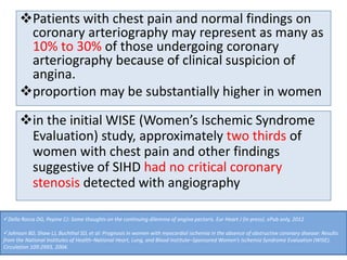
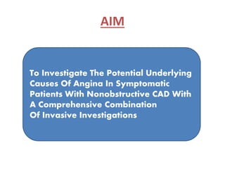
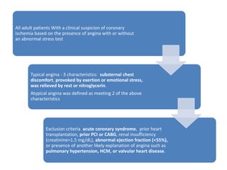
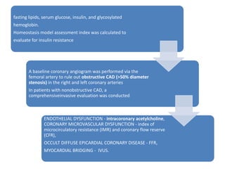
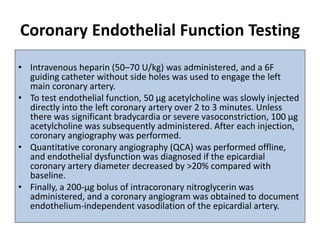
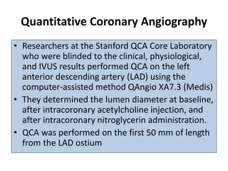
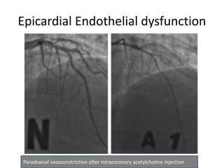
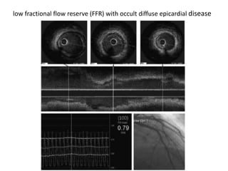
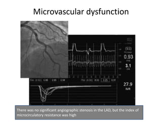
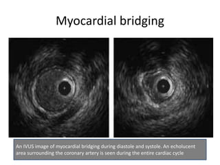
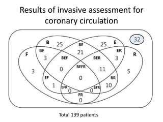
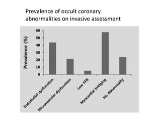
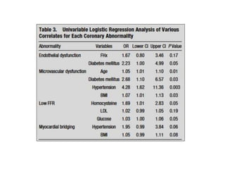
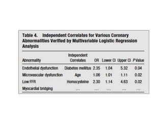
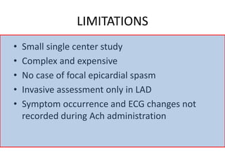
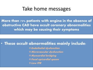
Circulation.
2015;131:1054-60
BACKGROUND
Patients with chest pain and normal findings on
coronary arteriography may represent as many as
10% to 30% of those undergoing coronary
arteriography because of clinical suspicion of
angina.
proportion may be substantially higher in women
in the initial WISE (Women’s Ischemic Syndrome
Evaluation) study, approximately two thirds of
women with chest pain and other findings
suggestive of SIHD had no critical coronary
stenosis detected with angiography
Della Rocca DG, Pepine CJ: Some thoughts on the continuing dilemma of angina pectoris. Eur Heart J (in press). ePub only, 2012
Johnson BD, Shaw LJ, Buchthal SD, et al: Prognosis in women with myocardial ischemia in the absence of obstructive coronary disease: Results
from the National Institutes of Health–National Heart, Lung, and Blood Institute–Sponsored Women’s Ischemia Syndrome Evaluation (WISE).
Circulation 109:2993, 2004.
?Coronary microcirculation
Coronary microvascular
dysfunction
Angina with normal coronary arteries,
a continuous dilemma
* Cardiac syndrome X : 1973
* Microvascular angina : 1985
Can we open the
“black box” ?
Finding the missing
piece to the puzzle
42 years
(1973-2015)
• Coronary Microcirculation
• Classification of CMVD
• Pathophysiologic mechanisms
• Clinical presentation
• Assessment
• Management
Coronary microvascular dysfunction(CMVD)
Muchdiscussed but little understood
What we can see is only 5% of the total coronary tree.
Coronary blood flow is driven by the pressure difference between the aorta and the capillary
bed and modulated further by various physical and neural factors, which affect the
microcirculation. Moreover, the different compartments of the microcirculation are
influenced by one main physiological mechanism to control their vascular tone with cardiac
metabolism as the final determining factor.
MICROVASCULAR DYSFUNCTION
• Pepine CJ, er al. Coronary microvascular reactivity to adenosine predicts adverse outcome in women evaluate for suspected
ischemia: results from the National Heart, Lung and Blood Institute WISE (Women’s Ischemia Syndrome Evaluation) study. J Am
Coll Cardiol. 2010;55:2825–2832
• Lerman A, Holmes DR, Herrmann J, Gersh BJ. Microcirculatory dysfunction in ST-elevation myocardial infarction: cause,
consequence, or both? Eur Heart J. 2007;28:788–797
EPICARDIAL ENDOTHELIAL DYSFUNCTION
• Halcox JP et al. Prognostic value of coronary vascular endothelial dysfunction. Circulation. 2002;106:653–658
FOCAL EPICARDIAL CORONARY SPASM
• Halcox JP et al. Prognostic value of coronary vascular endothelial dysfunction. Circulation. 2002;106:653–658
OCCULT DIFFUSE EPICARDIAL CORONARY DISEASE
• De Bruyne B, et al. Abnormal epicardial coronary resistance In patients with diffuse atherosclerosis but “normal” coronary
angiography. Circulation. 2001;104:2401–6
MYOCARDIAL BRIDGING
• Kramer JR, Kitazume H, Proudfit WL, Sones FM Jr. Clinical significance of isolated coronary bridges: benign and frequent condition
involving the left anterior descending artery. Am Heart J. 1982;103:283–288
The prevalence of these causes in
the same population is not defined.
The percentage of patients
without any of these abnormalities
and presumably noncardiac
symptoms is also unknown
Why important????
AIM
To Investigate The Potential Underlying
Causes Of Angina In Symptomatic
Patients With Nonobstructive CAD With
A Comprehensive Combination
Of Invasive Investigations
What for???
• Unnecessary overtreatment/ anxiety/ stress/
health expenditure
• Treatment deprivation when they really
need/ increasing risk of progression
METHODS
All adult patients With a clinical suspicion of coronary
ischemia based on the presence of angina with or without
an abnormal stress test
Typical angina - 3 characteristics: substernal chest
discomfort, provoked by exertion or emotional stress,
was relieved by rest or nitroglycerin.
Atypical angina was defined as meeting 2 of the above
characteristics
Exclusion criteria acute coronary syndrome, prior heart
transplantation, prior PCI or CABG, renal insufficiency
(creatinine>1.5 mg/dL), abnormal ejection fraction (<55%),
or presence of another likely explanation of angina such as
pulmonary hypertension, HCM, or valvular heart disease.
fasting lipids, serum glucose, insulin, and glycosylated
hemoglobin.
Homeostasis model assessment index was calculated to
evaluate for insulin resistance
A baseline coronary angiogram was performed via the
femoral artery to rule out obstructive CAD (>50% diameter
stenosis) in the right and left coronary arteries
In patients with nonobstructive CAD, a
comprehensiveinvasive evaluation was conducted
ENDOTHELIAL DYSFUNCTION - intracoronary acetylcholine,
CORONARY MICROVASCULAR DYSFUNCTION - index of
microcirculatory resistance (IMR) and coronary flow reserve
(CFR),
OCCULT DIFFUSE EPICARDIAL CORONARY DISEASE - FFR,
MYOCARDIAL BRIDGING - IVUS.
Coronary Endothelial Function Testing
• Intravenous heparin (50–70 U/kg) was administered, and a 6F
guiding catheter without side holes was used to engage the left
main coronary artery.
• To test endothelial function, 50 μg acetylcholine was slowly injected
directly into the left coronary artery over 2 to 3 minutes. Unless
there was significant bradycardia or severe vasoconstriction, 100 μg
acetylcholine was subsequently administered. After each injection,
coronary angiography was performed.
• Quantitative coronary angiography (QCA) was performed offline,
and endothelial dysfunction was diagnosed if the epicardial
coronary artery diameter decreased by >20% compared with
baseline.
• Finally, a 200-μg bolus of intracoronary nitroglycerin was
administered, and a coronary angiogram was obtained to document
endothelium-independent vasodilation of the epicardial artery.
Quantitative Coronary Angiography
• Researchers at the Stanford QCA Core Laboratory
who were blinded to the clinical, physiological,
and IVUS results performed QCA on the left
anterior descending artery (LAD) using the
computer-assisted method QAngio XA7.3 (Medis)
• They determined the lumen diameter at baseline,
after intracoronary acetylcholine injection, and
after intracoronary nitroglycerin administration.
• QCA was performed on the first 50 mm of length
from the LAD ostium
Coronary Physiology Measurements
• Within 10 minutes after endothelial function testing, CFR,
IMR, and FFR were measured.
• A pressure-temperature sensor guidewire (Certus Pressure
Wire, St. Jude Medical, St. Paul, MN) was used for physiology
measurements.
• With the sensor positioned at the tip of the catheter, the
pressure measurement from the wire was equalized with that
of the guiding catheter.
• The sensor was then positioned in the distal third of the LAD.
• Three injections of 3 mL of room-temperature saline were
made down the coronary artery, and the transit time was
measured after each and averaged to calculate the resting
mean transit time.
• An intravenous infusion of adenosine (140 μg/kg/min) was then
administered via a large peripheral or central vein to induce steady-
state maximal hyperemia, and 3 more injections of 3 mL of room
temperature saline were made.
• The transit time was measured after each and averaged to calculate
the hyperemic mean transit time.
• Simultaneous measurements of mean aortic pressure (by guiding
catheter) and mean distal coronary pressure (by pressure wire)
were also made during maximal hyperemia.
• IMR was calculated as the distal coronary pressure at maximal
hyperemia divided by the inverse of the hyperemic mean transit
time.
• CFR was calculated as resting mean transit time divided by
hyperemic mean transit time.
• FFR was calculated by the ratio of mean distal coronary pressure to
mean aortic pressure at maximal hyperemia.
• Microvascular dysfunction was defined as an IMR ≥25. An abnormal
FFR was defined as ≤0.80.
Intravascular Ultrasound
• IVUS was performed with a 40-MHz mechanical
transducer ultrasound catheter (Atlantis SR Pro2,
Boston Scientific Corp, Natick, MA) advanced
down the LAD so that the IVUS transducer was
positioned as close as possible to the pressure
transducer mounted on the pressure wire.
• An automated pullback at 0.5 mm/s was
performed, and the IVUS images were stored
onto DVD for offline analysis.
Intravascular Ultrasound
• The presence of a myocardial bridge was defined
by either the identification of an echolucent half-
moon sign or evidence of systolic compression
(≥10% systolic compression during the cardiac
cycle).
• Maximum percent systolic compression was
calculated by echo- Plaque software (Indec
Systems, Inc) and was defined as the change in
vessel area during the cardiac cycle divided by
vessel area during diastole.
RESULTS
Epicardial Endothelial dysfunction
Paradoxical vasoconstriction after intracoronary acetylcholine injection
low fractional flow reserve (FFR) with occult diffuse epicardial disease
Microvascular dysfunction
There was no significant angiographic stenosis in the LAD, but the index of
microcirculatory resistance was high
Myocardial bridging
An IVUS image of myocardial bridging during diastole and systole. An echolucent
area surrounding the coronary artery is seen during the entire cardiac cycle
Results of invasive assessment for
coronary circulation
Total 139 patients
Prevalence of occult coronary
abnormalities on invasive assessment
LIMITATIONS
• Small single center study
• Complex and expensive
• No case of focal epicardial spasm
• Invasive assessment only in LAD
• Symptom occurrence and ECG changes not
recorded during Ach administration
Take home messages
More than 75% patients with angina in the absence of
obstructive CAD have occult coronary abnormalities
which may be causing their symptoms
• These occult abnormalities mainly include:
Endothelial dysfunction
Microvascular dysfunction
Myocardial bridging
Focal epicardial spasm
Low FFR
Take home messages
A comprehensive invasive assessment of these
problems at the time of CART provides important
diagnostic info that may affect treatment and
outcomes
Large RCTs are warranted to check their cost
effectiveness and benefits in changing disease
outcome
ThankYou

Journal club april 2015

  • 1.
  • 2.
  • 3.
    Patients with chestpain and normal findings on coronary arteriography may represent as many as 10% to 30% of those undergoing coronary arteriography because of clinical suspicion of angina. proportion may be substantially higher in women in the initial WISE (Women’s Ischemic Syndrome Evaluation) study, approximately two thirds of women with chest pain and other findings suggestive of SIHD had no critical coronary stenosis detected with angiography Della Rocca DG, Pepine CJ: Some thoughts on the continuing dilemma of angina pectoris. Eur Heart J (in press). ePub only, 2012 Johnson BD, Shaw LJ, Buchthal SD, et al: Prognosis in women with myocardial ischemia in the absence of obstructive coronary disease: Results from the National Institutes of Health–National Heart, Lung, and Blood Institute–Sponsored Women’s Ischemia Syndrome Evaluation (WISE). Circulation 109:2993, 2004.
  • 5.
    ?Coronary microcirculation Coronary microvascular dysfunction Anginawith normal coronary arteries, a continuous dilemma * Cardiac syndrome X : 1973 * Microvascular angina : 1985 Can we open the “black box” ? Finding the missing piece to the puzzle 42 years (1973-2015)
  • 6.
    • Coronary Microcirculation •Classification of CMVD • Pathophysiologic mechanisms • Clinical presentation • Assessment • Management Coronary microvascular dysfunction(CMVD) Muchdiscussed but little understood
  • 7.
    What we cansee is only 5% of the total coronary tree.
  • 8.
    Coronary blood flowis driven by the pressure difference between the aorta and the capillary bed and modulated further by various physical and neural factors, which affect the microcirculation. Moreover, the different compartments of the microcirculation are influenced by one main physiological mechanism to control their vascular tone with cardiac metabolism as the final determining factor.
  • 9.
    MICROVASCULAR DYSFUNCTION • PepineCJ, er al. Coronary microvascular reactivity to adenosine predicts adverse outcome in women evaluate for suspected ischemia: results from the National Heart, Lung and Blood Institute WISE (Women’s Ischemia Syndrome Evaluation) study. J Am Coll Cardiol. 2010;55:2825–2832 • Lerman A, Holmes DR, Herrmann J, Gersh BJ. Microcirculatory dysfunction in ST-elevation myocardial infarction: cause, consequence, or both? Eur Heart J. 2007;28:788–797 EPICARDIAL ENDOTHELIAL DYSFUNCTION • Halcox JP et al. Prognostic value of coronary vascular endothelial dysfunction. Circulation. 2002;106:653–658 FOCAL EPICARDIAL CORONARY SPASM • Halcox JP et al. Prognostic value of coronary vascular endothelial dysfunction. Circulation. 2002;106:653–658 OCCULT DIFFUSE EPICARDIAL CORONARY DISEASE • De Bruyne B, et al. Abnormal epicardial coronary resistance In patients with diffuse atherosclerosis but “normal” coronary angiography. Circulation. 2001;104:2401–6 MYOCARDIAL BRIDGING • Kramer JR, Kitazume H, Proudfit WL, Sones FM Jr. Clinical significance of isolated coronary bridges: benign and frequent condition involving the left anterior descending artery. Am Heart J. 1982;103:283–288 The prevalence of these causes in the same population is not defined. The percentage of patients without any of these abnormalities and presumably noncardiac symptoms is also unknown
  • 10.
  • 13.
    AIM To Investigate ThePotential Underlying Causes Of Angina In Symptomatic Patients With Nonobstructive CAD With A Comprehensive Combination Of Invasive Investigations
  • 14.
    What for??? • Unnecessaryovertreatment/ anxiety/ stress/ health expenditure • Treatment deprivation when they really need/ increasing risk of progression
  • 15.
  • 16.
    All adult patientsWith a clinical suspicion of coronary ischemia based on the presence of angina with or without an abnormal stress test Typical angina - 3 characteristics: substernal chest discomfort, provoked by exertion or emotional stress, was relieved by rest or nitroglycerin. Atypical angina was defined as meeting 2 of the above characteristics Exclusion criteria acute coronary syndrome, prior heart transplantation, prior PCI or CABG, renal insufficiency (creatinine>1.5 mg/dL), abnormal ejection fraction (<55%), or presence of another likely explanation of angina such as pulmonary hypertension, HCM, or valvular heart disease.
  • 17.
    fasting lipids, serumglucose, insulin, and glycosylated hemoglobin. Homeostasis model assessment index was calculated to evaluate for insulin resistance A baseline coronary angiogram was performed via the femoral artery to rule out obstructive CAD (>50% diameter stenosis) in the right and left coronary arteries In patients with nonobstructive CAD, a comprehensiveinvasive evaluation was conducted ENDOTHELIAL DYSFUNCTION - intracoronary acetylcholine, CORONARY MICROVASCULAR DYSFUNCTION - index of microcirculatory resistance (IMR) and coronary flow reserve (CFR), OCCULT DIFFUSE EPICARDIAL CORONARY DISEASE - FFR, MYOCARDIAL BRIDGING - IVUS.
  • 18.
    Coronary Endothelial FunctionTesting • Intravenous heparin (50–70 U/kg) was administered, and a 6F guiding catheter without side holes was used to engage the left main coronary artery. • To test endothelial function, 50 μg acetylcholine was slowly injected directly into the left coronary artery over 2 to 3 minutes. Unless there was significant bradycardia or severe vasoconstriction, 100 μg acetylcholine was subsequently administered. After each injection, coronary angiography was performed. • Quantitative coronary angiography (QCA) was performed offline, and endothelial dysfunction was diagnosed if the epicardial coronary artery diameter decreased by >20% compared with baseline. • Finally, a 200-μg bolus of intracoronary nitroglycerin was administered, and a coronary angiogram was obtained to document endothelium-independent vasodilation of the epicardial artery.
  • 19.
    Quantitative Coronary Angiography •Researchers at the Stanford QCA Core Laboratory who were blinded to the clinical, physiological, and IVUS results performed QCA on the left anterior descending artery (LAD) using the computer-assisted method QAngio XA7.3 (Medis) • They determined the lumen diameter at baseline, after intracoronary acetylcholine injection, and after intracoronary nitroglycerin administration. • QCA was performed on the first 50 mm of length from the LAD ostium
  • 20.
    Coronary Physiology Measurements •Within 10 minutes after endothelial function testing, CFR, IMR, and FFR were measured. • A pressure-temperature sensor guidewire (Certus Pressure Wire, St. Jude Medical, St. Paul, MN) was used for physiology measurements. • With the sensor positioned at the tip of the catheter, the pressure measurement from the wire was equalized with that of the guiding catheter. • The sensor was then positioned in the distal third of the LAD. • Three injections of 3 mL of room-temperature saline were made down the coronary artery, and the transit time was measured after each and averaged to calculate the resting mean transit time.
  • 21.
    • An intravenousinfusion of adenosine (140 μg/kg/min) was then administered via a large peripheral or central vein to induce steady- state maximal hyperemia, and 3 more injections of 3 mL of room temperature saline were made. • The transit time was measured after each and averaged to calculate the hyperemic mean transit time. • Simultaneous measurements of mean aortic pressure (by guiding catheter) and mean distal coronary pressure (by pressure wire) were also made during maximal hyperemia. • IMR was calculated as the distal coronary pressure at maximal hyperemia divided by the inverse of the hyperemic mean transit time. • CFR was calculated as resting mean transit time divided by hyperemic mean transit time. • FFR was calculated by the ratio of mean distal coronary pressure to mean aortic pressure at maximal hyperemia. • Microvascular dysfunction was defined as an IMR ≥25. An abnormal FFR was defined as ≤0.80.
  • 22.
    Intravascular Ultrasound • IVUSwas performed with a 40-MHz mechanical transducer ultrasound catheter (Atlantis SR Pro2, Boston Scientific Corp, Natick, MA) advanced down the LAD so that the IVUS transducer was positioned as close as possible to the pressure transducer mounted on the pressure wire. • An automated pullback at 0.5 mm/s was performed, and the IVUS images were stored onto DVD for offline analysis.
  • 23.
    Intravascular Ultrasound • Thepresence of a myocardial bridge was defined by either the identification of an echolucent half- moon sign or evidence of systolic compression (≥10% systolic compression during the cardiac cycle). • Maximum percent systolic compression was calculated by echo- Plaque software (Indec Systems, Inc) and was defined as the change in vessel area during the cardiac cycle divided by vessel area during diastole.
  • 24.
  • 27.
    Epicardial Endothelial dysfunction Paradoxicalvasoconstriction after intracoronary acetylcholine injection
  • 28.
    low fractional flowreserve (FFR) with occult diffuse epicardial disease
  • 29.
    Microvascular dysfunction There wasno significant angiographic stenosis in the LAD, but the index of microcirculatory resistance was high
  • 30.
    Myocardial bridging An IVUSimage of myocardial bridging during diastole and systole. An echolucent area surrounding the coronary artery is seen during the entire cardiac cycle
  • 31.
    Results of invasiveassessment for coronary circulation Total 139 patients
  • 32.
    Prevalence of occultcoronary abnormalities on invasive assessment
  • 35.
    LIMITATIONS • Small singlecenter study • Complex and expensive • No case of focal epicardial spasm • Invasive assessment only in LAD • Symptom occurrence and ECG changes not recorded during Ach administration
  • 36.
    Take home messages Morethan 75% patients with angina in the absence of obstructive CAD have occult coronary abnormalities which may be causing their symptoms • These occult abnormalities mainly include: Endothelial dysfunction Microvascular dysfunction Myocardial bridging Focal epicardial spasm Low FFR
  • 37.
    Take home messages Acomprehensive invasive assessment of these problems at the time of CART provides important diagnostic info that may affect treatment and outcomes Large RCTs are warranted to check their cost effectiveness and benefits in changing disease outcome ThankYou