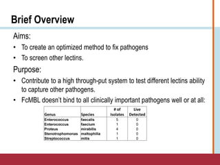
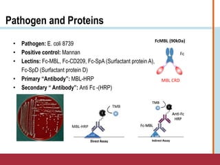
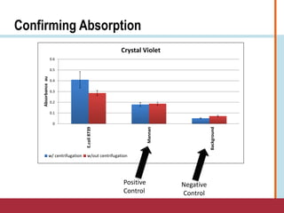
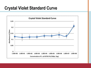
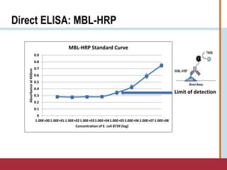
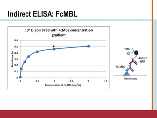
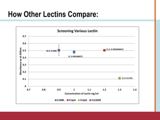
Optimizing Pathogen Fixation
Method to Screen Lectins
Keturah Kiper
August 11th, 2016
2016 Harvard Catalyst Clinical and Translational Research Program
University of Arkansas, Fayetteville
Don Ingber, MD, PhD
Michael Super, MSc, PhD
Alex Watters, PhD
Anna Waterhouse, PhD
Wyss Institute for Biologically Inspired Engineering
What is Sepsis?
What we know:
• It is an infection of the blood
• Some causes:
• Trauma
• Surgery
• Infection: skin, urinary, etc.
• Sepsis affects 18 million people worldwide every year,
with a 30-50% mortality rate
• Mortality rates increase as much as 9% for every
hour before the correct antibiotic therapy is administered
What is Sepsis? (cont.)
• Sepsis has been defined as a result of systemic inflammatory
response syndrome (SIRS) which can progress to severe sepsis
and eventually septic shock
•To be diagnosed with SIRS the patient would meet two or more of the criteria:
― Fever of more than 38°C (100.4°F) or less than 36°C (96.8°F)
― Heart rate of more than 90 beats per minute
― Respiratory rate of more than 20 breaths per minute or arterial carbon dioxide tension
(PaCO 2) of less than 32 mm Hg
― Abnormal white blood cell count >12,000/µL=mm3 or < 4,000/µL=mm3 or >10%
immature bands
•Important to understand that these are clinical observations and the definition
of sepsis is constantly changing as more information becomes available.
Connection to the Wyss Institute
• DARPA-BAA-11-30
•The goal of the Dialysis Like Therapeutics (DLT) program is to develop a
portable device capable of controlling relevant components in the entire
blood volume on clinically relevant time scales.
• DARPA-BAA-12-36
•The focus is to integrate key component technologies (sensing, complex
fluid manipulation architectures, separation technologies, and closed-loop
controller) that are being developed under DARPA-BAA-11-30 into a
functioning device that can effectively treat sepsis across the military
health system.
At the Wyss Institute
FcMBL (90kDa)
MBL CRD
Fc
FcMBL Magnetic opsonin
Hollow Fiber (HF)
Dialyzer
FcMBL
???
Treatment Diagnostics
Pathogen
Infected
Blood
Pathogen PAMP Capture
Detection
Agent
+TMB
Infection
No
Infection
Cartwright, M., et al., A broad spectrum infection diagnostic that detects pathogen-associated molecular patterns (PAMPS in whole blood,
EBioMedicine (2016)
Brief Overview
Aims:
• To create an optimized method to fix pathogens
• To screen other lectins.
Purpose:
• Contribute to a high through-put system to test different lectins ability
to capture other pathogens.
• FcMBL doesn’t bind to all clinically important pathogens well or at all:
Genus Species
# of
Isolates
Live
Detected
Enterococcus faecalis 5 0
Enterococcus faecium 1 0
Proteus mirabilis 4 0
Stenotrophomonas maltophilia 1 0
Streptococcus mitis 1 0
Experimental design
Using information provided on: sepsis, FcMBL, microbiology &
enzyme-linked immunosorbent assays (ELISA), etc.
1. Create a protocol
I. Fix pathogen for absorption
2. Use standard test to support functionality of protocol
I. Confirm absorption: Crystal Violet
II. Lectin Screening Assay: Direct ELISA & Indirect ELISA
3. Collect & Analyze data
Optimized Fixation Protocol
1. Prepare bacteria on plate—using streaking method
2. Incubate plate overnight at room temperature
3. Collect single colony of bacteria for liquid culture from
plate using inoculating loop
4. Make 5ml vial of liquid culture using RPMI/glucose
media
5. Grow to .5 optical density using incubator
6. Transfer total volume to centrifuge tube
7. Centrifuge liquid culture and collect supernatant
•@ 2250 x g for 10 minutes @ 4℃
8. Wash pellet X3
•Use 5ml PBS-low endotoxin
•Suspend cells in PBS-low endotoxin each time (keep volume
constant)
•Centrifuge @2250 x g for 5 minutes @ 4℃
•Collect waste in appropriate container
9. Add .5% PFA 1X PBS-low endotoxin (5ml)
10. Suspend cells into fixative solution
•Mix with vortex
11. Let incubate for 20 minutes
• Use HulaMixer
12. Centrifuge to collect supernatant
• @ 2250 x g for 5 minutes @ 4℃
13. Wash pellet X3
•Use 5ml PBS-low endotoxin
•Suspend cells in PBS-low endotoxin each time (keep volume
constant)
•Centrifuge @2250 x g for 5 minutes @ 4℃
•Collect waste in appropriate container
14. Prepare a 50ml stock solution of .1M bicarbonate
15. Suspend bacteria in 5ml of .1M bicarbonate solution
16. Coat columns with 100 μl/well of fixed cells
17. Centrifuge 96 well plate for 5 minutes at 2500 x g @
4℃
18. Incubate overnight at 4℃
Crystal Violet
Modified crystal violet procedure:
1. Wash plates with low endotoxin PBS 3X
2. Stain with 0.1% crystal violet for 1 hour
•Wear a lab coat, safety goggles and face mask when handling crystal violet powder and spray bench
top with 70% ethanol.
•Crystal violet – TOXIC! Put all tips and pipettes into bleach until crystal violet turns yellow
•Stain all samples simultaneously to reduce variation (ie. stain all time points of one experiment at the
same time)
3. Take crystal violet off
•Crystal violet can be reused
4. Wash plates under RO water tap 5-6 times
•Tap plates on benching to drain—residual water will dilute acetic acid
5. Add 10% acetic acid (to cover the well, 200 μl in 24-well plate) for 20 min
•Gently tap plates; releases/solubilizes dye
6. Read Absorbency at 570nm
Enzyme-linked
immunosorbent assay (ELISA)
1. Wash plate 5x 175 μl/well with TBS-T
2. Make blocking solution: 2.5% milk in 1x TBS
3. Block plate with 2.5% milk at 200 μl/well, incubate at 37C for 1-2 hours, shaking at 300RPM
4. Complete 8 point protein dilutions in HEPES incubation buffer starting at 100 μg/ml
5. Wash plate 5x 175 μl/well with TBS-T
6. Aliquot protein dilutions, 100 μl/well, in quadruples (wells with and without E. coli 8739). Incubate
at 37C for 1 hour, shaking at 300RPM
7. Dilute anti-Fc antibody 1:10k in 0.5% milk/TBS solution
8. Wash plate 5x 175 μl/well with TBS-T
9. Aliquot antibody to each well at 100 μl/well, incubate at 37C for 1 hour, shaking at 300RPM
10. Wash plate 5x 175 μl/well with TBS-T
11. Develop ELISA with TMB at 100 μl/well for 3-5 minutes in dark space
12. Stop reaction with sulfuric acid at 50ul/well
13. Read plate at 450nm
14. Subtract background signal from sample signal
Steps only for
indirect ELISA
Equipment
• Biotek Synergy H1 Multi-Mode Reader
• Thermo Fisher, Sorvall™ Legend™ XT
XF Centrifuge
• RO Water tap
Eppendorf Centrifuge
High binding 96 well plates
Multi-channel pipet
Pathogen and Proteins
• Pathogen: E. coli 8739
• Positive control: Mannan
• Lectins: Fc-MBL, Fc-CD209, Fc-SpA (Surfactant protein A),
Fc-SpD (Surfactant protein D)
• Primary “Antibody”: MBL-HRP
• Secondary “ Antibody”: Anti Fc -(HRP)
FcMBL (90kDa)
Fc
MBL CRD
E.coli
Hey
MBL-HRP
TMB
Confirming Absorption
0
0.1
0.2
0.3
0.4
0.5
0.6
E.coli8739
Mannan
Background
Absorbanceau
Crystal Violet
w/ centrifugation w/out centrifugation
Positive
Control
Negative
Control
Crystal Violet Standard Curve
0
0.05
0.1
0.15
0.2
0.25
1.00E+00 1.00E+01 1.00E+02 1.00E+03 1.00E+04 1.00E+05 1.00E+06 1.00E+07 1.00E+08
Absorbanceat570nm
Concentration of E. coli 8739 CFU/100μl (log)
Crystal Violet Standard Curve
Direct ELISA: MBL-HRP
0
0.1
0.2
0.3
0.4
0.5
0.6
0.7
0.8
0.9
1.00E+00 1.00E+01 1.00E+02 1.00E+03 1.00E+04 1.00E+05 1.00E+06 1.00E+07 1.00E+08
Absorbanceat450nm
Concentration of E. coli 8739 (log)
MBL-HRP Standard Curve
Limit of detection
Indirect ELISA: FcMBL
0
0.1
0.2
0.3
0.4
0.5
0.6
0 0.5 1 1.5 2 2.5
AbsorbanceAu
Concentration of Fc-MBL (mg/ml)
108 E. coli 8739 with FcMBL concentration
gradient
*
How Other Lectins Compare:
(1, 0.48166667)
(1.2, 0.505666667)
(1.3, 0.115)
(0.9, 0.506)
0
0.1
0.2
0.3
0.4
0.5
0.6
0.7
0.7 0.8 0.9 1 1.1 1.2 1.3 1.4
Absorbanceauat450nm
Concentration of Lectin mg/ml
Screening Various Lectin
FcMBL FcSpA FcSpD FcCd209
Conclusion
• Evidence supports that the optimized fixation
method is working
• Definitely more optimization to be done:
•Blocking agent:
―BSA versus 2.5% MILK 1x TBS-T-20
•Concentration of Lectin (mg/ml)
Acknowledgements
Lee Nadler, MD—Dean for Clinical and Translational Research
Joan Y. Reede, MD, MPH, MS, MBA—Dean for Diversity and Community Partnership
Don Ingber, MD, PhD—Founding Director, Core Faculty & Primary Investigator
Mike Super, PhD—Senior Staff Scientist and Mentor
Alex Watters, PhD—Staff Scientist
Anna Waterhouse, PhD—Research Scientist
Sepsis Lab Group—Wyss Institute
Mary Frances Lopez, PhD—Academic Advisor
Carol Martin, MPA—Program Manager
Danyellé Thorpe—Program Coordinator

KIPER_SCTRP_Presentation

  • 1.
    Optimizing Pathogen Fixation Methodto Screen Lectins Keturah Kiper August 11th, 2016 2016 Harvard Catalyst Clinical and Translational Research Program University of Arkansas, Fayetteville Don Ingber, MD, PhD Michael Super, MSc, PhD Alex Watters, PhD Anna Waterhouse, PhD Wyss Institute for Biologically Inspired Engineering
  • 2.
    What is Sepsis? Whatwe know: • It is an infection of the blood • Some causes: • Trauma • Surgery • Infection: skin, urinary, etc. • Sepsis affects 18 million people worldwide every year, with a 30-50% mortality rate • Mortality rates increase as much as 9% for every hour before the correct antibiotic therapy is administered
  • 3.
    What is Sepsis?(cont.) • Sepsis has been defined as a result of systemic inflammatory response syndrome (SIRS) which can progress to severe sepsis and eventually septic shock •To be diagnosed with SIRS the patient would meet two or more of the criteria: ― Fever of more than 38°C (100.4°F) or less than 36°C (96.8°F) ― Heart rate of more than 90 beats per minute ― Respiratory rate of more than 20 breaths per minute or arterial carbon dioxide tension (PaCO 2) of less than 32 mm Hg ― Abnormal white blood cell count >12,000/µL=mm3 or < 4,000/µL=mm3 or >10% immature bands •Important to understand that these are clinical observations and the definition of sepsis is constantly changing as more information becomes available.
  • 4.
    Connection to theWyss Institute • DARPA-BAA-11-30 •The goal of the Dialysis Like Therapeutics (DLT) program is to develop a portable device capable of controlling relevant components in the entire blood volume on clinically relevant time scales. • DARPA-BAA-12-36 •The focus is to integrate key component technologies (sensing, complex fluid manipulation architectures, separation technologies, and closed-loop controller) that are being developed under DARPA-BAA-11-30 into a functioning device that can effectively treat sepsis across the military health system.
  • 5.
    At the WyssInstitute FcMBL (90kDa) MBL CRD Fc FcMBL Magnetic opsonin Hollow Fiber (HF) Dialyzer FcMBL ??? Treatment Diagnostics Pathogen Infected Blood Pathogen PAMP Capture Detection Agent +TMB Infection No Infection Cartwright, M., et al., A broad spectrum infection diagnostic that detects pathogen-associated molecular patterns (PAMPS in whole blood, EBioMedicine (2016)
  • 6.
    Brief Overview Aims: • Tocreate an optimized method to fix pathogens • To screen other lectins. Purpose: • Contribute to a high through-put system to test different lectins ability to capture other pathogens. • FcMBL doesn’t bind to all clinically important pathogens well or at all: Genus Species # of Isolates Live Detected Enterococcus faecalis 5 0 Enterococcus faecium 1 0 Proteus mirabilis 4 0 Stenotrophomonas maltophilia 1 0 Streptococcus mitis 1 0
  • 7.
    Experimental design Using informationprovided on: sepsis, FcMBL, microbiology & enzyme-linked immunosorbent assays (ELISA), etc. 1. Create a protocol I. Fix pathogen for absorption 2. Use standard test to support functionality of protocol I. Confirm absorption: Crystal Violet II. Lectin Screening Assay: Direct ELISA & Indirect ELISA 3. Collect & Analyze data
  • 8.
    Optimized Fixation Protocol 1.Prepare bacteria on plate—using streaking method 2. Incubate plate overnight at room temperature 3. Collect single colony of bacteria for liquid culture from plate using inoculating loop 4. Make 5ml vial of liquid culture using RPMI/glucose media 5. Grow to .5 optical density using incubator 6. Transfer total volume to centrifuge tube 7. Centrifuge liquid culture and collect supernatant •@ 2250 x g for 10 minutes @ 4℃ 8. Wash pellet X3 •Use 5ml PBS-low endotoxin •Suspend cells in PBS-low endotoxin each time (keep volume constant) •Centrifuge @2250 x g for 5 minutes @ 4℃ •Collect waste in appropriate container 9. Add .5% PFA 1X PBS-low endotoxin (5ml) 10. Suspend cells into fixative solution •Mix with vortex 11. Let incubate for 20 minutes • Use HulaMixer 12. Centrifuge to collect supernatant • @ 2250 x g for 5 minutes @ 4℃ 13. Wash pellet X3 •Use 5ml PBS-low endotoxin •Suspend cells in PBS-low endotoxin each time (keep volume constant) •Centrifuge @2250 x g for 5 minutes @ 4℃ •Collect waste in appropriate container 14. Prepare a 50ml stock solution of .1M bicarbonate 15. Suspend bacteria in 5ml of .1M bicarbonate solution 16. Coat columns with 100 μl/well of fixed cells 17. Centrifuge 96 well plate for 5 minutes at 2500 x g @ 4℃ 18. Incubate overnight at 4℃
  • 9.
    Crystal Violet Modified crystalviolet procedure: 1. Wash plates with low endotoxin PBS 3X 2. Stain with 0.1% crystal violet for 1 hour •Wear a lab coat, safety goggles and face mask when handling crystal violet powder and spray bench top with 70% ethanol. •Crystal violet – TOXIC! Put all tips and pipettes into bleach until crystal violet turns yellow •Stain all samples simultaneously to reduce variation (ie. stain all time points of one experiment at the same time) 3. Take crystal violet off •Crystal violet can be reused 4. Wash plates under RO water tap 5-6 times •Tap plates on benching to drain—residual water will dilute acetic acid 5. Add 10% acetic acid (to cover the well, 200 μl in 24-well plate) for 20 min •Gently tap plates; releases/solubilizes dye 6. Read Absorbency at 570nm
  • 10.
    Enzyme-linked immunosorbent assay (ELISA) 1.Wash plate 5x 175 μl/well with TBS-T 2. Make blocking solution: 2.5% milk in 1x TBS 3. Block plate with 2.5% milk at 200 μl/well, incubate at 37C for 1-2 hours, shaking at 300RPM 4. Complete 8 point protein dilutions in HEPES incubation buffer starting at 100 μg/ml 5. Wash plate 5x 175 μl/well with TBS-T 6. Aliquot protein dilutions, 100 μl/well, in quadruples (wells with and without E. coli 8739). Incubate at 37C for 1 hour, shaking at 300RPM 7. Dilute anti-Fc antibody 1:10k in 0.5% milk/TBS solution 8. Wash plate 5x 175 μl/well with TBS-T 9. Aliquot antibody to each well at 100 μl/well, incubate at 37C for 1 hour, shaking at 300RPM 10. Wash plate 5x 175 μl/well with TBS-T 11. Develop ELISA with TMB at 100 μl/well for 3-5 minutes in dark space 12. Stop reaction with sulfuric acid at 50ul/well 13. Read plate at 450nm 14. Subtract background signal from sample signal Steps only for indirect ELISA
  • 11.
    Equipment • Biotek SynergyH1 Multi-Mode Reader • Thermo Fisher, Sorvall™ Legend™ XT XF Centrifuge • RO Water tap Eppendorf Centrifuge High binding 96 well plates Multi-channel pipet
  • 12.
    Pathogen and Proteins •Pathogen: E. coli 8739 • Positive control: Mannan • Lectins: Fc-MBL, Fc-CD209, Fc-SpA (Surfactant protein A), Fc-SpD (Surfactant protein D) • Primary “Antibody”: MBL-HRP • Secondary “ Antibody”: Anti Fc -(HRP) FcMBL (90kDa) Fc MBL CRD E.coli Hey MBL-HRP TMB
  • 13.
  • 14.
    Crystal Violet StandardCurve 0 0.05 0.1 0.15 0.2 0.25 1.00E+00 1.00E+01 1.00E+02 1.00E+03 1.00E+04 1.00E+05 1.00E+06 1.00E+07 1.00E+08 Absorbanceat570nm Concentration of E. coli 8739 CFU/100μl (log) Crystal Violet Standard Curve
  • 15.
    Direct ELISA: MBL-HRP 0 0.1 0.2 0.3 0.4 0.5 0.6 0.7 0.8 0.9 1.00E+001.00E+01 1.00E+02 1.00E+03 1.00E+04 1.00E+05 1.00E+06 1.00E+07 1.00E+08 Absorbanceat450nm Concentration of E. coli 8739 (log) MBL-HRP Standard Curve Limit of detection
  • 16.
    Indirect ELISA: FcMBL 0 0.1 0.2 0.3 0.4 0.5 0.6 00.5 1 1.5 2 2.5 AbsorbanceAu Concentration of Fc-MBL (mg/ml) 108 E. coli 8739 with FcMBL concentration gradient *
  • 17.
    How Other LectinsCompare: (1, 0.48166667) (1.2, 0.505666667) (1.3, 0.115) (0.9, 0.506) 0 0.1 0.2 0.3 0.4 0.5 0.6 0.7 0.7 0.8 0.9 1 1.1 1.2 1.3 1.4 Absorbanceauat450nm Concentration of Lectin mg/ml Screening Various Lectin FcMBL FcSpA FcSpD FcCd209
  • 18.
    Conclusion • Evidence supportsthat the optimized fixation method is working • Definitely more optimization to be done: •Blocking agent: ―BSA versus 2.5% MILK 1x TBS-T-20 •Concentration of Lectin (mg/ml)
  • 19.
    Acknowledgements Lee Nadler, MD—Deanfor Clinical and Translational Research Joan Y. Reede, MD, MPH, MS, MBA—Dean for Diversity and Community Partnership Don Ingber, MD, PhD—Founding Director, Core Faculty & Primary Investigator Mike Super, PhD—Senior Staff Scientist and Mentor Alex Watters, PhD—Staff Scientist Anna Waterhouse, PhD—Research Scientist Sepsis Lab Group—Wyss Institute Mary Frances Lopez, PhD—Academic Advisor Carol Martin, MPA—Program Manager Danyellé Thorpe—Program Coordinator

Editor's Notes

  • #3 Second bullet point: Sepsis is a leading cause of mortality and critical illness in the world.
  • #4 First Bullet Point: A 1991 consensus conference developed the criteria for diagnosing SIRS. Third Bullet Point: With the 2011 withdrawal of the Eli Lilly drugs Xigris (drotrecogin alpha)
  • #5 Closed loop controller is a system that has a sensor placed in the system that loops back from output to the controller at the stage of input in order to make adjustments to the current input
  • #6 Then: Bioinspired fluidic device design--Spleen-on-a-chip with magnetic beads—FDA not happy about magnetic beads Second Bullet Point: This raises the need for a rapid test to diagnose sepsis to decrease the number of incidences, but also to have “guided” treatment instead of a broad spectrum treatment approach they currently use
  • #7 All this in efforts to assist in the diagnostics developed for sepsis.
  • #16 This graphs shows us that the sensitivity of this assay begins at the concentration of E. coli at 10^5 -this is a good looking graph to see the limit of detection This is the combination of 1 colony of E.coli, 2 plates with three columns-three wells per concentration So what is next? An indirect assay.