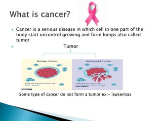
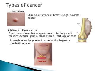
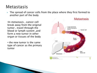
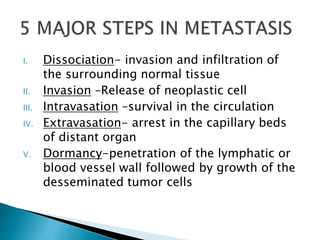
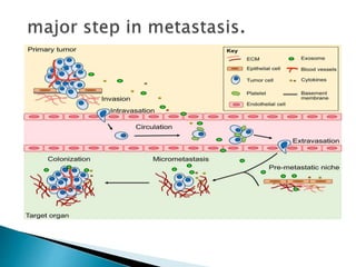
Metastasis is a pathogenic agent's spread from an initial or primary site to a different or secondary site within the host's body; the term is typically used when referring to metastasis by a cancerous tumor.
Cancer metastasis is the process where cancer cells spread from the primary tumor to distant sites in the body. There are 5 major steps in metastasis: 1) invasion of surrounding tissue, 2) release into circulation, 3) survival in circulation, 4) arrest in capillaries of distant organs, 5) penetration of vessel walls and growth. Metastasis can occur through the bloodstream (hematogenous), lymphatic system, or body cavities. Cancer cells degrade the extracellular matrix using enzymes to migrate, and can die from detachment, damage in vessels, or immune system attack. Some cancers have preferential metastatic sites thought to be due to anatomical proximity or a compatible tissue environment.
This document summarizes the molecular mechanisms of cancer metastasis. It begins with an overview of metastasis and defines it as the process by which tumor cells spread from the primary site to distant organs via the circulatory system. It then discusses the major steps in metastasis, including invasion, intravasation, survival in circulation, arrest at secondary sites, and extravasation. The key molecular components that drive metastasis are matrix-degrading enzymes like MMPs, cell adhesion molecules like integrins and E-cadherin, and cytoskeletal regulators like Rho GTPases that control cell motility. Signaling pathways activated by HGF/c-Met and other motility factors also promote invasion and metastasis. Understanding the molecular underpinnings of
The document summarizes the process of cancer metastasis through the invasion-metastasis cascade. It involves 6 key steps: 1) Localized invasion of primary tumor cells aided by loss of cell adhesion molecules and matrix metalloproteinases. 2) Intravasation of tumor cells into blood vessels assisted by tumor-associated macrophages. 3) Transport of circulating tumor cells protected by platelet emboli. 4) Extravasation of tumor cells from vessels into distant tissues. 5) Formation of dormant micrometastases. 6) Rare colonization of micrometastases into macroscopic tumors limited by the foreign tissue environment. Metastasis suppressor genes and strategies targeting multiple steps simultaneously show promise for preventing cancer spread.
Thrombosis is the formation of a blood clot within a blood vessel or cavity of the heart. Virchow identified three main factors that contribute to thrombosis: endothelial injury, changes in blood flow, and hypercoagulability. Thrombi can propagate or embolize, becoming lodged in another vessel and resulting in infarction of downstream tissue. Infarctions appear pale/white in solid organs and red/hemorrhagic in lungs/other tissues. Over time, infarcted tissue progresses from coagulative necrosis to phagocytosis and scar formation.
Cancer arises from mutations in genes that regulate cell growth and division. These mutations can cause cells to grow uncontrollably and form tumors. There are two main types of cancer genes - oncogenes which promote cell growth when mutated, and tumor suppressor genes which normally inhibit cell growth but cannot when mutated in both copies of the gene. Most cancers are caused by multiple mutations that accumulate over time due to environmental exposures, random errors in cell division, or inherited genetic syndromes.
Laboratory diagnosis of cancer involves cytology, histopathology, and other techniques. Fine needle aspiration cytology (FNAC) is a simple, rapid, and economical technique to obtain cells for cancer diagnosis. It has high accuracy but loses tissue architecture. Histopathology examines diseased tissue microscopically and macroscopically. Cancer is graded based on cell differentiation and staged based on tumor size, lymph node involvement, and metastasis. Immunohistochemistry aids in cancer classification and detection of molecules with prognostic value.
Mutistep carcinogenesis refers to the process by which normal cells transform into cancerous cells through the accumulation of multiple genetic mutations over time. These mutations can be caused by environmental or inherited factors and affect genes that regulate cell growth (oncogenes) or cell cycle arrest (tumor suppressor genes). The accumulation of mutations in genes that control processes like apoptosis, cell proliferation, and DNA repair enable cells to proliferate uncontrollably and form malignant tumors.
This document discusses tumor metastasis and the pathways involved. It covers:
1. Metastasis occurs when tumor cells spread discontinuously from the primary site to distant organs. More aggressive tumors are more likely to metastasize.
2. Tumor cells can spread via lymphatic vessels, hematogenous spread through the bloodstream, or by seeding body cavities. The most common route is lymphatic spread for carcinomas and hematogenous spread for sarcomas.
3. The metastatic process involves tumor progression, invasion of surrounding tissue, entry and survival in the bloodstream, arrest at distant sites, extravasation, and colonization of secondary tumors. Specific adhesion molecules and chemotactic
This document provides an overview of neoplasia (tumors), including definitions, nomenclature used to classify benign and malignant tumors, the biology of tumor growth, epidemiology, molecular basis of cancer and carcinogenesis. It discusses the natural history of malignant tumors from malignant change to growth, invasion and metastasis. Key concepts covered include differentiation, dysplasia, tumor growth rates, features that distinguish benign from malignant tumors, predisposing geographic, environmental and genetic factors for cancer development.
Metaplasia is a reversible change where one adult cell type replaces another in response to stress. For example, in smokers the ciliated trachea cells are often replaced with squamous cells. This change arises from the reprogramming of stem cells due to signals from cytokines, growth factors and the extracellular matrix. If the stress persists, metaplasia can predispose the tissue to malignant transformation.
This document discusses methods for cancer diagnosis including clinical examination, imaging, biochemical assays, and morphological and molecular techniques. Histological diagnosis through microscopic examination of tissue specimens is the gold standard for cancer diagnosis. Several sampling methods can be used including excision or biopsy, fine-needle aspiration, and cytologic smears. Immunocytochemistry, flow cytometry, and molecular techniques like PCR and FISH can also provide diagnostic information. Proper sampling and preservation of specimens is important for accurate histological diagnosis of cancer.
This document defines neoplasia and provides details on the classification and nomenclature of tumors. It begins by defining a neoplasm as an abnormal mass of tissue that grows in an uncoordinated manner. Benign tumors are non-invasive and localized, while malignant tumors are invasive and spreading. Tumors are classified based on the tissue of origin, such as carcinomas arising from epithelial tissue and sarcomas from connective tissue. The document further describes features of benign versus malignant tumors and provides examples of tumor classifications and histological slides.
This document provides an overview of amyloidosis, including:
- Amyloidosis is characterized by extracellular deposition of misfolded proteins that form insoluble fibrils, damaging tissues.
- There are different types classified by the misfolded protein involved, including AL, AA, and rare forms.
- Organs commonly affected include the kidney, heart, GI tract, and nerves.
- Diagnosis involves biopsy of affected tissues and staining with Congo red to identify amyloid deposits.
- Prognosis depends on type and organ involvement, with generalized amyloidosis having a poor prognosis of around 2 years.
Neoplasia refers to new abnormal growth of tissue. A neoplasm is an abnormal mass of tissue that grows in an uncontrolled manner. Neoplasms can be benign (non-cancerous) or malignant (cancerous). The growth of neoplasms is driven by genetic changes that allow the cells to proliferate autonomously. Tumors are monoclonal in origin, arising from a single cell. The hallmarks of neoplasms include persistent and purposeless proliferation, a parasitic nature, progression, and clonal expansion. Tumors are classified based on their tissue of origin and behavior. Malignant tumors can invade surrounding tissues and metastasize to distant sites. The development of neoplasms involves genetic and environmental factors
Necrosis is the morphologic changes that follow cell death in living tissues. It can be caused by ischemia, physical agents, chemical agents, or immunological injury. The main types of necrosis include coagulative necrosis, liquefactive necrosis, caseous necrosis, fat necrosis, and fibrinoid necrosis. Coagulative necrosis preserves tissue architecture with denatured proteins and enzymatic digestion of cells. Liquefactive necrosis results in a liquid viscous mass. Caseous necrosis is a type of coagulative necrosis seen in tuberculosis that leaves cheesy white tissue.
Metastasis is the spread of cancer cells from the primary tumor to other parts of the body. Cancer cells can metastasize through direct extension, lymphatic spread, or hematogenous spread through the bloodstream. The process of metastasis involves local invasion of tissue, entry and survival in the circulatory or lymphatic system, arrest in capillaries, extravasation into new organs, and growth of microscopic tumor deposits into macroscopic secondary tumors. Common sites of metastasis include the lungs, liver, brain, and bones. The mechanisms of metastasis are complex and involve changes in cancer cell adhesion and signaling to encourage migration, invasion of surrounding tissues, angiogenesis, and survival at distant sites.
1) Cancer begins as abnormal cells that grow uncontrollably and can become invasive, spreading to other parts of the body.
2) There are several types of cancer including carcinomas, sarcomas, leukemias, lymphomas, and central nervous system cancers.
3) Metastasis is when cancer cells spread from the original tumor site through the bloodstream or lymphatic system and form new tumors in other parts of the body. The major steps in metastasis include invasion, circulation, arrest in distant organs, and growth of new tumors.
Tumors can spread through direct extension into surrounding tissues, through the lymphatic or vascular systems, or by transcoelomic spread through body cavities. Lymphatic spread is common for carcinomas and follows drainage routes to regional lymph nodes. Vascular spread occurs via blood vessels either as tumor emboli or growing along vessel walls. Certain primary tumors have tendencies to metastasize to specific organs, such as prostate cancer spreading to bone. Transcoelomic spread involves cavities such as the peritoneal, pleural, or pericardial spaces, potentially causing malignant ascites or pleural effusions. Perineural spread grows along nerve bundles.
There are two types of tumors - benign and malignant. Benign tumors are non-cancerous, grow slowly, and do not spread to other parts of the body, though if left untreated can become problematic due to their size. Malignant tumors are cancerous, grow quickly, can spread to other areas, and are often resistant to treatment. While benign tumors are generally localized, malignant tumors infiltrate surrounding tissues and metastasize to distant regions.
Carcinogenesis is the process by which normal cells are transformed into cancer cells. It occurs through genetic mutations, usually involving oncogenes and tumor suppressor genes such as p53. Carcinogens like chemicals and radiation can cause these mutations by damaging DNA. Carcinogenesis involves initiation of the DNA damage and promotion of the abnormal cell growth. It is a multi-step process that takes place over many years and can involve genetic and epigenetic changes in cells. Environmental toxins, diet, and lifestyle factors can influence cancer risk by affecting carcinogenesis.
The document provides information on the classification of tumours, including:
1. Tumours are classified based on their presumed cell/tissue of origin or predicted behavior. Malignant tumours are classified as epithelial, connective tissue, lymphoid/hematological, or mixtures.
2. Benign tumours are generally slow growing, remain localized, and do not invade or metastasize. Malignant tumours are generally fast growing, invade surrounding tissues, and can metastasize via lymphatics or blood vessels.
3. Malignant tumours are staged based on factors like tumor size, lymph node involvement, and presence of distant metastases using systems like TNM. Higher
Necrosis is irreversible injury and death of cells and living tissue. There are several patterns of necrosis that occur in tissues including coagulative, liquefactive, caseous, fat and gangrenous necrosis. Coagulative necrosis involves the maintenance of cell outlines but loss of cellular details. Liquefactive necrosis results in the disintegration and liquefaction of dead cells. Caseous necrosis converts dead tissue into a granular mass resembling cottage cheese.
This document discusses neoplasia and provides definitions and classifications of different types of tumors. Some key points include:
- Neoplasms can be classified as benign or malignant based on their ability to invade surrounding tissues and metastasize. Malignant tumors are less differentiated.
- Epithelial tumors are further classified based on cell of origin and growth pattern (e.g. adenoma, papilloma, polyp). Mesenchymal tumors are classified as sarcomas.
- Environmental and genetic factors can affect cancer risk and distribution. Cancer incidence generally increases with age. Certain inherited syndromes confer higher cancer susceptibility.
- Precancerous conditions like dysplasia are characterized by cellular
White Blood Cell Disorders can affect neutrophils, eosinophils, basophils and mast cells. Neutropenia is classified by severity based on absolute neutrophil count and risk of infection. Causes include acquired conditions like drugs/infections or congenital disorders. Hypereosinophilic syndrome is a broad condition caused by primary or secondary eosinophilia leading to tissue damage. Diagnosis involves ruling out secondary causes and identifying organ involvement. Treatment depends on etiology and includes steroids, hydroxyurea, interferon-alpha, imatinib or anti-IL-5 antibodies.
The document discusses neoplasia (abnormal growths) and cancer biology. It provides definitions of key terms like neoplasia, benign and malignant tumors. It describes factors involved in carcinogenesis like oncogenes, growth factors and tumor suppressor genes. It also summarizes cancer features including differentiation, growth rate, invasion, metastasis and staging/grading systems. Overall, the document provides a comprehensive overview of the pathogenesis and classification of neoplasms.
This document discusses tumors and cancer classification. It defines benign and malignant tumors, with malignant tumors being able to invade surrounding tissues and metastasize. Tumor genesis is described as a multi-step process involving genetic alterations that transform normal cells into cancerous cells. Cancers are classified by their site of origin, tissue type, grade which describes cell differentiation, and stage which describes tumor size, node involvement and metastasis. The main types of cancer are carcinoma, sarcoma, leukemia, lymphoma and mixed types.
The document provides information about Hematoxylin and Eosin staining. It states that H&E staining is widely used because of its simplicity, ability to clearly demonstrate tissue structures, and how it specifically stains cell nuclei blue-black and cytoplasm pink to red. Hematoxylin stains nuclei while Eosin stains cytoplasm. The document then discusses the chemistry and preparation of hematoxylin solutions, including how it is extracted from logwood and oxidized to its active form hematein. It also covers the use of mordants like aluminum, iron, and tungsten to improve hematoxylin's staining ability.
Metastasis is the spread of cancer cells from the primary tumor to other parts of the body. Cancer cells can metastasize through direct extension, lymphatic spread, or hematogenous spread through the bloodstream. The process of metastasis involves local invasion of tissue, entry and survival in the circulatory or lymphatic system, arrest in capillaries, extravasation into new organs, and growth of microscopic tumor deposits into macroscopic secondary tumors. Common sites of metastasis include the lungs, liver, brain, and bones depending on the location of the primary tumor. Once cancer has metastasized, treatment options aim to slow the growth and spread of tumors.
This document provides information about cancer (malignant tumors) including:
- Definitions of key terms like neoplasm, benign vs malignant tumors, and metastasis.
- Cancer is abnormal cell growth that is uncontrolled, able to invade other tissues, and spread to other parts of the body.
- Cancers are classified by site of origin, cell type, grade, and stage. The stage considers tumor size, node involvement, and metastasis.
- Cancers are caused by genetic, environmental, and lifestyle factors and can be treated through surgery, radiation, chemotherapy, biotherapy, and hormonal therapy depending on the cancer type and stage.
about tumors:
Epithelial tumors are a type of tumor that originates from cells in the epithelium, which is a tissue that lines the surfaces and cavities of the body, such as the skin, organs, and glands. Epithelial tumors are also known as epithelial cell tumors or epitheliomas.
Cancer results from abnormal cell growth and differentiation. The four main steps in carcinogenesis are initiation, promotion, progression, and metastasis. Initiation involves DNA damage from factors like chemicals, radiation, or viruses. Promotion enhances the growth of mutated cells through exposure to factors like hormones or food additives. Progression refers to further growth and spread within the tissue. Metastasis is the spread of cancer to other organs. Genetic mutations in oncogenes, tumor suppressor genes, and DNA repair genes can drive uncontrolled cell growth. Cancer manifests through local effects on tissues and systemic effects like fatigue, weight loss, and abnormal hormone production. Tumor markers are substances released by cancer cells or in response to cancer that can
Cancer [medical surgical nursing] basic informationTejalRathva1
This document discusses cancer and its stages. It defines cancer as uncontrolled cell growth that can spread to other parts of the body. The World Health Organization defines cancer as abnormal cells that grow beyond their usual boundaries and invade other tissues or spread to other organs. The document then discusses carcinogenesis, the process by which normal cells are transformed into cancerous cells, as well as the stages of cancer including initiation, promotion, progression, and metastasis. It concludes by explaining the TNM staging system used to describe the extent of solid tumors.
Multicellular organisms coordinate their functions for the proper survival of the individual. Cells send, receive and interpret an elaborate set of extracellular signals that serve as social controls. Resting, growing dividing, differentiating and dying all depend on these signals.
Mutations tend to disrupt these social controls. Mutations give single cells a selective advantage of more prolific growth and division as compared to other cells.
Cancer develops when an individual mutant clone of cells begins prospering at the expense of its neighbours.
So, Cancer is a genetic disease characterized by uncontrolled division of abnormal cells leading to a malignant and invasive growth or tumor.
TUMORS may be benign or malignant.
BENIGN TUMORS: These, such as a common skin wart, remains confined to their original location, neither invade surrounding normal tissue nor spread to distant body sites.
MALIGNANT TUMORS: These are capable of both invading surrounding normal tissue and spreading throughout the body via the circulatory or lymphatic systems (Metastasis).
spread of tumour, gradding ; stagging.tumor immunology, effects of tumor on t...Uday Shanker Pandey
This document discusses various aspects of tumor spread and the host immune response against tumors. It covers mechanisms of local and distant tumor spread including direct invasion, lymphatic spread, hematogenous spread, and spread along body cavities. It also discusses grading and staging of tumors and defines different grading systems. Regarding tumor immunology, it outlines evidence for tumor immunity and host response against tumors. It describes different types of tumor antigens that can elicit an immune response.
Types of Cancer its intro and difference between normal and cancerous cell.pptxSachinKumar945617
This document discusses cancer, including its characteristics, types, causes, and spread. Cancer is defined as abnormal cell growth that can invade other tissues. The main types of cancer are carcinomas, sarcomas, leukemia, lymphomas, and multiple myeloma. Carcinomas make up most cancers and affect epithelial tissues. Sarcomas develop in bone and soft tissues. Leukemia originates in bone marrow and causes abnormal blood cells. Lymphomas involve lymphocytes. Multiple myeloma affects plasma cells. Cancers can spread via the lymphatic system or bloodstream to other parts of the body. Common causes of cancer include carcinogens like tobacco, viruses, radiation, and genetic mutations.
Malignant tumors can spread locally through direct invasion of surrounding tissues and through the lymphatic or blood vessels to distant sites (metastasis). The main routes of metastasis are: (1) Lymphatic spread, where cancer cells travel to nearby lymph nodes; (2) Hematogenous spread, where cancer cells travel through the bloodstream to organs like the lungs, liver, bones, and brain; (3) Spread through body cavities like the peritoneum or along epithelial surfaces in rare cases. Malignant tumors are distinguished from benign tumors by their ability to invade surrounding tissues and metastasize to distant organs.
This document summarizes key concepts related to tumors and metastasis. It defines a tumor as abnormal cell proliferation. Metastasis occurs when tumor cells spread from the primary site to other parts of the body through routes like lymphatic or blood vessels. Common sites of metastasis include the liver, lungs, bones and brain. Carcinogens and genetic factors can cause carcinogenesis, the process of tumor formation. Grading and staging provide information on tumor characteristics and spread.
This document provides an overview of cancer mechanisms for researchers. It describes:
1) The histology and pathology of cancerous tumors, how they differ from normal tissue, and tumor staging.
2) The multistep process of carcinogenesis from initiation by carcinogens to metastasis, and how cancer hijacks normal processes like tissue renewal.
3) The cancer stem cell model which suggests that only a small subset of cancer cells can self-renew and may be responsible for tumor growth; this provides opportunities for new therapies.
4) The importance of understanding cancer as a systemic process rather than just targeting genes or pathways in isolation.
This document defines neoplasia and discusses the classification and characteristics of benign and malignant tumors. Some key points:
- Neoplasia refers to abnormal and uncontrolled cell growth. Benign tumors are non-cancerous, while malignant tumors are cancers that can invade nearby tissue and metastasize.
- Tumors are named based on the cell/tissue type and growth pattern. Malignant tumors ending in "-carcinoma" involve epithelial cells, while "-sarcomas" involve connective tissue.
- The characteristics that distinguish benign and malignant tumors are differentiation, growth rate, invasion, and metastasis. Malignant tumors tend to be less differentiated, grow faster, invade surrounding tissue, and metastas
Cancer is caused by uncontrolled cell growth. Cancer cells can invade nearby healthy tissue and spread to other parts of the body through a process called metastasis. There are over 200 types of cancer that form when genes controlling cell growth become mutated. Cancers are broadly classified as carcinomas, sarcomas, or hematopoietic cancers depending on the tissue they originate from. Key genes involved in cancer development include oncogenes, tumor suppressor genes, and genes that control cell division like p53 and RB. Mutations in these genes can cause cells to proliferate uncontrollably and form tumors.
This document discusses cancer and tumors from several perspectives. It begins by defining key terms like neoplasm, tumor, and cancer. It then covers the classification of tumors based on tissue of origin and behavior. Benign and malignant tumors are compared in terms of characteristics like growth rate, invasion, and metastasis. The metastatic cascade is described as a multi-step process. The major sites of cancer metastasis are outlined. Risk factors for cancer and models of carcinogenesis on the genetic and molecular level are reviewed.
Cancer is caused by uncontrolled cell growth and can spread throughout the body. There are two main types of tumors - benign tumors which are noncancerous and do not spread, and malignant tumors which are cancerous and can invade nearby tissues and spread to distant parts of the body through a process called metastasis. Cancer is diagnosed through histological examination of biopsied tissue under a microscope. Differences between benign and malignant tumors include rate of growth, ability to invade surrounding tissue, and potential to metastasize to other locations.
The document discusses neoplasia (abnormal cell growth), including:
1. Malignant tumors cause over 1 million cancer deaths worldwide each year from cancers like gastric, lung, and breast. The highest frequency of cancers is seen in countries like Italy, France, and the US.
2. Tumors are classified based on structure, location, benign vs malignant characteristics, and organ of origin. There are over 200 tumor types grouped into 7 categories.
3. Tumors can be benign (noncancerous), malignant (cancerous), or have local destructive growth. Malignant tumors spread via metastasis and affect the body, while benign tumors do not spread or affect the body.
4.
Epithelial and mesenchymal transition in invasion and metastasisAshwini Gowda
This document discusses neoplasia and the process of metastasis. It defines neoplasia as new, uncontrolled growth and describes the hallmarks of cancer cells, including autonomous growth, loss of differentiation, invasion and metastasis. It explains the multi-step process of metastasis, beginning with local invasion of tumor cells into surrounding tissue facilitated by degradation of the extracellular matrix and migration of cells. The document then discusses the vascular dissemination of tumor cells and colonization at distant sites, outlining several theories for how metastatic potential arises in tumors. Key genes and pathways involved in epithelial-mesenchymal transition and the generation of cancer stem cells are also reviewed.
This particular slides consist of- what is hypotension,what are it's causes and it's effect on body, risk factors, symptoms,complications, diagnosis and role of physiotherapy in it.
This slide is very helpful for physiotherapy students and also for other medical and healthcare students.
Here is the summary of hypotension:
Hypotension, or low blood pressure, is when the pressure of blood circulating in the body is lower than normal or expected. It's only a problem if it negatively impacts the body and causes symptoms. Normal blood pressure is usually between 90/60 mmHg and 120/80 mmHg, but pressures below 90/60 are generally considered hypotensive.
INFECTION OF THE BRAIN -ENCEPHALITIS ( PPT)blessyjannu21
Neurological system includes brain and spinal cord. It plays an important role in functioning of our body. Encephalitis is the inflammation of the brain. Causes include viral infections, infections from insect bites or an autoimmune reaction that affects the brain. It can be life-threatening or cause long-term complications. Treatment varies, but most people require hospitalization so they can receive intensive treatment, including life support.
Can Allopathy and Homeopathy Be Used Together in India.pdfDharma Homoeopathy
This article explores the potential for combining allopathy and homeopathy in India, examining the benefits, challenges, and the emerging field of integrative medicine.
Rate Controlled Drug Delivery Systems, Activation Modulated Drug Delivery Systems, Mechanically activated, pH activated, Enzyme activated, Osmotic activated Drug Delivery Systems, Feedback regulated Drug Delivery Systems systems are discussed here.
Can coffee help me lose weight? Yes, 25,422 users in the USA use it for that ...nirahealhty
The South Beach Coffee Java Diet is a variation of the popular South Beach Diet, which was developed by cardiologist Dr. Arthur Agatston. The original South Beach Diet focuses on consuming lean proteins, healthy fats, and low-glycemic index carbohydrates. The South Beach Coffee Java Diet adds the element of coffee, specifically caffeine, to enhance weight loss and improve energy levels.
About this webinar: This talk will introduce what cancer rehabilitation is, where it fits into the cancer trajectory, and who can benefit from it. In addition, the current landscape of cancer rehabilitation in Canada will be discussed and the need for advocacy to increase access to this essential component of cancer care.
Feeding plate for a newborn with Cleft Palate.pptxSatvikaPrasad
A feeding plate is a prosthetic device used for newborns with a cleft palate to assist in feeding and improve nutrition intake. From a prosthodontic perspective, this plate acts as a barrier between the oral and nasal cavities, facilitating effective sucking and swallowing by providing a more normal anatomical structure. It helps to prevent milk from entering the nasal passage, thereby reducing the risk of aspiration and enhancing the infant's ability to feed efficiently. The feeding plate also aids in the development of the oral muscles and can contribute to better growth and weight gain. Its custom fabrication and proper fitting by a prosthodontist are crucial for ensuring comfort and functionality, as well as for minimizing potential complications. Early intervention with a feeding plate can significantly improve the quality of life for both the infant and the parents.
Dr. David Greene R3 stem cell Breakthroughs: Stem Cell Therapy in CardiologyR3 Stem Cell
Dr. David Greene, founder and CEO of R3 Stem Cell, is at the forefront of groundbreaking research in the field of cardiology, focusing on the transformative potential of stem cell therapy. His latest work emphasizes innovative approaches to treating heart disease, aiming to repair damaged heart tissue and improve heart function through the use of advanced stem cell techniques. This research promises not only to enhance the quality of life for patients with chronic heart conditions but also to pave the way for new, more effective treatments. Dr. Greene's work is notable for its focus on safety, efficacy, and the potential to significantly reduce the need for invasive surgeries and long-term medication, positioning stem cell therapy as a key player in the future of cardiac care.
Trauma Outpatient Center is a comprehensive facility dedicated to addressing mental health challenges and providing medication-assisted treatment. We offer a diverse range of services aimed at assisting individuals in overcoming addiction, mental health disorders, and related obstacles. Our team consists of seasoned professionals who are both experienced and compassionate, committed to delivering the highest standard of care to our clients. By utilizing evidence-based treatment methods, we strive to help our clients achieve their goals and lead healthier, more fulfilling lives.
Our mission is to provide a safe and supportive environment where our clients can receive the highest quality of care. We are dedicated to assisting our clients in reaching their objectives and improving their overall well-being. We prioritize our clients' needs and individualize treatment plans to ensure they receive tailored care. Our approach is rooted in evidence-based practices proven effective in treating addiction and mental health disorders.
Gemma Wean- Nutritional solution for Artemiasmuskaan0008
GEMMA Wean is a high end larval co-feeding and weaning diet aimed at Artemia optimisation and is fortified with a high level of proteins and phospholipids. GEMMA Wean provides the early weaned juveniles with dedicated fish nutrition and is an ideal follow on from GEMMA Micro or Artemia.
GEMMA Wean has an optimised nutritional balance and physical quality so that it flows more freely and spreads readily on the water surface. The balance of phospholipid classes to- gether with the production technology based on a low temperature extrusion process improve the physical aspect of the pellets while still retaining the high phospholipid content.
GEMMA Wean is available in 0.1mm, 0.2mm and 0.3mm. There is also a 0.5mm micro-pellet, GEMMA Wean Diamond, which covers the early nursery stage from post-weaning to pre-growing.
Hypertension and it's role of physiotherapy in it.Vishal kr Thakur
This particular slides consist of- what is hypertension,what are it's causes and it's effect on body, risk factors, symptoms,complications, diagnosis and role of physiotherapy in it.
This slide is very helpful for physiotherapy students and also for other medical and healthcare students.
Here is summary of hypertension -
Hypertension, also known as high blood pressure, is a serious medical condition that occurs when blood pressure in the body's arteries is consistently too high. Blood pressure is the force of blood pushing against the walls of blood vessels as the heart pumps it. Hypertension can increase the risk of heart disease, brain disease, kidney disease, and premature death.
Chandrima Spa Ajman is one of the leading Massage Center in Ajman, which is open 24 hours exclusively for men. Being one of the most affordable Spa in Ajman, we offer Body to Body massage, Kerala Massage, Malayali Massage, Indian Massage, Pakistani Massage Russian massage, Thai massage, Swedish massage, Hot Stone Massage, Deep Tissue Massage, and many more. Indulge in the ultimate massage experience and book your appointment today. We are confident that you will leave our Massage spa feeling refreshed, rejuvenated, and ready to take on the world.
Visit : https://massagespaajman.com/
Call : 052 987 1315
2. Introduction of cancer.
Introduction on metastasis.
Stages of metastatic progression.
Prerequisites for metastasis.
Epithelial-mesenchymal transition.
Biochemical parameters acquired by
metastatic cell.
3. Cancer is a serious disease in which cell in one part of the
body start uncontrol growing and form lumps also called
tumor
Tumor
Some type of cancer do not form a tumor ex:- leukemias
4. 1. carcinoma
Skin ,solid tumor ex- breast ,lungs, prostate
cancer
2.lukemias-blood cancer
3.sacroma- tissue that support connect the body ex-fat
muscles , tendon, joints , blood vessels ,cartilage or bone
4. lymphomas- lymphoma is a cancer that begins in
lymphatic system .
5. The spread of cancer cells from the place where they first formed to
another part of the body
•In metastasis , cancer cell
break away from the original
tumor , travel through the
blood or lymph system ,and
form a new tumor in other
organ or tissues of the body
• the new tumor is the same
type of cancer as the primary
tumor
6. I. Dissociation- invasion and infiltration of
the surrounding normal tissue
II. Invasion –Release of neoplastic cell
III. Intravasation –survival in the circulation
IV. Extravasation- arrest in the capillary beds
of distant organ
V. Dormancy-penetration of the lymphatic or
blood vessel wall followed by growth of the
desseminated tumor cells
7.
8. There are three prerequisites necessaey for
metastasis
1. The tumor cell must migrate away from the
primary tumor
2. The neoplastic cell must reach a transport system
such lymphatic to carry them to a distant site
3. And the tumor cell must be able to establish and
grow at the site where they are arrested.
9. Spread via lymphatic channels
Spread via blood vessels [ Hematogenous spread]
Spread via body cavity and natural passage
Transplantation of the cancer needle or surgical instruments
to other part of the body during surgery
10. A process where cells can do
transition from epithelial
state to mesenchymal state is
called EMT
The reverse process of EMT
is called as mesenchymal-
epithelial transition MET
Epithelial state
of the cell
Mesenchymal
state of the cell
EMT
MET
Cells loose cell to cell intractions specific of epithelial cell system
Cells achieve migratory and invasive properties of mesenchymal cell
system