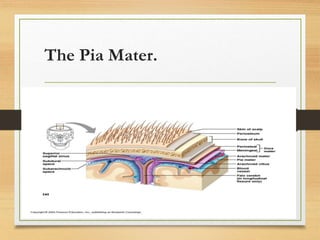
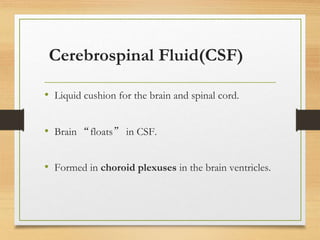
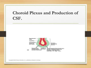
ACUTE CNS INFECTIONS.
(BACTERIAL MENINGITIS
AND CEREBRAL MALARIA).
DR.ODONG RICHARD J.
Paediatrician.
Department of Pediatrics and Child Health –KIUWC.
Definition:
Meningitis:- Inflammation of the leptomeninges.
Leptomeninges are the two innermost layers of tissue
that cover the brain and the spinal cord.
The two layers are called the arachnoid and pia mater.
RELEVANT CNS ANATOMY.
Protection of the Brain.
• Brain is protected from injury by:
• The skull.
• Meninges.
• Cerebrospinal fluid.
• Blood-brain barrier.
Meninges.
• Three meninges.
• Dura mater.
• Arachnoid mater.
• Pia mater.
• Cover and protect the CNS
• Enclose and protect the vessels that supply the CNS
• Contain the cerebrospinal fluid
The Dura Mater.
• Strongest of the meninges.
• Composed of two layers.
• Periosteal layer.
• Meningeal layer.
The Arachnoid Mater.
• Located beneath the Dura mater
• Subdural space
• Potential space between Dura and arachnoid mater
• Subarachnoid space.
• Filled with CSF.
• Contains the blood vessels that supply the brain.
• Arachnoid villi.
• Allow CSF to pass into the dural blood sinuses.
The Pia Mater.
• Delicate connective tissue.
• Clings tightly to the surface of the brain.
• Follows all convolutions of the cortex.
The Pia Mater.
Cerebrospinal Fluid(CSF)
• Liquid cushion for the brain and spinal cord.
• Brain “floats” in CSF.
• Formed in choroid plexuses in the brain ventricles.
Choroid Plexus and Production of
CSF.
Protection of the Brain (Blood-Br
ain Barrier).
• Prevents most blood-borne toxins from entering the
brain.
• Impermeable capillaries
• Not an absolute barrier.
• Nutrients such as oxygen pass through
• Allows alcohol, nicotine, and anesthetics through.
Epidemiology.
• But incidence of meningitis is presumed to be higher in developi
ng countries.
• Because of less access to preventative services such as vaccinatio
n.
• Age- Due to lack of immunity to specific pathogens
associated with young age.
• Sex-Male infants >>>> incidence of gram-negative
neonatal meningitis.
• Female infants >>>> L. monocytogenes infection(Neon
ates).
MENINGITIS BELT.
Importance:
Significant cause of morbidity and mortality amon
g children.
Overall mortality rate is 5 – 10%.
15 – 20% in neonates.
3 -10% in older children.
Mortality rate from S.pneumoniae is 26.3 – 30%,
H. influenza type B is 7.7 – 10.3.
N. meningitidis is 3.5 – 10.3%
High frequency of neurologic sequelae: up to 30%, highest with S pn
eumonia.
Most important causes are preventable through immunization.
(S. pneumonia, Hib, N. meningitides, Mtb, and some viral causes e.g. Measles,
Rubella).
Aetiology.
a) 0 - 2 months.
- Group B streptococci.
- Escherichia coli.
- Listeria monocytogenes.
- Klebsiella, Salmonella.
b) 2 months – 2 years.
- Streptococcus pneumoniae.
- Haemophilus influenza type b.
- Neisseria meningitidis.
c) 3 years and above.
- S. pneumonia.
- N. meningitis.
- Hib.
- Mycobacterium tuberculosis.
d) Unusual bacteria.
- Staphylococcus aureus.
- Pasteurella multocida.
- Mycoplasma.
Predisposing/ Risk factors.
• Age: prematures, neonates.
• Intrauterine infection.
• Maternal infection and pyrexia at delivery.
• Open head trauma (with skull fracture or CSF leakage).
• Contiguous focus of infection e.g. sinusitis, otitis medi
a, mastoiditis, osteomyelitis of skull, periorbital and fac
ial cellulitis, septic arthritis,
Predisposing Factors.
• Open neural tube defects.
• Neurosurgical procedures and patients with ventriculoperitoneal
shunts.
• Immune deficiency (primary or secondary).
• Sickle cell anaemia or asplenia.
• Overcrowding.
• Immunization status.
Pathogenesis.
• Colonization of the nasopharnyx with a potentially pathogenic micr
oorganism.
• Respiratory viruses enhance the entry of bacterial agents into the int
ravascular compartment, presumably by damaging mucosal defenses.
• Leads to systemic invasion and development of a bacteremia.
• Bacterial encapsulation facilitates bacteremia by inhibiting neutrophil
phagocytosis and resisting classic complement-mediated bactericidal
activity.
• Bacteria gain entrance to the CSF through the choroid plexus.
• Rapidly multiply in the CSF (complement and antibody inadequate).
• Production and/or release of virulence factors (peptidoglycans an
d lipopolysaccharides) into CSF.
• Stimulation of formation of inflammatory cytokines(IL-1,TNF-α).
• Net result is vascular endothelial injury and ↑BBB permeabil
ity
• Leading to the entry of many blood components into the subarac
hnoid space.
• PECOLATION OF FLUIDS→ vasogenic edema and ↑ CSF pro
tein.
• In response to the cytokines and chemotactic molecules, neutrop
hils migrate from the bloodstream and penetrate the damaged BB
B, → profound neutrophilic pleocytosis characteristic of bacterial
meningitis.
• Influx of plasma components into the subarachnoid space leads ↑
CSF viscosity & ↓ venous outflow lead to interstitial edema, &
the products of bacterial degradation, neutrophils, and other cellu
lar activation lead to cytotoxic edema.
• Cerebral edema (ie, vasogenic, cytotoxic, interstitial) → intracran
ial hypertension and a consequent ↓ in cerebral blood flow.
• Anaerobic metabolism ensues, → ↑sed lactate concentration and
↓ CSF Glucose. In addition, ↓ CSF Glucose results from ↓glucos
e transport into the spinal fluid compartment.
• If process uncontrolled , transient neuronal dysfunction or perma
nent neuronal injury results.
Clinical features.
• Neonate:
Change in feeding or sleeping habits, irritability, lethargy, vomiting,
high pitched cry, seizures.
Paradoxical irritability (quiet at rest, cries when moved or comforte
d).
Infants:
Fever, lethargy, irritability, change in behaviour, restlessness, seizure
s, coma.
After 2 - 3 years:
Headache, irritability, nausea, vomiting, anorexia, nuchal rigidity, ph
otophobia, confusion, back pain, seizures, coma.
Physical Examination.
Young infant:
Irritable, unconscious.
Febrile, hypothermic.
Bulging fontanelle.
Diastasis of the sutures.
+/- Nuchal rigidity.
Older child:
Meningeal signs: Neck stiffness, +ve Kerning and Brudzinski signs.
Bulging fontanelle.
Ptosis, Sixth nerve palsy, diplopia.
Bradycardia, hypertension and apnea = Cushing’s triad – brain he
rniation.
Focal neurological signs in 15% of patients
Seizures in up to 30% patients.
Altered consciousness and coma 15 – 20%.
DDx: Bacterial Meningitis.
• Cerebral malaria.
• Encephalitis/Viral. Meningoencephalitis.
• Tuberculous meningitis.
• Fungal meningitis.
• Brain abscess.
• Bacterial endocarditis with embolism.
• Intracranial or spinal epidural abscesses.
• Subdural empyema.
• Subarachnoid hemorrhage.
• Brain tumors.
VIRALMENINGITIS/ENCEPHALITS.
Herpesviruses
• Herpes simplex
• Varicella zoster
• Epstein Barr
• Cytomegalovirus
Enteroviruses
• Polio
• Coxsackie
• Echovirus
Para/myoxoviruses
• Para/influenza
• Mumps
• Measles
Togaviruses
• Eastern, Western equine
• Venezuelan,
Clinical Features.
Onset variable;
Fever, general malaise, anorexia, vomiting.
Features of pharyngitis, conjunctivitis, myositis.
Seizures and evidence of encephalitis.
BRAIN ABSCESS.
• Infrequent but not uncommon.
• Cause often polymicrobial.
• Strep, Enteric GNR, anaerobes, Staph in associated with IE.
• Less commonly Nocardia, fungi, Mtb, T.gondii, Neurocysticercosis.
BRAIN ABSCESS
• CF: Headache, fever, mental status changes, hemipar
esis, papillo-edema.
• Diagnosis often suggested by neuro-imaging.
• LP contraindicated due to risk of herniation.
• Drainage often a necessary component of manag
ement.
CERERAL MALARIA.
Encephalopathy that presents with impaired
consciousness, delirium, and seizures.
Onset may be gradual or sudden following a
convulsion.
STANDARD CASE DEFINITION.
Blantyre coma score ≤2.
 Asexual forms of P. falciparum parasitemia (any
density).
No other identifiable cause of coma (eg,
hypoglycemia, meningitis, or a post-ictal state) .
OR.
• Signs and symptoms of malaria.
• Ability to demonstrate presence of asexual forms of
p.falciparum using a peripheral blood film.
• An arousable coma for 30minutes to 1 hour.
• Normal CSF analysis.
Histologic hallmark - cerebral sequestration of
parasitized erythrocytes.
Presence of one or more elements of malaria
retinopathy: white-centered hemorrhages, vessel
changes, and whitening in areas of the retina,
papilledema .( above + this, most reliable).
NOTE.
In the absence of malaria retinopathy, other
causes for coma should be pursued and treated,
even in the presence of established malaria
infestation.
Pathophysiology.
• Seizures and coma:
Intracranial sequestration of metabolically active para
sites, cerebral hypoxia, increased intracranial pressure,
cerebral edema, hypoglycemia, hyponatremia.
MANAGEMENT.
• INVESTIGATIONS
• COMPLICATIONS
• TREATMENT
BLOOD TESTS:
CBC: WBC (4.0-11.0×10ˆ9/L)+ Diff cells predominately neutr
ophils & Plts counts (150-400×10ˆ9/L).
2 aerobic blood cultures; appropriate volumes.
Serum electrolytes & glucose (60-100mg/dL).
Clotting profile (PT (11-15sec), aPTT (33-47sec), TT(11-15sec)). in
case of petechiae or purpuric lesions.
.
Lumbar Puncture for CSF analysis.
1. Opening pressure (11.5-28cmH2O) & Appearance: yellow for ex
cess proteins, turbid, blood staining. Clear and colourless.
2. Cell count: WBC (0-7/mL) and differential cell counts.
3. Chemistry: glucose (40-80mg/dL)and proteins(5-40mg/dL), that is
½-2/3 of serum glucose.
4. Microbiological tests: Gram stain and culture.
Indications:
Suspected CNS infection.
Sudden severe headaches/ worsens when lying down
and improves on standing.
Suspected subarachnoid hemorrhage, DO CT- SCA
N first for all suspects.
Removal of CSF in treatment of idiopathic intracranial h
ypertension.
.
Contraindications: (absolute/relative)
Increased ICP (risk of cerebral herniation Do CT- SCAN)
Bleeding diathesis.
Cardiopulmonary instability.
Soft tissue infection at site.
.
Indications of imaging before LP in children with suspecte
d meningitis.
Coma.
Presence of CSF shunt.
Recent h/o CNS trauma or neurosurgery.
H/o hydrocephalus.
Papilledema.
Focal neurological deficit (except CN VI &VII palsies).
PROTOCAL.
Rapidly progressing disease < 24hrs.
I. No increased ICP; Give antibiotics immediately after LP.
II.With signs of increased ICP/ focal neurological findings; give b
road spectrum antibiotics without LP.
Protracted subacute course- over 1-7 days.
I. Evaluate for increased ICP + focal neurological deficits.
II.Broad spectrum antibiotic initiated before LP & CT- scan.
Interpretation of CSF.
1. Opening pressure & Appearance.
2. WBC >1000/microL, with predominance of neutrophil
s. Traumatic tap; we subtract 1WBC for every 1000 RB
Cs/microL.
3. Glucose : <40mg/dl. (ratio CSF: Blood glucose concen
tration < 0.6).
4. Proteins: CSF protein ranges from100-500mg/dl. Trau
matic tap; its raised coz of RBC lysis. 1mg/dl for every
1000 RB
BACTERIAL MENINGITIS SCORE (BMS).
1-Positive CSF Gm-stain( 2points).
2-CSF absolute neutrophils (ANC) ≥10,000cells/microL.(1p
oint)
3-CSF protein of ≥80mg/dl.(1point)
4-Peripheral blood ANC of ≥10,000cells/microL.(1point)
5- H/O seizure before or at time of presentation.(1 point)
TOTAL CRETERIA POINT C
OUNT.
• RISK OF BACTERIAL MENINGITIS.
0 POINTS. ASEPTIC MENINGITIS.
1 POINT. ASEPTIC MENINGITIS LESS LIKELY.
2-6 POINTS. BACTERIAL MENINGITIS MORE LIKELY.
• BMS as a clinical prediction rule should not be used
in < 2mo, immunocompromised, ill-appearing,
pretreated with antibiotics, have petechiae or purpura
on examination.
• Model used to distinguish bacterial from aseptic
meningitis in children in the post Haemophilus
influenza era.
4.Gram stain/culture:
MORPHOLOGY PATHOGENS.
•Gram- positive diplococcic. S. pneumoniae
•Gram- negative diplococcic. N. meningitidis
•Small pleomorphic gram negative coccobacill
i.
Hib
•Gram-positive cocci or coccobacilli. GBS
•Gram positive rod and coccobacilli. L. monocytogenes
For negative cultures & gram stain; PCR of CSF
is most helpful for documenting meningococcal
disease in patients with Negative cultures.
TREATMENT : emergency
SUPORTIVE CARE.
1. Repeated medical and neurological assessment.
2. Initially nil per os.
3. IV fluids- ½- 2/3 of maintenance (or 800-1000ml/m2/24hr) until its establish
ed that no increased ICP/SIADH. When Na- levels normalise; maintain 15
00-1700ml/m2/24hr.
4. Antibiotics.
5. Corticosteroids. IV dexamethasone 0.15mg/kg 6hly* 2/7
NB: fluid restriction is inappropriate in presence of systemic hypotension th
at may result in reduced cerebral perfusion pressure and CNS ischemia.
Rapid killing of bacteria in the CSF effectively sterilises th
e meningeal infection but releases toxic cell products after
cell lysis( cell wall endotoxin) that preciptate the cytokine-
mediated inflammatory response.
Admit in pead ICU shock, markedly elevated ICP and re
fractory seizures.
Septic shock needs resuscitation with vasoactive agent( dopamine, epi
nephrine & sodium nitroprusside).
Neurological complications; increased ICP resulting into he
rniation, seizures, hydrocephalus.
• Emergently treat signs of increased ICP with endotrac
heal intubation and hyperventilation (maintain PCo2~25m
mHg) + IV frusemide 1mg/kg and mannitol 0.5-1g/kg os
motherapy.
• Seizures; Immediate mgt- IV diazepam 0.1-0.2mg/kg/d
ose.
• IV Phenytoin 15-20mg/kg loading dose, then 5mg/kg/24hr.
INFECTIONS RISK PATIENTS INTIAL/ EMPIRICAL TREATMEN
T
L. monocytogenes 1-2mo
T-lymphocyte def
IV Ampicillin 200mg/kg/24hr; given 6hly +
ceftriaxone/ cefotaxime.
ALT… IV Trimethoprim sulphameth
oxasole 20mg/kg/24hrgiven 6-8hly
Gram negative organisms Immunocompromised pts Ceftazine 150mg/kg/24hr; given 8hly. Max
6g/24hrs + Gentamycin 7.5mg/kg/24h
r; given 8hhly
S. pneumonia Vancomycin 60mg/kg/24hr; given 6hly. +
Cefotaxime 200mg/kg/24hrs; Given 6hly. or
+
Ceftriaxone 100mg/kg/24hrs.
If allegic: chloraphenicol 100mg/kg/24hrs.
N. meningitidis IV PEN 400,000U/Kg/24hr ; given 4-6hly
Cephalospoins e.g ceftriaxone/ cefotaxime.
.
INFECTIONS CONTINUATION
TREATMENT
UNCOMPLICATED PENICILLIN SENSITIV
E S. pneumonia infection.
3rd gen ceph, e.g. ceftriaxone/ cefotaxime.
IV PEN 400,000U/Kg/24hr; given 4-6hly * 10-14
/7
UNCOMPLICATED PENICILLIN RESISTA
NT S. pneumonia infection.
Vancomycin 20mg/kg/ dose * 6hly
UNCOMPLICATED N. meningitidis infectio
n.
IV PEN 400,000U/Kg/24hr ; given 4-6hly * 10-1
4/7
UNCOMPLICATED H. Influenzae type b inf
xn
IV Ampicillin 200-400mg/kg/24hrs; 4-6hrly* 7-10
/7
ESCHERICHIA COLI / P. aeruginosa infxn 3rd gen ceph e.g ceftriaxone/ cefotaxime * 3/52
PREVENTION.
INFECTION RISK CHEMOPROPHYLAX
IS:
VACCINATION:
NEISSERIA MENIN
GITIS
All close contacts of men
ingococcal pts regardless
of age and immunisation
status
Tabs Rifampin 10mg/kg/dos
e 12hly * 2/7 (max 600mg)
Meningococcal quadrivalent
vaccine against serogrou
ps A, C, Y and W135.
HAEMOPHILUS INF
LUENZAE
All household contacts, Tabs Rifampin 20mg/kg/24
hr.(600mg) for only <48m
o/immunocompromise
d family members. CI:
pregnancy
Hemophilus-type b vac
cine(2-6mo of age).
STREPTOCOCCUS P
NEUMONIAE
pts with anatomical/
fxnal asplenia, HIV,
1o immunodef, & th
ose on immunosupp
ressant.
Pentavalent Conjugate
pneumococcal vaccine-
(<2yrs)
COMPLICATIONS.
ACUTE ( during treatment). CHRONIC.
•Seizures. subdural effusions /empyema , hyd
rocephalus , cerebral palsy
•Increased ICP, Cerebral edema. SIADH ( secretion of inappropriate anti-di
uretic hormone)
•CN-palsies Fever>10days
•Stroke anemia, thrombocytosis, eosinophilia.
•Thrombosis of the dural venous sinuses DIC ( disseminated intravascular coagulop
athy)
•Cerebral/cerebellar herniation, Septic shock
.
• Collections of fluid- SUBDURAL EFFUSIONS (10-30% of pts). Bulging f
ontanel, diastasis of sutures, enlarging HC, emesis, seizures, fever, abn cranial transillu
mination. NB: Do ventricular tap for symptomatic subdural effusions, if GC
S<8, raised ICP)
• SIADH; results in hyponatremia & reduced serum osmolarity (30-50
%). This may exacerbate cerebral edema or independently lead to hypona
tremic seizures.
• FEVER: usually resoles 5-7days of onset of therapy. if >10days fever-10%. (Inter
current viral infxn, nosocomial / 2o bacterial infxn, thrombophlebitis, drug rxn).
‘
• THROMBOCYTOSIS, EOSINOPHILIA, & A
NEMIA: anemia 2o hemolysis, BM suppression.
• DIC ( disseminated intravascular coagulopathy): noted in pt
s with rapidly progressive pattern of presentation, sh
ock and purpura. NB: the combination of endotoxemia and
severe hypotension initiates the coagulation cascade> thrombosi
s>symmetric peripheral gangrene.
Cerebral malaria(CM) Vs Acute bacteri
al meningitis(ABM) in children with i
mpaired consciousness.
• According to an Article in QJM: Monthly journal of the
association of physicians 92(3):151-7 April 1999.
• CM and ABM are the two common causes of impaired
consciousness in children presenting to hospitals in sub-
Saharan Africa.
• Features of the two diseases may be very similar, treatment is
often guided by the initial laboratory findings.
CM vs ABM
Stiff neck
Bulging Fontanelle
Arching of the back
Convulsions
Pallor
Confusion
• Data from 555 children with impaired consciousness
admitted to Kilifi district hospital Kenya.
• Defined groups were established based on malaria
slide, cerebral spinal fluids, leucocyte count, results of
blood and CSF culture and CSF bacterial antigen
testing.
• Data suggested significant overlap in the initial
findings between CM and ABM.
• Absolute minimum proportion of children with
impaired consciousness and malaria parasitemia
who also had definite bacterial meningitis were
4% of all children and 14% of children under 1
year of age.
• Estimated maximum proportion of all children with
impaired consciousness and malaria parasitemia in
whom the diagnosis was dual or unclear was at least
13%.
• Findings of malaria parasites in blood of an
unconscious child in sub-Saharan Africa is not
sufficient to establish a diagnosis of cerebral malaria.
• Acute bacterial meningitis must be actively excluded
in all cases.
Rules.
• You cannot distinguish cerebral malaria and meningitis
clinically.
• A febrile child with altered consciousness and / or neur
ological signs must be assessed with both;
• Malaria slide.
• Lumbar puncture.
THANKS.

Bacterial meningitis

  • 1.
    ACUTE CNS INFECTIONS. (BACTERIALMENINGITIS AND CEREBRAL MALARIA). DR.ODONG RICHARD J. Paediatrician. Department of Pediatrics and Child Health –KIUWC.
  • 2.
    Definition: Meningitis:- Inflammation ofthe leptomeninges. Leptomeninges are the two innermost layers of tissue that cover the brain and the spinal cord. The two layers are called the arachnoid and pia mater.
  • 3.
    RELEVANT CNS ANATOMY. Protectionof the Brain. • Brain is protected from injury by: • The skull. • Meninges. • Cerebrospinal fluid. • Blood-brain barrier.
  • 4.
    Meninges. • Three meninges. •Dura mater. • Arachnoid mater. • Pia mater. • Cover and protect the CNS • Enclose and protect the vessels that supply the CNS • Contain the cerebrospinal fluid
  • 5.
    The Dura Mater. •Strongest of the meninges. • Composed of two layers. • Periosteal layer. • Meningeal layer.
  • 6.
    The Arachnoid Mater. •Located beneath the Dura mater • Subdural space • Potential space between Dura and arachnoid mater
  • 7.
    • Subarachnoid space. •Filled with CSF. • Contains the blood vessels that supply the brain. • Arachnoid villi. • Allow CSF to pass into the dural blood sinuses.
  • 8.
    The Pia Mater. •Delicate connective tissue. • Clings tightly to the surface of the brain. • Follows all convolutions of the cortex.
  • 9.
  • 10.
    Cerebrospinal Fluid(CSF) • Liquidcushion for the brain and spinal cord. • Brain “floats” in CSF. • Formed in choroid plexuses in the brain ventricles.
  • 11.
    Choroid Plexus andProduction of CSF.
  • 12.
    Protection of theBrain (Blood-Br ain Barrier). • Prevents most blood-borne toxins from entering the brain. • Impermeable capillaries • Not an absolute barrier. • Nutrients such as oxygen pass through • Allows alcohol, nicotine, and anesthetics through.
  • 13.
    Epidemiology. • But incidenceof meningitis is presumed to be higher in developi ng countries. • Because of less access to preventative services such as vaccinatio n.
  • 14.
    • Age- Dueto lack of immunity to specific pathogens associated with young age. • Sex-Male infants >>>> incidence of gram-negative neonatal meningitis. • Female infants >>>> L. monocytogenes infection(Neon ates).
  • 15.
  • 16.
    Importance: Significant cause ofmorbidity and mortality amon g children. Overall mortality rate is 5 – 10%. 15 – 20% in neonates. 3 -10% in older children. Mortality rate from S.pneumoniae is 26.3 – 30%, H. influenza type B is 7.7 – 10.3. N. meningitidis is 3.5 – 10.3%
  • 17.
    High frequency ofneurologic sequelae: up to 30%, highest with S pn eumonia. Most important causes are preventable through immunization. (S. pneumonia, Hib, N. meningitides, Mtb, and some viral causes e.g. Measles, Rubella).
  • 18.
    Aetiology. a) 0 -2 months. - Group B streptococci. - Escherichia coli. - Listeria monocytogenes. - Klebsiella, Salmonella. b) 2 months – 2 years. - Streptococcus pneumoniae. - Haemophilus influenza type b. - Neisseria meningitidis.
  • 19.
    c) 3 yearsand above. - S. pneumonia. - N. meningitis. - Hib. - Mycobacterium tuberculosis. d) Unusual bacteria. - Staphylococcus aureus. - Pasteurella multocida. - Mycoplasma.
  • 20.
    Predisposing/ Risk factors. •Age: prematures, neonates. • Intrauterine infection. • Maternal infection and pyrexia at delivery. • Open head trauma (with skull fracture or CSF leakage). • Contiguous focus of infection e.g. sinusitis, otitis medi a, mastoiditis, osteomyelitis of skull, periorbital and fac ial cellulitis, septic arthritis,
  • 21.
    Predisposing Factors. • Openneural tube defects. • Neurosurgical procedures and patients with ventriculoperitoneal shunts. • Immune deficiency (primary or secondary). • Sickle cell anaemia or asplenia. • Overcrowding. • Immunization status.
  • 22.
    Pathogenesis. • Colonization ofthe nasopharnyx with a potentially pathogenic micr oorganism. • Respiratory viruses enhance the entry of bacterial agents into the int ravascular compartment, presumably by damaging mucosal defenses. • Leads to systemic invasion and development of a bacteremia.
  • 23.
    • Bacterial encapsulationfacilitates bacteremia by inhibiting neutrophil phagocytosis and resisting classic complement-mediated bactericidal activity. • Bacteria gain entrance to the CSF through the choroid plexus. • Rapidly multiply in the CSF (complement and antibody inadequate).
  • 24.
    • Production and/orrelease of virulence factors (peptidoglycans an d lipopolysaccharides) into CSF. • Stimulation of formation of inflammatory cytokines(IL-1,TNF-α). • Net result is vascular endothelial injury and ↑BBB permeabil ity • Leading to the entry of many blood components into the subarac hnoid space. • PECOLATION OF FLUIDS→ vasogenic edema and ↑ CSF pro tein.
  • 25.
    • In responseto the cytokines and chemotactic molecules, neutrop hils migrate from the bloodstream and penetrate the damaged BB B, → profound neutrophilic pleocytosis characteristic of bacterial meningitis. • Influx of plasma components into the subarachnoid space leads ↑ CSF viscosity & ↓ venous outflow lead to interstitial edema, & the products of bacterial degradation, neutrophils, and other cellu lar activation lead to cytotoxic edema.
  • 26.
    • Cerebral edema(ie, vasogenic, cytotoxic, interstitial) → intracran ial hypertension and a consequent ↓ in cerebral blood flow. • Anaerobic metabolism ensues, → ↑sed lactate concentration and ↓ CSF Glucose. In addition, ↓ CSF Glucose results from ↓glucos e transport into the spinal fluid compartment. • If process uncontrolled , transient neuronal dysfunction or perma nent neuronal injury results.
  • 27.
    Clinical features. • Neonate: Changein feeding or sleeping habits, irritability, lethargy, vomiting, high pitched cry, seizures. Paradoxical irritability (quiet at rest, cries when moved or comforte d).
  • 28.
    Infants: Fever, lethargy, irritability,change in behaviour, restlessness, seizure s, coma. After 2 - 3 years: Headache, irritability, nausea, vomiting, anorexia, nuchal rigidity, ph otophobia, confusion, back pain, seizures, coma.
  • 29.
    Physical Examination. Young infant: Irritable,unconscious. Febrile, hypothermic. Bulging fontanelle. Diastasis of the sutures. +/- Nuchal rigidity.
  • 30.
    Older child: Meningeal signs:Neck stiffness, +ve Kerning and Brudzinski signs. Bulging fontanelle. Ptosis, Sixth nerve palsy, diplopia. Bradycardia, hypertension and apnea = Cushing’s triad – brain he rniation. Focal neurological signs in 15% of patients Seizures in up to 30% patients. Altered consciousness and coma 15 – 20%.
  • 31.
    DDx: Bacterial Meningitis. •Cerebral malaria. • Encephalitis/Viral. Meningoencephalitis. • Tuberculous meningitis. • Fungal meningitis. • Brain abscess.
  • 32.
    • Bacterial endocarditiswith embolism. • Intracranial or spinal epidural abscesses. • Subdural empyema. • Subarachnoid hemorrhage. • Brain tumors.
  • 33.
    VIRALMENINGITIS/ENCEPHALITS. Herpesviruses • Herpes simplex •Varicella zoster • Epstein Barr • Cytomegalovirus Enteroviruses • Polio • Coxsackie • Echovirus Para/myoxoviruses • Para/influenza • Mumps • Measles Togaviruses • Eastern, Western equine • Venezuelan,
  • 34.
    Clinical Features. Onset variable; Fever,general malaise, anorexia, vomiting. Features of pharyngitis, conjunctivitis, myositis. Seizures and evidence of encephalitis.
  • 35.
    BRAIN ABSCESS. • Infrequentbut not uncommon. • Cause often polymicrobial. • Strep, Enteric GNR, anaerobes, Staph in associated with IE. • Less commonly Nocardia, fungi, Mtb, T.gondii, Neurocysticercosis.
  • 36.
    BRAIN ABSCESS • CF:Headache, fever, mental status changes, hemipar esis, papillo-edema. • Diagnosis often suggested by neuro-imaging. • LP contraindicated due to risk of herniation. • Drainage often a necessary component of manag ement.
  • 37.
    CERERAL MALARIA. Encephalopathy thatpresents with impaired consciousness, delirium, and seizures. Onset may be gradual or sudden following a convulsion.
  • 38.
    STANDARD CASE DEFINITION. Blantyrecoma score ≤2.  Asexual forms of P. falciparum parasitemia (any density). No other identifiable cause of coma (eg, hypoglycemia, meningitis, or a post-ictal state) .
  • 39.
    OR. • Signs andsymptoms of malaria. • Ability to demonstrate presence of asexual forms of p.falciparum using a peripheral blood film. • An arousable coma for 30minutes to 1 hour. • Normal CSF analysis.
  • 40.
    Histologic hallmark -cerebral sequestration of parasitized erythrocytes. Presence of one or more elements of malaria retinopathy: white-centered hemorrhages, vessel changes, and whitening in areas of the retina, papilledema .( above + this, most reliable).
  • 41.
    NOTE. In the absenceof malaria retinopathy, other causes for coma should be pursued and treated, even in the presence of established malaria infestation.
  • 42.
    Pathophysiology. • Seizures andcoma: Intracranial sequestration of metabolically active para sites, cerebral hypoxia, increased intracranial pressure, cerebral edema, hypoglycemia, hyponatremia.
  • 43.
  • 44.
    BLOOD TESTS: CBC: WBC(4.0-11.0×10ˆ9/L)+ Diff cells predominately neutr ophils & Plts counts (150-400×10ˆ9/L). 2 aerobic blood cultures; appropriate volumes.
  • 45.
    Serum electrolytes &glucose (60-100mg/dL). Clotting profile (PT (11-15sec), aPTT (33-47sec), TT(11-15sec)). in case of petechiae or purpuric lesions.
  • 46.
    . Lumbar Puncture forCSF analysis. 1. Opening pressure (11.5-28cmH2O) & Appearance: yellow for ex cess proteins, turbid, blood staining. Clear and colourless. 2. Cell count: WBC (0-7/mL) and differential cell counts. 3. Chemistry: glucose (40-80mg/dL)and proteins(5-40mg/dL), that is ½-2/3 of serum glucose. 4. Microbiological tests: Gram stain and culture.
  • 47.
    Indications: Suspected CNS infection. Suddensevere headaches/ worsens when lying down and improves on standing. Suspected subarachnoid hemorrhage, DO CT- SCA N first for all suspects. Removal of CSF in treatment of idiopathic intracranial h ypertension.
  • 48.
    . Contraindications: (absolute/relative) Increased ICP(risk of cerebral herniation Do CT- SCAN) Bleeding diathesis. Cardiopulmonary instability. Soft tissue infection at site.
  • 49.
    . Indications of imagingbefore LP in children with suspecte d meningitis. Coma. Presence of CSF shunt. Recent h/o CNS trauma or neurosurgery. H/o hydrocephalus. Papilledema. Focal neurological deficit (except CN VI &VII palsies).
  • 50.
    PROTOCAL. Rapidly progressing disease< 24hrs. I. No increased ICP; Give antibiotics immediately after LP. II.With signs of increased ICP/ focal neurological findings; give b road spectrum antibiotics without LP. Protracted subacute course- over 1-7 days. I. Evaluate for increased ICP + focal neurological deficits. II.Broad spectrum antibiotic initiated before LP & CT- scan.
  • 51.
    Interpretation of CSF. 1.Opening pressure & Appearance. 2. WBC >1000/microL, with predominance of neutrophil s. Traumatic tap; we subtract 1WBC for every 1000 RB Cs/microL. 3. Glucose : <40mg/dl. (ratio CSF: Blood glucose concen tration < 0.6). 4. Proteins: CSF protein ranges from100-500mg/dl. Trau matic tap; its raised coz of RBC lysis. 1mg/dl for every 1000 RB
  • 52.
    BACTERIAL MENINGITIS SCORE(BMS). 1-Positive CSF Gm-stain( 2points). 2-CSF absolute neutrophils (ANC) ≥10,000cells/microL.(1p oint) 3-CSF protein of ≥80mg/dl.(1point) 4-Peripheral blood ANC of ≥10,000cells/microL.(1point) 5- H/O seizure before or at time of presentation.(1 point)
  • 53.
    TOTAL CRETERIA POINTC OUNT. • RISK OF BACTERIAL MENINGITIS. 0 POINTS. ASEPTIC MENINGITIS. 1 POINT. ASEPTIC MENINGITIS LESS LIKELY. 2-6 POINTS. BACTERIAL MENINGITIS MORE LIKELY.
  • 54.
    • BMS asa clinical prediction rule should not be used in < 2mo, immunocompromised, ill-appearing, pretreated with antibiotics, have petechiae or purpura on examination. • Model used to distinguish bacterial from aseptic meningitis in children in the post Haemophilus influenza era.
  • 55.
    4.Gram stain/culture: MORPHOLOGY PATHOGENS. •Gram-positive diplococcic. S. pneumoniae •Gram- negative diplococcic. N. meningitidis •Small pleomorphic gram negative coccobacill i. Hib •Gram-positive cocci or coccobacilli. GBS •Gram positive rod and coccobacilli. L. monocytogenes
  • 56.
    For negative cultures& gram stain; PCR of CSF is most helpful for documenting meningococcal disease in patients with Negative cultures.
  • 57.
    TREATMENT : emergency SUPORTIVECARE. 1. Repeated medical and neurological assessment. 2. Initially nil per os. 3. IV fluids- ½- 2/3 of maintenance (or 800-1000ml/m2/24hr) until its establish ed that no increased ICP/SIADH. When Na- levels normalise; maintain 15 00-1700ml/m2/24hr. 4. Antibiotics. 5. Corticosteroids. IV dexamethasone 0.15mg/kg 6hly* 2/7 NB: fluid restriction is inappropriate in presence of systemic hypotension th at may result in reduced cerebral perfusion pressure and CNS ischemia.
  • 58.
    Rapid killing ofbacteria in the CSF effectively sterilises th e meningeal infection but releases toxic cell products after cell lysis( cell wall endotoxin) that preciptate the cytokine- mediated inflammatory response. Admit in pead ICU shock, markedly elevated ICP and re fractory seizures. Septic shock needs resuscitation with vasoactive agent( dopamine, epi nephrine & sodium nitroprusside). Neurological complications; increased ICP resulting into he rniation, seizures, hydrocephalus.
  • 59.
    • Emergently treatsigns of increased ICP with endotrac heal intubation and hyperventilation (maintain PCo2~25m mHg) + IV frusemide 1mg/kg and mannitol 0.5-1g/kg os motherapy. • Seizures; Immediate mgt- IV diazepam 0.1-0.2mg/kg/d ose. • IV Phenytoin 15-20mg/kg loading dose, then 5mg/kg/24hr.
  • 60.
    INFECTIONS RISK PATIENTSINTIAL/ EMPIRICAL TREATMEN T L. monocytogenes 1-2mo T-lymphocyte def IV Ampicillin 200mg/kg/24hr; given 6hly + ceftriaxone/ cefotaxime. ALT… IV Trimethoprim sulphameth oxasole 20mg/kg/24hrgiven 6-8hly Gram negative organisms Immunocompromised pts Ceftazine 150mg/kg/24hr; given 8hly. Max 6g/24hrs + Gentamycin 7.5mg/kg/24h r; given 8hhly S. pneumonia Vancomycin 60mg/kg/24hr; given 6hly. + Cefotaxime 200mg/kg/24hrs; Given 6hly. or + Ceftriaxone 100mg/kg/24hrs. If allegic: chloraphenicol 100mg/kg/24hrs. N. meningitidis IV PEN 400,000U/Kg/24hr ; given 4-6hly Cephalospoins e.g ceftriaxone/ cefotaxime.
  • 61.
    . INFECTIONS CONTINUATION TREATMENT UNCOMPLICATED PENICILLINSENSITIV E S. pneumonia infection. 3rd gen ceph, e.g. ceftriaxone/ cefotaxime. IV PEN 400,000U/Kg/24hr; given 4-6hly * 10-14 /7 UNCOMPLICATED PENICILLIN RESISTA NT S. pneumonia infection. Vancomycin 20mg/kg/ dose * 6hly UNCOMPLICATED N. meningitidis infectio n. IV PEN 400,000U/Kg/24hr ; given 4-6hly * 10-1 4/7 UNCOMPLICATED H. Influenzae type b inf xn IV Ampicillin 200-400mg/kg/24hrs; 4-6hrly* 7-10 /7 ESCHERICHIA COLI / P. aeruginosa infxn 3rd gen ceph e.g ceftriaxone/ cefotaxime * 3/52
  • 62.
    PREVENTION. INFECTION RISK CHEMOPROPHYLAX IS: VACCINATION: NEISSERIAMENIN GITIS All close contacts of men ingococcal pts regardless of age and immunisation status Tabs Rifampin 10mg/kg/dos e 12hly * 2/7 (max 600mg) Meningococcal quadrivalent vaccine against serogrou ps A, C, Y and W135. HAEMOPHILUS INF LUENZAE All household contacts, Tabs Rifampin 20mg/kg/24 hr.(600mg) for only <48m o/immunocompromise d family members. CI: pregnancy Hemophilus-type b vac cine(2-6mo of age). STREPTOCOCCUS P NEUMONIAE pts with anatomical/ fxnal asplenia, HIV, 1o immunodef, & th ose on immunosupp ressant. Pentavalent Conjugate pneumococcal vaccine- (<2yrs)
  • 63.
    COMPLICATIONS. ACUTE ( duringtreatment). CHRONIC. •Seizures. subdural effusions /empyema , hyd rocephalus , cerebral palsy •Increased ICP, Cerebral edema. SIADH ( secretion of inappropriate anti-di uretic hormone) •CN-palsies Fever>10days •Stroke anemia, thrombocytosis, eosinophilia. •Thrombosis of the dural venous sinuses DIC ( disseminated intravascular coagulop athy) •Cerebral/cerebellar herniation, Septic shock
  • 64.
    . • Collections offluid- SUBDURAL EFFUSIONS (10-30% of pts). Bulging f ontanel, diastasis of sutures, enlarging HC, emesis, seizures, fever, abn cranial transillu mination. NB: Do ventricular tap for symptomatic subdural effusions, if GC S<8, raised ICP) • SIADH; results in hyponatremia & reduced serum osmolarity (30-50 %). This may exacerbate cerebral edema or independently lead to hypona tremic seizures. • FEVER: usually resoles 5-7days of onset of therapy. if >10days fever-10%. (Inter current viral infxn, nosocomial / 2o bacterial infxn, thrombophlebitis, drug rxn).
  • 65.
    ‘ • THROMBOCYTOSIS, EOSINOPHILIA,& A NEMIA: anemia 2o hemolysis, BM suppression. • DIC ( disseminated intravascular coagulopathy): noted in pt s with rapidly progressive pattern of presentation, sh ock and purpura. NB: the combination of endotoxemia and severe hypotension initiates the coagulation cascade> thrombosi s>symmetric peripheral gangrene.
  • 66.
    Cerebral malaria(CM) VsAcute bacteri al meningitis(ABM) in children with i mpaired consciousness. • According to an Article in QJM: Monthly journal of the association of physicians 92(3):151-7 April 1999. • CM and ABM are the two common causes of impaired consciousness in children presenting to hospitals in sub- Saharan Africa. • Features of the two diseases may be very similar, treatment is often guided by the initial laboratory findings.
  • 67.
    CM vs ABM Stiffneck Bulging Fontanelle Arching of the back Convulsions Pallor Confusion
  • 68.
    • Data from555 children with impaired consciousness admitted to Kilifi district hospital Kenya. • Defined groups were established based on malaria slide, cerebral spinal fluids, leucocyte count, results of blood and CSF culture and CSF bacterial antigen testing. • Data suggested significant overlap in the initial findings between CM and ABM.
  • 69.
    • Absolute minimumproportion of children with impaired consciousness and malaria parasitemia who also had definite bacterial meningitis were 4% of all children and 14% of children under 1 year of age. • Estimated maximum proportion of all children with impaired consciousness and malaria parasitemia in whom the diagnosis was dual or unclear was at least 13%.
  • 70.
    • Findings ofmalaria parasites in blood of an unconscious child in sub-Saharan Africa is not sufficient to establish a diagnosis of cerebral malaria. • Acute bacterial meningitis must be actively excluded in all cases.
  • 71.
    Rules. • You cannotdistinguish cerebral malaria and meningitis clinically. • A febrile child with altered consciousness and / or neur ological signs must be assessed with both; • Malaria slide. • Lumbar puncture.
  • 72.

Editor's Notes

  • #52 NB. Older than 3yrs abn >6 WBC/microL, 29-90mo abn >9 WBC/microL.
  • #53 BMS is aclinical prediction rule for chlidren with pleocytosis(WBC count≥10cells/microL) not used in < 2mo, immunocompromiesd, ill-appearing, pretreated with antibiotics, have petechiae or purpura on examination.
  • #58 Nb. Rapid killing of bacteria in the CSF effectively sterilises the meningeal infection but releases toxic cell products after cell lysis( cell wall endotoxin) that preciptate the cytokine- mediated inflammatory response.
  • #59 Patients with shock, markedly elevated ICP and refractory seizures require intensive monitoring with cerebral arterial & venous access, vital signs & admit in pead ICU. Septic shock needs resuscitation with vasoactive agent( dopamine, epinephrine & sodium nitroprusside).
  • #65 Secndary fever refers to the recrudescence of elevated temp after an afebrile interval.