MENINGITIS
COVERING OF THE BRAIN
1. PIA MATTER
2. Arachonid matter
3. Duramatter
Routes of entry
Introduction
• Meningitis is usually caused by a bacterial
infection but can also be viral or fungal.
Vaccines can prevent some forms of
meningitis.
Introduction
• Rare
• Fewer than 1 million cases per year (India)
• Some types preventable by vaccine
• Treatable by a medical professional
• Spreads by airborne droplets
• Requires a medical diagnosis
• Lab tests or imaging always required
• Short-term: resolves within days to weeks
• Critical: needs emergency care
DEFINITION
• Meningitis is an acute inflammation of the
meningeal tissues surrounding the brain and
the spinal cord (meninges) OR
• inflammation (swelling) of the protective
membranes covering the brain and spinal
cord.
TYPES
• Viral meningitis
• Bacterial meningitis
• Fungal meningitis
• Parasitic meningitis
Viral Meningitis
• It is more common than bacterial Meningitis
and usually less serious.
• Less likely to have permanent brain damage
after the infection resolves.
• Treatment: No specific treatment available.
• Most patients recover completely on their
own
Causative organism
• Enterovirus
• Adenovirus
• Arbovirus
• Measles virus
• Herpes simplex virus
• Varicella
Bacterial Meningitis
• Bacterial Meningitis also known as septic
meningitis.
• Extremely serious that requires immediate
care.
• It Can lead to permanent damage of brain or
disability and death.
• Spreads by: -coughing or sneezing
Causative Agents:
• Streptococcus Pneumonia 30-80
• Neisseria meningitis 15- 40%
• Hemophilus Influenza 2-7%
Treatment
• Antibiotics as per causative organism. •
• Tubercular Meningitis.
• TB meningitis is caused by Mycobacterium
tuberculi.
• Infection with this bacterium begins usually in
the lungs
• 1 – 2% of cases the bacteria travel via the
bloodstream.
•
Fungal Meningitis
• It is much less common than the other two
infections.
• It is rare in healthy people but it is more likely
in persons who have impaired immune
system.
Pathophysiology
• Bacteria enters blood stream/ trauma
• Enters the mucosal surface/ cavity
• Breakdown of normal barriers
• Crosses the blood brain barrier
• Proliferates in the CSF
• Inflammation of the meninges
• Increase in ICP
Clinical Manifestations
• a high temperature.
• cold hands and feet.
• vomiting.
• confusion.
• breathing quickly.
• muscle and joint pain.
• pale, mottled or blotchy skin (this may be harder
to see on brown or black skin)
• spots or a rash (this may be harder to see on
brown or black skin)
Clinical Manifestations
KERIG’S SIGN
Severe stiffness of the hamstrings (The
muscles in the posterior compartment of
the thigh are collectively known as the
hamstrings )causes an inability to
straighten the leg when the hip is flexed to
90 degrees.
BRUDZINKI’SIGN
Severe neck stiffness causes a patient's
hips and knees to flex when the neck is
flexed.
Assessment and Diagnosis
• • History taking
• • Physical assessment
• • CT and MRI
• • Blood culture and sensitivity
• • Lumbar Puncture
Medical Management
Bacterial meningitis:
• Third-generation cefalosporin such as
cefotaxime or ceftriaxone
• Vancomycin is added in the regime in case of
resistance.
• Dexamethasone
• Dehydration and shock can be treated with
fluid therapy.
• Phenytoin for seizure management.
Tubercular Meningitis:
 ATT medications are started: Isoniazid; rifampacin;
pyrazinamide and streptomycin.
 Second line drugs:
Aminoglycosides(Amikacine.Gentamicine.Kanamycine.
Néomycine.Plazomicine.Streptomycine.Tobramycine.)
Fluroquinolones
(Ciprofloxacine,Lévofloxaxine,Ofloxacine,Norfloxacine,
Moxifloxacine ,Loméfloxacine)
 Conventional therapy is given for 6-9 months
 In children BCG vaccine offers (approx 64%) protective
effect
Viral Meningitis:
• Treatment is mostly supportive and no
medicines are prescribed.
• Seizure prophylaxis: Lorazepam or phenytoin
or barbiturate.
• Increased ICP: Inj. Mannitol 1g/kg followed by
0.25- 0.5g/kg Q6H or/and dexamethasone
• Rest is advised
• In case hydrocephalus is present VP shunt is
required.
• Adequate hydration is to be maintained
• Antipyretics
• Anti emetics
NURSING MANAGEMENT
• ASSESSMENT
• Obtain a history of recent infections such as
upper respiratory infection, and exposure to
causative agents.
• Assess neurologic status and vital signs.
• Evaluate for signs of meningeal irritation.
• Assess sensorineural hearing loss (vision and
hearing), cranial nerve damage (eg, facial nerve
palsy), and diminished cognitive function.
NURSING DIAGNOSIS
• Ineffective Tissue Perfusion (cerebral) related
to infectious process and cerebral edema
• Hyperthermia related to the infectious
process and cerebral edema
• Fluid Volume Deficit related to fever and
decreased intake
• Acute Pain related to meningeal irritation
• Impaired Physical Mobility related to
prolonged bed rest
• GOAL :
Nursing diagnosis
Ineffective Tissue Perfusion (cerebral) related to infectious
process and cerebral edema.
Goal; To Enhanced Cerebral Tissue Perfusion
NURSING INTERVENTIONS
• Enhancing Cerebral Perfusion
• Assess LOC, vital signs, and neurologic parameters
frequently. Observe for signs and symptoms of ICP (e.g.
decreased LOC, dilated pupils, widening pulse pressure).
• Maintain a quiet, calm environment to prevent agitation,
which may cause an increased ICP.
• Prepare patient for a lumbar puncture for CSF evaluation,
and repeat spinal tap, if indicated. Lumbar puncture
typically precedes neuroimaging
• Notify the health care provider of signs of deterioration:
increasing temperature, decreasing LOC, seizure activity, or
altered respirations.
• I/V mannitol is administered.
Nursing diagnosis
• Hyperthermia related to the infectious process
and cerebral edema
GOAL ;To Reduce Fever
Nursing intervention:
• Administer antimicrobial agents on time to
maintain optimal blood levels.
• Monitor temperature frequently or continuously.
• Institute other cooling measures, such as a
hypothermia blanket, as indicated.
• Administer antipyretics as ordered like
paracetamol.
Nursing diagnosis
Fluid Volume Deficit related to fever and
decreased intake
GOAL ; To Maintain Fluid Balance
Nursing intervention:
• Prevent I.V. fluid overload, which may worsen
cerebral edema.
• Monitor intake and output closely.
• Monitor CVP frequently.
• Administration of osmotic diuretic- mannitol
Nursing diagnosis
Acute Pain related to meningeal irritation
GOAL ; To Reduce Pain
Nursing intervention:
• Assess level, intensity, duration & location of pain.
• Darken the room if photophobia is present.
• Assist with position of comfort for neck stiffness, and
turn patient slowly and carefully with head and neck in
alignment.
• Elevate the head of the bed to decrease ICP and
reduce pain.
• Administer analgesics as ordered; monitor for
response and adverse reactions. Avoid opioids, which
may decreasing LOC.
• Nursing Diagnosis; Impaired Physical Mobility
related to prolonged bed rest
• Goal; To Return to Optimal Level of
Functioning/ mobility
• Nursing intervention
• Promoting Return to Optimal Level of
Functioning
• Implement rehabilitation interventions after
admission (eg, turning, positioning).
• Progress from passive to active exercises
based on the patient's neurologic status.
CONCLUSION
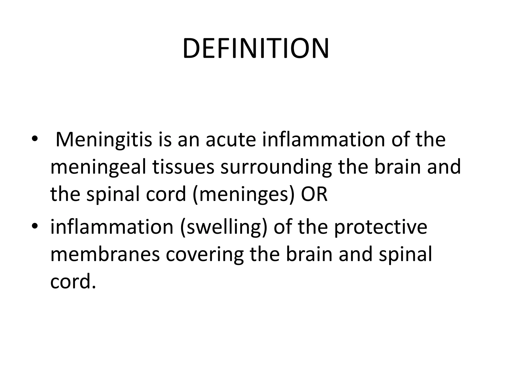
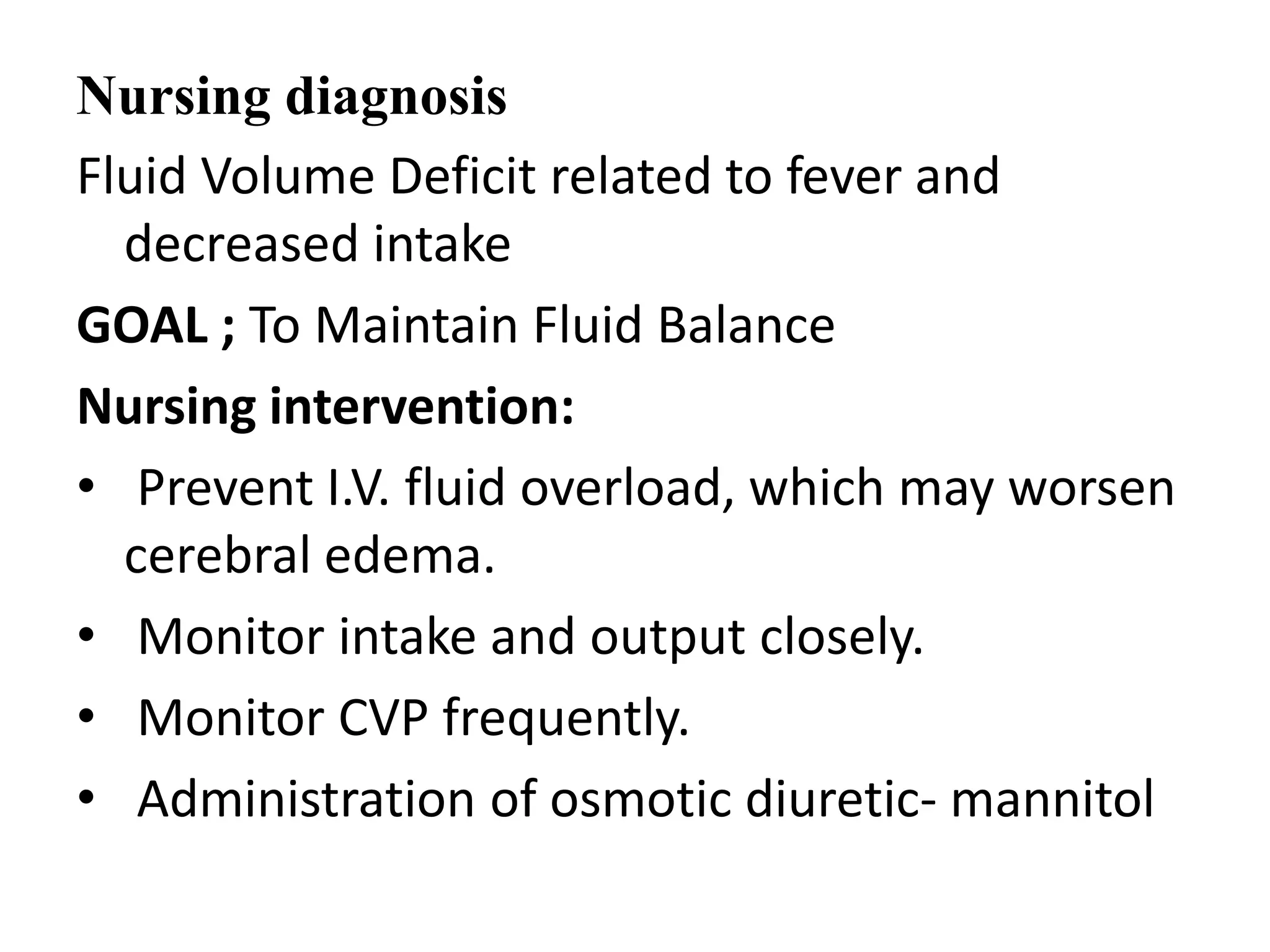
• Meningitis is an inflammation of the
meninges. The meninges are the three
membranes that cover the brain and spinal
cord. Meningitis can occur when fluid
surrounding the meninges becomes infected.
•
REFERENCES
• Boyer Jo Mary(2004), Textbook Of Medical
Surgical Nursing, Philadelphia, Lippincott
William & Wilkins.
• Lewis Mantik Sharon et. Al. (2000), Medical
Surgical Nursing, Assessment & Management
Of Clinical Problems, St. Louis, Missouri,
Mosby Publishers.
• Lippincott (2001), Manual of Nursing Practice,
J.P. Brothers, Philadelphia.

Menigitis final.ppt medical surgical nursing

  • 1.
  • 2.
    COVERING OF THEBRAIN 1. PIA MATTER 2. Arachonid matter 3. Duramatter
  • 3.
  • 4.
    Introduction • Meningitis isusually caused by a bacterial infection but can also be viral or fungal. Vaccines can prevent some forms of meningitis.
  • 5.
    Introduction • Rare • Fewerthan 1 million cases per year (India) • Some types preventable by vaccine • Treatable by a medical professional • Spreads by airborne droplets • Requires a medical diagnosis • Lab tests or imaging always required • Short-term: resolves within days to weeks • Critical: needs emergency care
  • 6.
    DEFINITION • Meningitis isan acute inflammation of the meningeal tissues surrounding the brain and the spinal cord (meninges) OR • inflammation (swelling) of the protective membranes covering the brain and spinal cord.
  • 7.
    TYPES • Viral meningitis •Bacterial meningitis • Fungal meningitis • Parasitic meningitis
  • 8.
    Viral Meningitis • Itis more common than bacterial Meningitis and usually less serious. • Less likely to have permanent brain damage after the infection resolves. • Treatment: No specific treatment available. • Most patients recover completely on their own
  • 9.
    Causative organism • Enterovirus •Adenovirus • Arbovirus • Measles virus • Herpes simplex virus • Varicella
  • 10.
    Bacterial Meningitis • BacterialMeningitis also known as septic meningitis. • Extremely serious that requires immediate care. • It Can lead to permanent damage of brain or disability and death. • Spreads by: -coughing or sneezing
  • 11.
    Causative Agents: • StreptococcusPneumonia 30-80 • Neisseria meningitis 15- 40% • Hemophilus Influenza 2-7%
  • 12.
    Treatment • Antibiotics asper causative organism. • • Tubercular Meningitis. • TB meningitis is caused by Mycobacterium tuberculi. • Infection with this bacterium begins usually in the lungs • 1 – 2% of cases the bacteria travel via the bloodstream. •
  • 13.
    Fungal Meningitis • Itis much less common than the other two infections. • It is rare in healthy people but it is more likely in persons who have impaired immune system.
  • 14.
    Pathophysiology • Bacteria entersblood stream/ trauma • Enters the mucosal surface/ cavity • Breakdown of normal barriers • Crosses the blood brain barrier • Proliferates in the CSF • Inflammation of the meninges • Increase in ICP
  • 16.
    Clinical Manifestations • ahigh temperature. • cold hands and feet. • vomiting. • confusion. • breathing quickly. • muscle and joint pain. • pale, mottled or blotchy skin (this may be harder to see on brown or black skin) • spots or a rash (this may be harder to see on brown or black skin)
  • 17.
    Clinical Manifestations KERIG’S SIGN Severestiffness of the hamstrings (The muscles in the posterior compartment of the thigh are collectively known as the hamstrings )causes an inability to straighten the leg when the hip is flexed to 90 degrees. BRUDZINKI’SIGN Severe neck stiffness causes a patient's hips and knees to flex when the neck is flexed.
  • 18.
    Assessment and Diagnosis •• History taking • • Physical assessment • • CT and MRI • • Blood culture and sensitivity • • Lumbar Puncture
  • 19.
    Medical Management Bacterial meningitis: •Third-generation cefalosporin such as cefotaxime or ceftriaxone • Vancomycin is added in the regime in case of resistance. • Dexamethasone • Dehydration and shock can be treated with fluid therapy. • Phenytoin for seizure management.
  • 20.
    Tubercular Meningitis:  ATTmedications are started: Isoniazid; rifampacin; pyrazinamide and streptomycin.  Second line drugs: Aminoglycosides(Amikacine.Gentamicine.Kanamycine. Néomycine.Plazomicine.Streptomycine.Tobramycine.) Fluroquinolones (Ciprofloxacine,Lévofloxaxine,Ofloxacine,Norfloxacine, Moxifloxacine ,Loméfloxacine)  Conventional therapy is given for 6-9 months  In children BCG vaccine offers (approx 64%) protective effect
  • 21.
    Viral Meningitis: • Treatmentis mostly supportive and no medicines are prescribed. • Seizure prophylaxis: Lorazepam or phenytoin or barbiturate. • Increased ICP: Inj. Mannitol 1g/kg followed by 0.25- 0.5g/kg Q6H or/and dexamethasone • Rest is advised
  • 22.
    • In casehydrocephalus is present VP shunt is required. • Adequate hydration is to be maintained • Antipyretics • Anti emetics
  • 23.
    NURSING MANAGEMENT • ASSESSMENT •Obtain a history of recent infections such as upper respiratory infection, and exposure to causative agents. • Assess neurologic status and vital signs. • Evaluate for signs of meningeal irritation. • Assess sensorineural hearing loss (vision and hearing), cranial nerve damage (eg, facial nerve palsy), and diminished cognitive function.
  • 24.
    NURSING DIAGNOSIS • IneffectiveTissue Perfusion (cerebral) related to infectious process and cerebral edema • Hyperthermia related to the infectious process and cerebral edema • Fluid Volume Deficit related to fever and decreased intake • Acute Pain related to meningeal irritation • Impaired Physical Mobility related to prolonged bed rest
  • 25.
  • 26.
    Nursing diagnosis Ineffective TissuePerfusion (cerebral) related to infectious process and cerebral edema. Goal; To Enhanced Cerebral Tissue Perfusion NURSING INTERVENTIONS • Enhancing Cerebral Perfusion • Assess LOC, vital signs, and neurologic parameters frequently. Observe for signs and symptoms of ICP (e.g. decreased LOC, dilated pupils, widening pulse pressure). • Maintain a quiet, calm environment to prevent agitation, which may cause an increased ICP. • Prepare patient for a lumbar puncture for CSF evaluation, and repeat spinal tap, if indicated. Lumbar puncture typically precedes neuroimaging • Notify the health care provider of signs of deterioration: increasing temperature, decreasing LOC, seizure activity, or altered respirations. • I/V mannitol is administered.
  • 27.
    Nursing diagnosis • Hyperthermiarelated to the infectious process and cerebral edema GOAL ;To Reduce Fever Nursing intervention: • Administer antimicrobial agents on time to maintain optimal blood levels. • Monitor temperature frequently or continuously. • Institute other cooling measures, such as a hypothermia blanket, as indicated. • Administer antipyretics as ordered like paracetamol.
  • 28.
    Nursing diagnosis Fluid VolumeDeficit related to fever and decreased intake GOAL ; To Maintain Fluid Balance Nursing intervention: • Prevent I.V. fluid overload, which may worsen cerebral edema. • Monitor intake and output closely. • Monitor CVP frequently. • Administration of osmotic diuretic- mannitol
  • 29.
    Nursing diagnosis Acute Painrelated to meningeal irritation GOAL ; To Reduce Pain Nursing intervention: • Assess level, intensity, duration & location of pain. • Darken the room if photophobia is present. • Assist with position of comfort for neck stiffness, and turn patient slowly and carefully with head and neck in alignment. • Elevate the head of the bed to decrease ICP and reduce pain. • Administer analgesics as ordered; monitor for response and adverse reactions. Avoid opioids, which may decreasing LOC.
  • 30.
    • Nursing Diagnosis;Impaired Physical Mobility related to prolonged bed rest • Goal; To Return to Optimal Level of Functioning/ mobility • Nursing intervention • Promoting Return to Optimal Level of Functioning • Implement rehabilitation interventions after admission (eg, turning, positioning). • Progress from passive to active exercises based on the patient's neurologic status.
  • 31.
    CONCLUSION • Meningitis isan inflammation of the meninges. The meninges are the three membranes that cover the brain and spinal cord. Meningitis can occur when fluid surrounding the meninges becomes infected. •
  • 32.
    REFERENCES • Boyer JoMary(2004), Textbook Of Medical Surgical Nursing, Philadelphia, Lippincott William & Wilkins. • Lewis Mantik Sharon et. Al. (2000), Medical Surgical Nursing, Assessment & Management Of Clinical Problems, St. Louis, Missouri, Mosby Publishers. • Lippincott (2001), Manual of Nursing Practice, J.P. Brothers, Philadelphia.