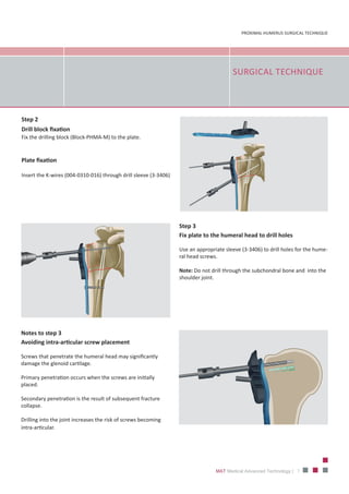
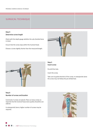
PROXIMALHUMERUSSURGICALTECHNIQUE
FOR TITANIUM AND STEEL IMPLANT SYSTEM
PROXIMAL HUMERUS SURGICAL TECHNIQUE
2 | MAT Medical Advanced Technology
MAT– a committed specialist
that cares for clinical resources
Specialized in the fields of orthopedics, traumatology and spinal
column, MAT innovative products and solutions, from which you as
a dealer benefit just like the respectively responsible doctor or the
OP personnel and - beyond the economic and administrative aspect -
clinic management as well.
The excellent quality and precision of our products, as well as the
willingness to adapt our developments exclusively to your
requirements, are the most important elements of our offered
products and services.
Our product ideas help you to preserve the most precious resources
of daily clinic operations: Nerves, time and money.
3
PROXIMAL HUMERUS SURGICAL TECHNIQUE
MAT Medical Advanced Technology |
SMALL FRAGMENT
LOCKING INSTRUMENT
SET OVERVIEW
PROXIMAL HUMERUS SURGICAL TECHNIQUE
4 | MAT Medical Advanced Technology
INDICATIONS
I. Proximal Humerus Plate Short Plate Version
•	 Dislocated fractures of the proximal humerus (2, 3 and 4), including fractures involving osteopenic bone
•	 Pseudarthroses
•	 Osteotomies
II. Proximal Humerus Plate Long Plate Version
•	 As for short version, but for fractures extending to the shaft or without medial support
5
PROXIMAL HUMERUS SURGICAL TECHNIQUE
MAT Medical Advanced Technology |
SCREW ANGLE
PROXIMAL HUMERUS SURGICAL TECHNIQUE
6 | MAT Medical Advanced Technology
PATIENT
POSITIONING
Step 1
Plate position
The correct plate position is between 5-8 mm distal to the top
of the greater tuberosity.
Aligned the plate along the axis of the humeral shaft slightly
posterior to the bicipital grove (2-4 mm).
SURGICAL TECHNIQUE
Place the patient in the supine position on a radiolucent table.
Ensure the fluoroscope is positioned in a way that allows visualization
of the proximal humerus in two axes (AP and lateral/axial). 
Prepare the patient’s arm so that it can be mobilized intraoperatively.
7
PROXIMAL HUMERUS SURGICAL TECHNIQUE
MAT Medical Advanced Technology |
SURGICAL TECHNIQUE
Step 3
Fix plate to the humeral head to drill holes
Use an appropriate sleeve (3‐3406) to drill holes for the hume-
ral head screws.
Note: Do not drill through the subchondral bone and  into the
shoulder joint.
Notes to step 3
Avoiding intra-articular screw placement
Screws that penetrate the humeral head may significantly
damage the glenoid cartilage.
Primary penetration occurs when the screws are initially
placed.
Secondary penetration is the result of subsequent fracture
collapse.
Drilling into the joint increases the risk of screws becoming
intra-articular.
Step 2
Drill block fixation
Fix the drilling block (Block‐PHMA‐M) to the plate.
Plate fixation
Insert the K‐wires (004‐0310‐016) through drill sleeve (3‐3406)
PROXIMAL HUMERUS SURGICAL TECHNIQUE
8 | MAT Medical Advanced Technology
SURGICAL TECHNIQUE
Step 5
Insert screw
Pre-drill the hole.
Insert the screw.
Take care to guide direction of the screw. In osteoporotic bone
the screw may not follow the pre drilled hole.
Step 4
Determine screw length
Check with the depth gauge whether the sub‐chondral bone 
is intact.
Ensure that the screw stays within the humeral head.
Choose a screw slightly shorter than the measured length.
Step 6
Number of screws and location
Commonly 5 screws are placed. Place as many screws as
required into the humeral head, bone quality should be con-
sidered.
In osteoporotic bone a higher number of screws may be
required.
9
PROXIMAL HUMERUS SURGICAL TECHNIQUE
MAT Medical Advanced Technology |
SURGICAL TECHNIQUE
Step 8
Insert additional screws into the humeral shaft
Insert one or two additional bi-cortical screws into the humeral
shaft.
All K-wires may now be removed.
Step 7
Calcar screws, 
are recommended in all varus displaced fractures, especial-
ly, if there is medial comminution as this adds mechanical
stability.
MAT GmbH & C o.KG
Friedrich-Wöhler Straße 10
78576 Emmingen-Liptingen | Germany
Phone: +49 7465 909-188 ∙ Fax : +49 7465 909-024
www.mat-medical.com · info@mat-medical.com
REV. 12.06.2017
MAT GmbH&Co.KG_Copyright_Germany

MAT - Proximal Humerus Plate

  • 1.
  • 2.
    PROXIMAL HUMERUS SURGICALTECHNIQUE 2 | MAT Medical Advanced Technology MAT– a committed specialist that cares for clinical resources Specialized in the fields of orthopedics, traumatology and spinal column, MAT innovative products and solutions, from which you as a dealer benefit just like the respectively responsible doctor or the OP personnel and - beyond the economic and administrative aspect - clinic management as well. The excellent quality and precision of our products, as well as the willingness to adapt our developments exclusively to your requirements, are the most important elements of our offered products and services. Our product ideas help you to preserve the most precious resources of daily clinic operations: Nerves, time and money.
  • 3.
    3 PROXIMAL HUMERUS SURGICALTECHNIQUE MAT Medical Advanced Technology | SMALL FRAGMENT LOCKING INSTRUMENT SET OVERVIEW
  • 4.
    PROXIMAL HUMERUS SURGICALTECHNIQUE 4 | MAT Medical Advanced Technology INDICATIONS I. Proximal Humerus Plate Short Plate Version • Dislocated fractures of the proximal humerus (2, 3 and 4), including fractures involving osteopenic bone • Pseudarthroses • Osteotomies II. Proximal Humerus Plate Long Plate Version • As for short version, but for fractures extending to the shaft or without medial support
  • 5.
    5 PROXIMAL HUMERUS SURGICALTECHNIQUE MAT Medical Advanced Technology | SCREW ANGLE
  • 6.
    PROXIMAL HUMERUS SURGICALTECHNIQUE 6 | MAT Medical Advanced Technology PATIENT POSITIONING Step 1 Plate position The correct plate position is between 5-8 mm distal to the top of the greater tuberosity. Aligned the plate along the axis of the humeral shaft slightly posterior to the bicipital grove (2-4 mm). SURGICAL TECHNIQUE Place the patient in the supine position on a radiolucent table. Ensure the fluoroscope is positioned in a way that allows visualization of the proximal humerus in two axes (AP and lateral/axial).  Prepare the patient’s arm so that it can be mobilized intraoperatively.
  • 7.
    7 PROXIMAL HUMERUS SURGICALTECHNIQUE MAT Medical Advanced Technology | SURGICAL TECHNIQUE Step 3 Fix plate to the humeral head to drill holes Use an appropriate sleeve (3‐3406) to drill holes for the hume- ral head screws. Note: Do not drill through the subchondral bone and  into the shoulder joint. Notes to step 3 Avoiding intra-articular screw placement Screws that penetrate the humeral head may significantly damage the glenoid cartilage. Primary penetration occurs when the screws are initially placed. Secondary penetration is the result of subsequent fracture collapse. Drilling into the joint increases the risk of screws becoming intra-articular. Step 2 Drill block fixation Fix the drilling block (Block‐PHMA‐M) to the plate. Plate fixation Insert the K‐wires (004‐0310‐016) through drill sleeve (3‐3406)
  • 8.
    PROXIMAL HUMERUS SURGICALTECHNIQUE 8 | MAT Medical Advanced Technology SURGICAL TECHNIQUE Step 5 Insert screw Pre-drill the hole. Insert the screw. Take care to guide direction of the screw. In osteoporotic bone the screw may not follow the pre drilled hole. Step 4 Determine screw length Check with the depth gauge whether the sub‐chondral bone  is intact. Ensure that the screw stays within the humeral head. Choose a screw slightly shorter than the measured length. Step 6 Number of screws and location Commonly 5 screws are placed. Place as many screws as required into the humeral head, bone quality should be con- sidered. In osteoporotic bone a higher number of screws may be required.
  • 9.
    9 PROXIMAL HUMERUS SURGICALTECHNIQUE MAT Medical Advanced Technology | SURGICAL TECHNIQUE Step 8 Insert additional screws into the humeral shaft Insert one or two additional bi-cortical screws into the humeral shaft. All K-wires may now be removed. Step 7 Calcar screws,  are recommended in all varus displaced fractures, especial- ly, if there is medial comminution as this adds mechanical stability.
  • 10.
    MAT GmbH &C o.KG Friedrich-Wöhler Straße 10 78576 Emmingen-Liptingen | Germany Phone: +49 7465 909-188 ∙ Fax : +49 7465 909-024 www.mat-medical.com · info@mat-medical.com REV. 12.06.2017 MAT GmbH&Co.KG_Copyright_Germany