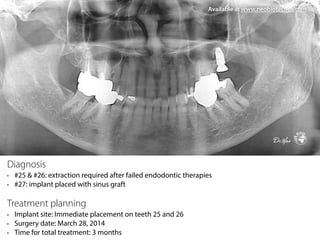
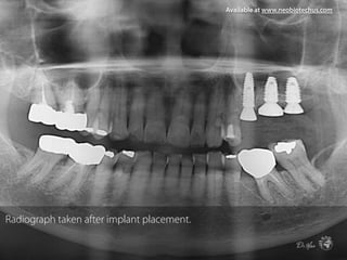
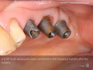
SCA Kit Clinical Case
Safe, Simple, Speedy Sinus crestal approach
No.1 Sales Sinus Approach Kit
Originality since 2007
Diagnosis
• #25 & #26: extraction required after failed endodontic therapies
• #27: implant placed with sinus graft
Treatment planning
• Implant site: Immediate placement on teeth 25 and 26
• Surgery date: March 28, 2014
• Time for total treatment: 3 months
Available at www.neobiotechus.com
Both extraction sockets 25 and 26 were carefully debrided with copious saline
solution after extractions. Clinical view of socket 26 clearly shows interraicular
bony septa.
Available at www.neobiotechus.com
The center of the interradicular septum of bone was prepared with S-reamer
to enable an implant to be placed in the middle of the socket without the
membrane tearing.
Available at www.neobiotechus.com
The remaining bone height was 3mm. Starting from 2mm stopper, the site
was drilled 1mm by 1mm until the sinus cavity opened without the
membrane tearing.
Available at www.neobiotechus.com
Occlusal view of the drilling site. Sinus membrane was seen intact.
Available at www.neobiotechus.com
In a reduced bone height less than 3mm, the maxillary sinus membrane can
be elevated using a micro-lateral sinus elevator in the SLA kit through the
crestal approach. This is #1 elevator.
Available at www.neobiotechus.com
#2 elevator. After elevation of the membrane, bone graft was performed with
an alloplast (Calpore)
Available at www.neobiotechus.com
CMI IS-II Active implant 4.5*11mm was placed in the site 25, achieving 30Ncm
of insertion torque value.
Available at www.neobiotechus.com
CMI IS-II Active implant 5.0*8.5mm was placed in the site 26, achieving
30Ncm of insertion torque value.
Available at www.neobiotechus.com
The gaps between the implants and the bony socket walls were filled with an
allogenic bone substitute material (RegenOss).
Available at www.neobiotechus.com
Healing abutments in place. Ø8.0 wide healing abutment was palced on
tooth 26.
Available at www.neobiotechus.com
Radiograph taken after implant placement.
Available at www.neobiotechus.com
Clinical view of the implant site after 3 months of healing.
Available at www.neobiotechus.com
A 3 unit full zirconia SCPR was prefabricated.
Available at www.neobiotechus.com
Occlusal view.
Available at www.neobiotechus.com
3 SCRP multi-abutments were connected in the implant 3 months after the
surgery.
Available at www.neobiotechus.com
The 3 unit SCRP full zirconia was cemented on the SCRP multi-abutments and
the abutment-prosthesis unit was removed and polished and reinserted in
the mouth.
Available at www.neobiotechus.com
2 years postloading radiograph.
Available at www.neobiotechus.com

SCA Kit case(Neobiotech)

  • 1.
    SCA Kit ClinicalCase Safe, Simple, Speedy Sinus crestal approach No.1 Sales Sinus Approach Kit Originality since 2007
  • 2.
    Diagnosis • #25 &#26: extraction required after failed endodontic therapies • #27: implant placed with sinus graft Treatment planning • Implant site: Immediate placement on teeth 25 and 26 • Surgery date: March 28, 2014 • Time for total treatment: 3 months Available at www.neobiotechus.com
  • 3.
    Both extraction sockets25 and 26 were carefully debrided with copious saline solution after extractions. Clinical view of socket 26 clearly shows interraicular bony septa. Available at www.neobiotechus.com
  • 4.
    The center ofthe interradicular septum of bone was prepared with S-reamer to enable an implant to be placed in the middle of the socket without the membrane tearing. Available at www.neobiotechus.com
  • 5.
    The remaining boneheight was 3mm. Starting from 2mm stopper, the site was drilled 1mm by 1mm until the sinus cavity opened without the membrane tearing. Available at www.neobiotechus.com
  • 6.
    Occlusal view ofthe drilling site. Sinus membrane was seen intact. Available at www.neobiotechus.com
  • 7.
    In a reducedbone height less than 3mm, the maxillary sinus membrane can be elevated using a micro-lateral sinus elevator in the SLA kit through the crestal approach. This is #1 elevator. Available at www.neobiotechus.com
  • 8.
    #2 elevator. Afterelevation of the membrane, bone graft was performed with an alloplast (Calpore) Available at www.neobiotechus.com
  • 9.
    CMI IS-II Activeimplant 4.5*11mm was placed in the site 25, achieving 30Ncm of insertion torque value. Available at www.neobiotechus.com
  • 10.
    CMI IS-II Activeimplant 5.0*8.5mm was placed in the site 26, achieving 30Ncm of insertion torque value. Available at www.neobiotechus.com
  • 11.
    The gaps betweenthe implants and the bony socket walls were filled with an allogenic bone substitute material (RegenOss). Available at www.neobiotechus.com
  • 12.
    Healing abutments inplace. Ø8.0 wide healing abutment was palced on tooth 26. Available at www.neobiotechus.com
  • 13.
    Radiograph taken afterimplant placement. Available at www.neobiotechus.com
  • 14.
    Clinical view ofthe implant site after 3 months of healing. Available at www.neobiotechus.com
  • 15.
    A 3 unitfull zirconia SCPR was prefabricated. Available at www.neobiotechus.com
  • 16.
    Occlusal view. Available atwww.neobiotechus.com
  • 17.
    3 SCRP multi-abutmentswere connected in the implant 3 months after the surgery. Available at www.neobiotechus.com
  • 18.
    The 3 unitSCRP full zirconia was cemented on the SCRP multi-abutments and the abutment-prosthesis unit was removed and polished and reinserted in the mouth. Available at www.neobiotechus.com
  • 19.
    2 years postloadingradiograph. Available at www.neobiotechus.com