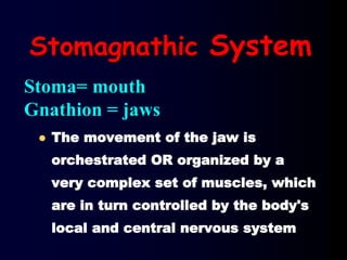
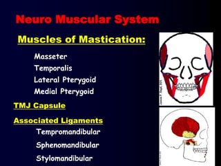
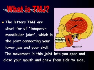
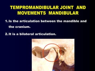
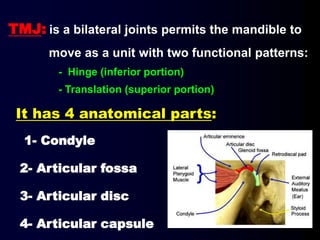
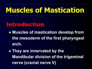
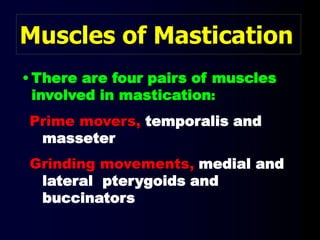
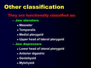
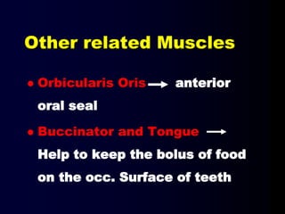
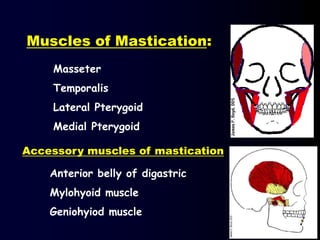
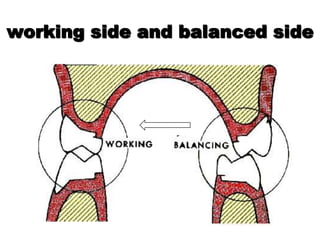
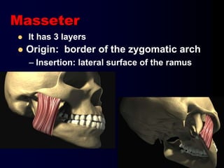
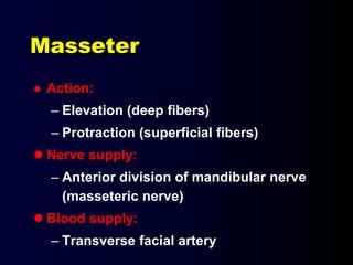
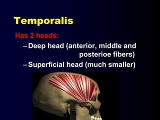
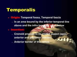
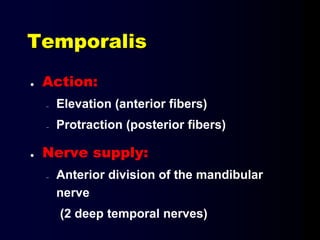
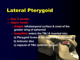
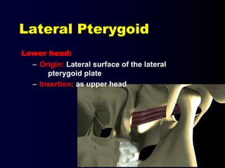
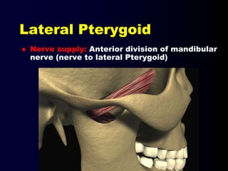
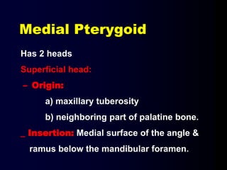
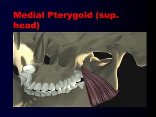
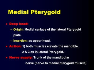
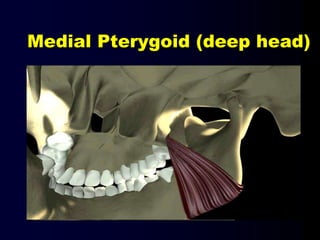
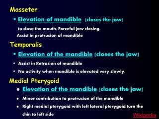
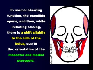
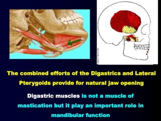
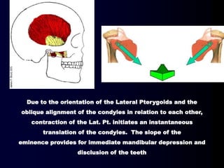
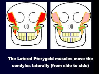
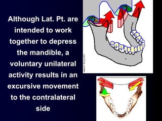
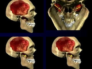
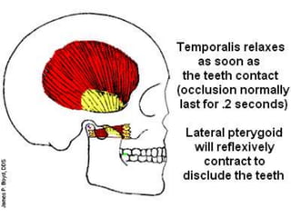
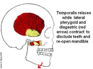
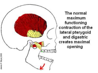
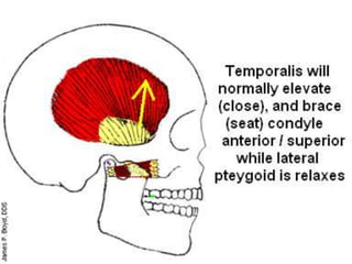
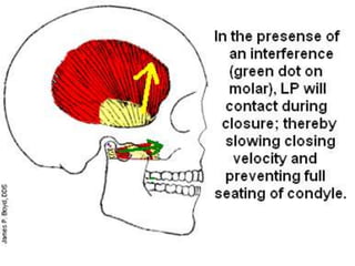
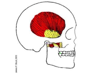
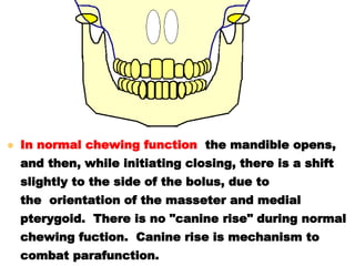
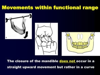
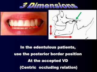
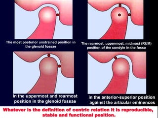
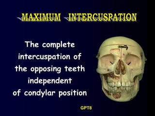
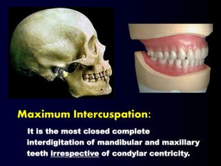
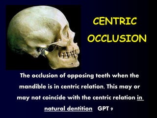
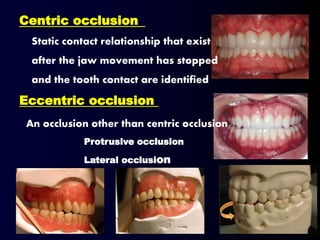
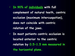
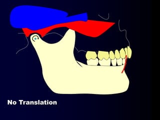
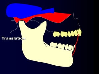
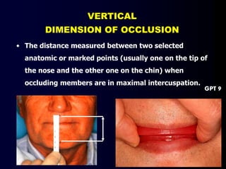
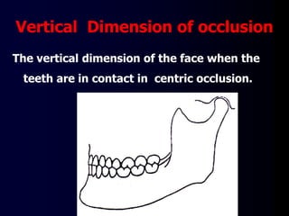
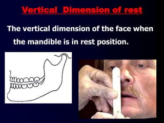
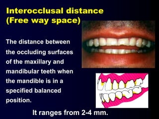
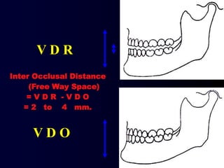
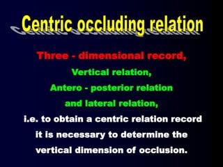
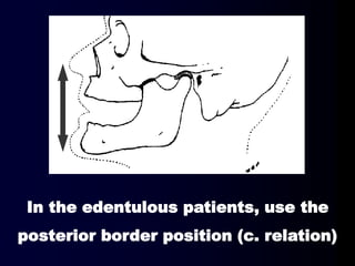
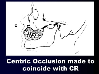
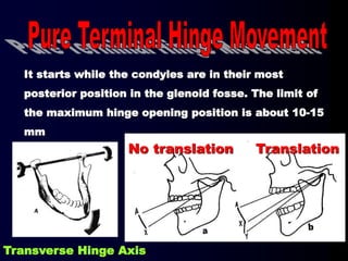
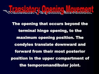
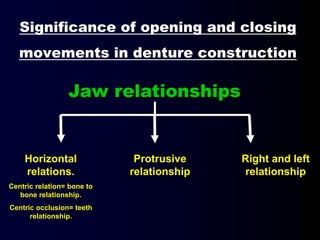
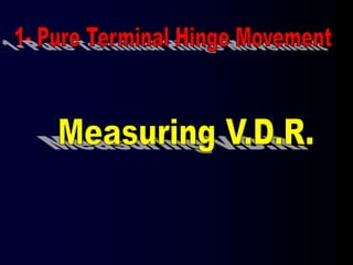
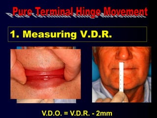
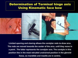
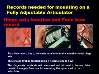
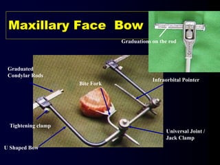
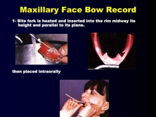
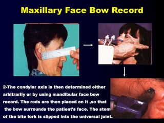
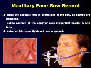
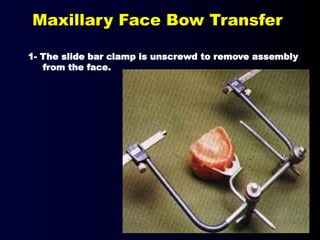
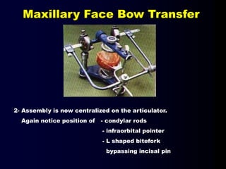
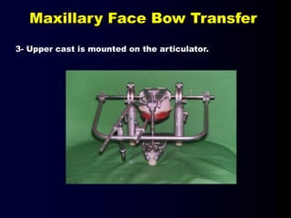
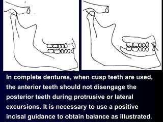
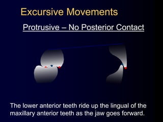
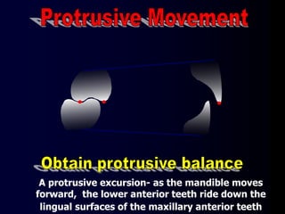
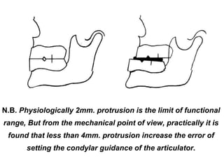
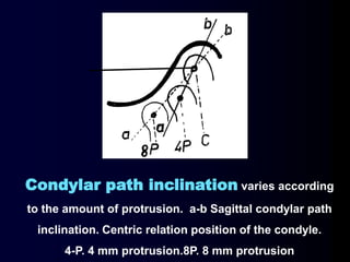
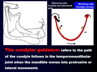
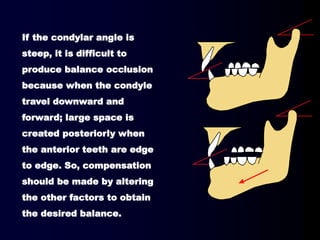
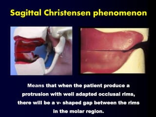
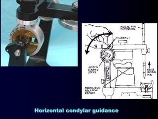
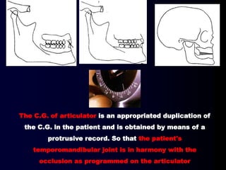
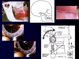
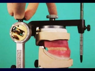
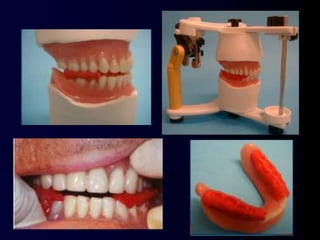
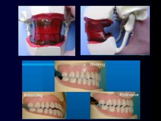
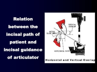
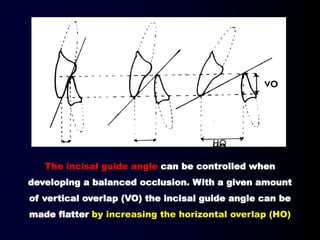
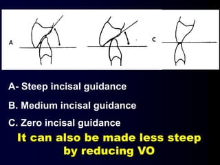
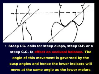
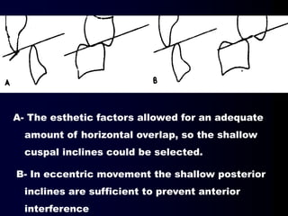
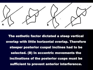
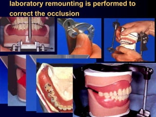
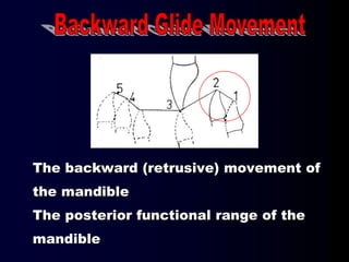
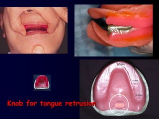
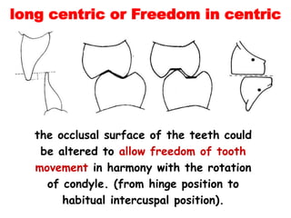
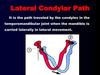
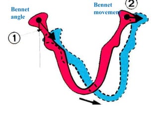
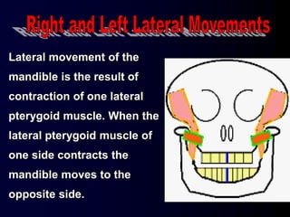
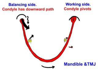
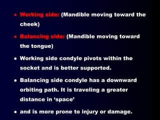
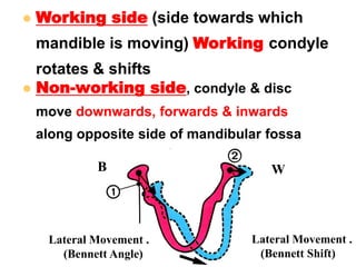
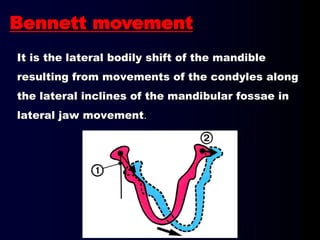
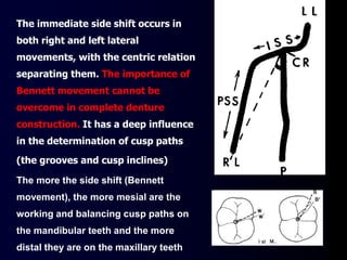
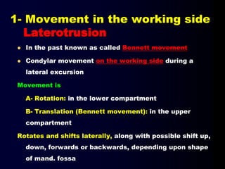
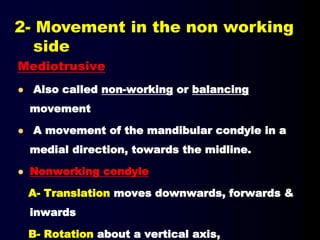
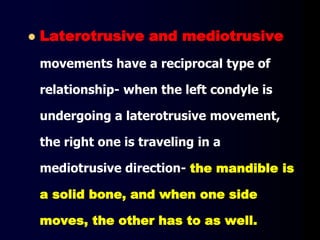
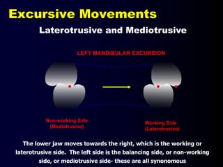
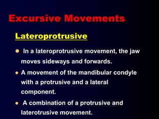
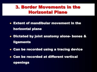
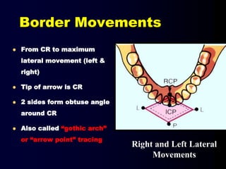
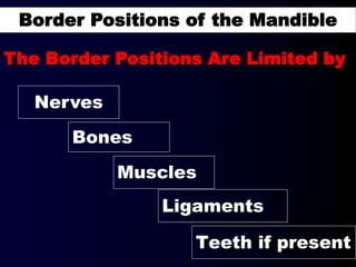
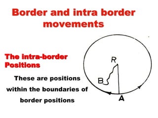
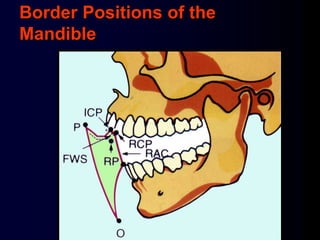
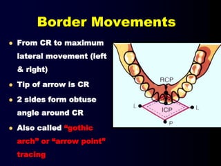
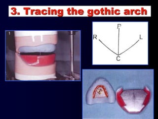
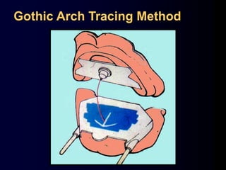
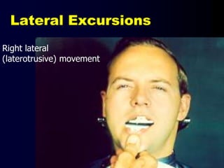
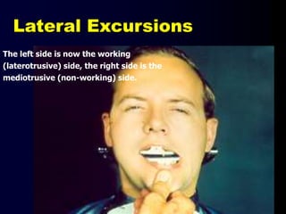
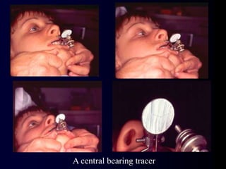
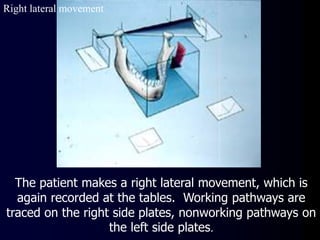
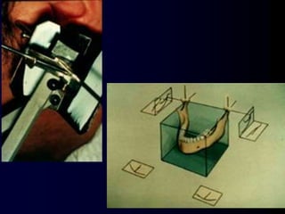
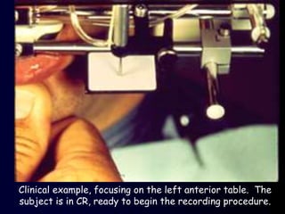
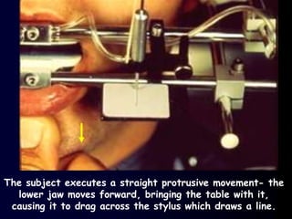
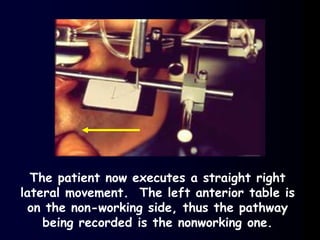
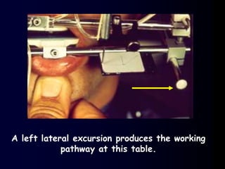
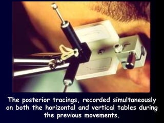
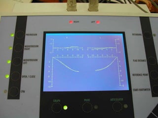
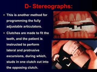
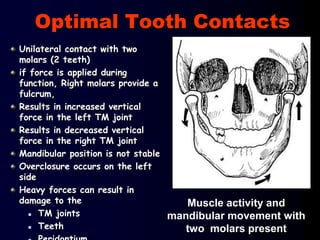
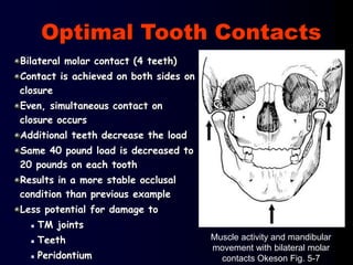
Neutral Zone
In dentistry, the neutral zone refers to that space in the oral cavity where the forces exerted by the musculature.of.the tongue are equal and balanced with the.forces exerted by the buccinators.muscle of the cheek.laterally and the orbicularis.oris muscle anteriorly
The human mandible can be related to the maxilla in several positions in the horizontal plane. Among these centric relation is a significant position, because of its usefulness in relating the dentulous and edentulous mandible to maxilla, where the teeth , muscles and temporomandibular joint function in harmony. It is a position of occluso-articular harmony.
8 - setting of teeth for class I, II and II arch relation ship (Edited)Amal Kaddah
Prosthetic Problems and possible solutions in Setting –up
of teeth for skeletal Class II and Class III arch relationship
of completely edentulous patients
Prof. Amal F. Kaddah
Anatomy and clinical significance of denture bearing areasOgundiran Temidayo
A presentation on the anatomy and clinical significance of the denture bearing areas by Ogundiran Temidayo who is a dental student at OBAFEMI AWOLOWO UNIVERSITY ILE-IFE
Indian Dental Academy: will be one of the most relevant and exciting training center with best faculty and flexible training programs for dental professionals who wish to advance in their dental practice,Offers certified courses in Dental implants,Orthodontics,Endodontics,Cosmetic Dentistry, Prosthetic Dentistry, Periodontics and General Dentistry.
Muscles of mastication /certified fixed orthodontic courses by Indian dental ...Indian dental academy
The Indian Dental Academy is the Leader in continuing dental education , training dentists in all aspects of dentistry and offering a wide range of dental certified courses in different formats.
Indian dental academy provides dental crown & Bridge,rotary endodontics,fixed orthodontics,
Dental implants courses.for details pls visit www.indiandentalacademy.com ,or call
0091-9248678078
This slide is about mechanism of mastication. it include muscles of mastication. disorders .of masticatory muscles also included. mastication in different condition is also presented
6- Prosthetic Problems and possible solutions in Setting –up of teeth for ske...Amal Kaddah
COMPLETE DENTURE CONSTRUCTION
1- Diagnosis and Treatment Planning for Removable Prosthodontics
2- Preliminary Maxillary and mandibular impression procedures
3- Final Maxillary and mandibular impression procedures
4- Jaw Relation Registration
a. Introduction and the stomatognathic system
What 'occlusion' is and why it is important
b. Definitions.
c. Check denture foundation and Establishment of facial contour.
d. Establishment of the occlusal plane.
e. Importance of mounting the maxillary cast using Maxillary face-bow record and transfer.
f. Determination of vertical dimension of centric occluding relation.
g. Determination of centric and eccentric relations at the accepted vertical dimension.
5- Selection of Artificial Teeth
6- Prosthetic Problems and possible solutions in Setting –up of teeth for skeletal Class I, II and Class III arch relationship of completely edentulous patients.
7- Try-in of the wax trial complete denture.
8- Complete denture insertion (Delivery)
9- Occlusal corrections.
10- Managements of Post Insertion Problems and Complaints.
11- Single denture and Kelly's Syndrome
12- Denture Processing and Laboratory Errors.
This is a presentation by Dada Robert in a Your Skill Boost masterclass organised by the Excellence Foundation for South Sudan (EFSS) on Saturday, the 25th and Sunday, the 26th of May 2024.
He discussed the concept of quality improvement, emphasizing its applicability to various aspects of life, including personal, project, and program improvements. He defined quality as doing the right thing at the right time in the right way to achieve the best possible results and discussed the concept of the "gap" between what we know and what we do, and how this gap represents the areas we need to improve. He explained the scientific approach to quality improvement, which involves systematic performance analysis, testing and learning, and implementing change ideas. He also highlighted the importance of client focus and a team approach to quality improvement.
The French Revolution, which began in 1789, was a period of radical social and political upheaval in France. It marked the decline of absolute monarchies, the rise of secular and democratic republics, and the eventual rise of Napoleon Bonaparte. This revolutionary period is crucial in understanding the transition from feudalism to modernity in Europe.
For more information, visit-www.vavaclasses.com
The Indian economy is classified into different sectors to simplify the analysis and understanding of economic activities. For Class 10, it's essential to grasp the sectors of the Indian economy, understand their characteristics, and recognize their importance. This guide will provide detailed notes on the Sectors of the Indian Economy Class 10, using specific long-tail keywords to enhance comprehension.
For more information, visit-www.vavaclasses.com
2024.06.01 Introducing a competency framework for languag learning materials ...Sandy Millin
http://sandymillin.wordpress.com/iateflwebinar2024
Published classroom materials form the basis of syllabuses, drive teacher professional development, and have a potentially huge influence on learners, teachers and education systems. All teachers also create their own materials, whether a few sentences on a blackboard, a highly-structured fully-realised online course, or anything in between. Despite this, the knowledge and skills needed to create effective language learning materials are rarely part of teacher training, and are mostly learnt by trial and error.
Knowledge and skills frameworks, generally called competency frameworks, for ELT teachers, trainers and managers have existed for a few years now. However, until I created one for my MA dissertation, there wasn’t one drawing together what we need to know and do to be able to effectively produce language learning materials.
This webinar will introduce you to my framework, highlighting the key competencies I identified from my research. It will also show how anybody involved in language teaching (any language, not just English!), teacher training, managing schools or developing language learning materials can benefit from using the framework.
Ethnobotany and Ethnopharmacology:
Ethnobotany in herbal drug evaluation,
Impact of Ethnobotany in traditional medicine,
New development in herbals,
Bio-prospecting tools for drug discovery,
Role of Ethnopharmacology in drug evaluation,
Reverse Pharmacology.
How to Make a Field invisible in Odoo 17Celine George
It is possible to hide or invisible some fields in odoo. Commonly using “invisible” attribute in the field definition to invisible the fields. This slide will show how to make a field invisible in odoo 17.
Model Attribute Check Company Auto PropertyCeline George
In Odoo, the multi-company feature allows you to manage multiple companies within a single Odoo database instance. Each company can have its own configurations while still sharing common resources such as products, customers, and suppliers.
We all have good and bad thoughts from time to time and situation to situation. We are bombarded daily with spiraling thoughts(both negative and positive) creating all-consuming feel , making us difficult to manage with associated suffering. Good thoughts are like our Mob Signal (Positive thought) amidst noise(negative thought) in the atmosphere. Negative thoughts like noise outweigh positive thoughts. These thoughts often create unwanted confusion, trouble, stress and frustration in our mind as well as chaos in our physical world. Negative thoughts are also known as “distorted thinking”.
Palestine last event orientationfvgnh .pptxRaedMohamed3
An EFL lesson about the current events in Palestine. It is intended to be for intermediate students who wish to increase their listening skills through a short lesson in power point.
5. Definitions.
What 'occlusion' is
The stomatognathic system
Why occlusion is important
The significance of 'ideal occlusion‘
Difference between natural and artificial Occ.
Mandibular Movements.
Articulators and Facebows.
Factors affecting B. Occlusion
Concepts of occlusion Nat. and Art. Occ.
Recording of Occ. for removable prosthodontics.
Occlusion
8. Stomagnathic System
The movement of the jaw is
orchestrated OR organized by a
very complex set of muscles, which
are in turn controlled by the body's
local and central nervous system
Stoma= mouth
Gnathion = jaws
9. The stomatognathic system
= the masticatory
system =
•Teeth
•Periodontium
•Jaws
•TMJ
•Associated muscles +
tongue & ms of the soft
palate
•Investing tissues
•Neural control
10. The stomatognathic system
• There is a complex, dynamic
balance between the way that the
teeth come together, the muscle
that work the jaws, the joints, and
their relationship to the posture of the head & neck. A
change in any part of the system affects all the others
and change the balance
The masticatory system
11. Is the functional unit of the body primary
responsible for chewing, speaking and
swallowing, as well as parafunctional
actions.
Disturbance of any part could disturb the
whole odontostomatognathic system and
subsequently the body as well.
The stomatognathic system
12. When opposing teeth are in
contact and mandibular movements
are made, the direction of the
movement is controlled by the
neuromuscular system as limited by
the movement
The stomatognathic system
13. Muscles of Mastication:
Neuro Muscular System
Masseter
Temporalis
Lateral Pterygoid
Medial Pterygoid
Tempromandibular
Sphenomandibular
Stylomandibular
TMJ Capsule
Associated Ligaments
14. The letters TMJ are
short for of 'temporo-
mandibular joint', which is
the joint connecting your
lower jaw and your skull.
The movement in this joint lets you open and
close your mouth and chew from side to side.
Temporalis
Masseter
15. 1.Is the articulation between the mandible and
the cranium.
2.It is a bilateral articulation.
16. It has 4 anatomical parts:
1- Condyle
2- Articular fossa
3- Articular disc
4- Articular capsule
TMJ: is a bilateral joints permits the mandible to
move as a unit with two functional patterns:
- Hinge (inferior portion)
- Translation (superior portion)
17. Condyle: The rounded
articular surface at the
end of the mandible
(lower jaw).
Glenoid fossa: A deep concavity in the temporal
bone at the root of the zygomatic arch that
receives the condyle of the mandible.
Tubercle: A slight elevation from the surface of
the bone giving attachment to a muscle or
ligament.
18. Biconcave
articular
disc
C.T. capsule
Dense fibrous connective tissue
Lacks blood vessels and nerves
Able to tolerate forces without damage or pain being
produced
Provides protection to condyle and fossa during
movements
19. The Synovial fluid:
Consist of small amounts of a clear, straw-
colored viscous fluid. It is an infiltrate of the
blood diffused out from the rich capillary
network of the Synovial membrane.
Function:
1- Lubrication
2- Nutrition.
3- Clear the tissue debris.
20. TMJ LIGAMENTS AND CAPSULE
1-Collateral(discal)
2-Capsular
3-Tempromandibular
4-Sphenomandibular
5-Stylomandibular
23. Fundamental characteristics
Variability (can change and
affect situation)
Rhythmicity
(regular pattern of beats )
Two dimensional frontal view of the
movement of the mandible data were
recorded with model 5 kinesiograph
24. Introduction
Muscles of mastication develop from
the mesoderm of the first pharyngeal
arch.
They are innervated by the
Mandibular division of the trigeminal
nerve (cranial nerve V)
Muscles of Mastication
25. Muscles of Mastication
• There are four pairs of muscles
involved in mastication:
Prime movers, temporalis and
masseter
Grinding movements, medial and
lateral pterygoids and
buccinators
26. Other classification
They are functionally classified as:
– Jaw elevators
Masseter
Temporalis
Medial pterygoid
Upper head of lateral pterygoid
– Jaw depressors
Lower head of lateral pterygoid
Anterior digastric
Geniohyoid
Mylohyoid
27. Other related Muscles
Orbicularis Oris anterior
oral seal
Buccinator and Tongue
Help to keep the bolus of food
on the occ. Surface of teeth
33. Temporalis
Origin: Temporal fossa, Temporal fascia
– In an area bound by the inferior temporal line
above and the infra temporal crest below
Insertion:
– Cronoid process in its medial aspect (apex,
anterior and posterior border)
– Anterior border of the ramus.
34. Temporalis
Action:
– Elevation (anterior fibers)
– Protraction (posterior fibers)
Nerve supply:
– Anterior division of the mandibular
nerve
(2 deep temporal nerves)
35. Lateral Pterygoid
Has 2 heads:
Upper head:
– Origin: infratemporal surface & crest of the
greater wing of sphenoid
– Insertion: enters the TMJ & inserted into:
a) Pterygoid fovea of the neck of the mandible
b) Articular disc
c) capsule of TMJ (anterior aspect)
37. Lateral Pterygoid
Action:
1. Both muscles produce
depression of the mandible.
2. Lat. & Med pterygoid on 1 side
protrude the mandible to the
opposite side.
3. Lat & Med pterygoid on the 2
sides cause side to side movement
38. Lateral Pterygoid
Nerve supply: Anterior division of mandibular
nerve (nerve to lateral Pterygoid)
39. Medial Pterygoid
Has 2 heads
Superficial head:
– Origin:
a) maxillary tuberosity
b) neighboring part of palatine bone.
_ Insertion: Medial surface of the angle &
ramus below the mandibular foramen.
41. Medial Pterygoid
Deep head:
– Origin: Medial surface of the lateral Pterygoid
plate.
– Insertion: as upper head.
Action: 1) both muscles elevate the mandible.
2 & 3 as in lateral Pterygoid.
Nerve supply: Trunk of the mandibular
nerve (nerve to medial pterygoid muscle)
43. Elevation of mandible (closes the jaw)
to close the mouth, Forceful jaw closing.
Masseter
Temporalis
Elevation of the mandible (closes the jaw)
Assist in Retrusion of mandible
No activity when mandible is elevated very slowly.
Assist in protrusion of mandible
Elevation of the mandible (closes the jaw)
Minor contribution to protrusion of the mandible
Right medial pterygoid with left lateral pterygoid turn the
chin to left side
Medial Pterygoid
Wikipedia
44. Protrusion of the mandible: The primary
function of the lateral pterygoid muscle is to pull the
head of the condyle out of the mandibular fossa along
the articular eminence to protrude the mandible.
Jaw opening (Depresses the mandible) it
is assisted by the digastric, mylohyoid and geniohyoid
muscles..
SIDE TO SIDE movements GRINDING MOVEMENT
Unilateral action of a lateral pterygoid produces
contralateral excursion (a form of mastication), usually
performed in concert with the medial pterygoids.
Lateral Pterygoid
Wikipedia
45. In normal chewing
function, the mandible
opens, and then, while
initiating closing,
there is a shift slightly
to the side of the
bolus, due to
the orientation of the
masseter and medial
pterygoid.
46. The Lat. Pterygo. advance the
condyles, thereby opening the
mouth (depressing the
mandible), with the
assistance of the Digastric.
The oblique orientation of the
Masseters and Med. Pterygo.
create a sling. The non-
working side Med. Pterygo.
contacts simultaneously with
the opposite side working
Masseter
normal reciprocal functioning of the Lateral Pterygoids
and Masseters/Med.Pteygoids/Temporalis
47. The combined efforts of the Digastrics and Lateral
Pterygoids provide for natural jaw opening
Digastric muscles is not a muscle of
mastication but it play an important role in
mandibular function
48. Due to the orientation of the Lateral Pterygoids and the
oblique alignment of the condyles in relation to each other,
contraction of the Lat. Pt. initiates an instantaneous
translation of the condyles. The slope of the
eminence provides for immediate mandibular depression and
disclusion of the teeth
50. Although Lat. Pt. are
intended to work
together to depress
the mandible, a
voluntary unilateral
activity results in an
excursive movement
to the contralateral
side
51.
52.
53.
54.
55.
56.
57.
58. In normal chewing function, the mandible opens,
and then, while initiating closing, there is a shift
slightly to the side of the bolus, due to
the orientation of the masseter and medial
pterygoid. There is no "canine rise" during normal
chewing fuction. Canine rise is mechanism to
combat parafunction.
59. The closure of the mandible does not occur in a
straight upward movement but rather in a curve
Movements within functional range
61. Joint Stability is achieved by
1- The position of the articular disc on
the condyle
2- Cervical and Masticatory Muscles in tonus
3- The occlusion of teeth provides the
necessary stability in maximum
intercuspation
4- Interarticular pressure between the condyle
and eminence
62. In the edentulous patients,
use the posterior border position
At the accepted VD
(Centric occluding relation)
65. The static relationship between the
incising or masticating surfaces of the
maxillary and mandibular teeth, or tooth
analogues.
66. The contact relationship
between the occlusal
surfaces of teeth during
function.
It is the DYNAMIC contacts relationship of
the teeth as the mandible moved to and from
eccentric relation.
67. EXCURSIVE MOVEMENT
Movement occurring when mandible
moves away from maximum
intercuspation
OCCLUSAL INTERFERENCES
Undesirable contacts
occurring during lateral
movements
68. Working side
Non working (balancing) side
Side that side of the
mandible that moves
toward the median line in
a lateral excursion.
The side toward which
the mandible moves in a
lateral excursion
69. The most retruded relation of the mandible to the
maxillae when the condyles are in the most
posterior unstrained position in the glenoid fossae
from which lateral movement can be made, (within
hinge movement).
70. The relation of the
mandible to the maxilla
with the mandible in its
most retruded position.
(GPT) 2005
71. A maxillomandibular relationship, independent
of tooth contact, in which the condyles
articulate in the anterior- superior position
against the posterior slopes of the articular
eminences; in this position, the mandible is
restricted to a purely rotary movements; from
this unstrained, physiologic, maxillomandibular
relationship, the patient can make vertical,
lateral or protrusive movements, it is a clinically
useful, repeatable reference position
(within functional range of movement). (GPT 9)
72. Dawson has defined this position
as the rearmost, uppermost,
midmost (RUM) position of the
condyle in the fossa at which the
medial pole of the condyle disc
assembly is braced against the
bony wall of the eminentia.
73. Whatever is the definition of centric relation it is reproducible,
stable and functional position.
The rearmost, uppermost, midmost (RUM)
position of the condyle in the fossa
The most posterior unstrained position in
the glenoid fossae
In the uppermost and rearmost
position in the glenoid fossae
in the anterior-superior position
against the articular eminences
74. Is the only constant repeatable
position within the functional
limit to start constructing a
stable occlusion
76. Maximum Intercuspation:
It is the most closed complete
interdigitation of mandibular and maxillary
teeth irrespective of condylar centricity.
77. CENTRIC
OCCLUSION
The occlusion of opposing teeth when the
mandible is in centric relation, This may or
may not coincide with the centric relation in
natural dentition GPT 9
78. Centric occlusion
Static contact relationship that exist
after the jaw movement has stopped
and the tooth contact are identified
Eccentric occlusion
An occlusion other than centric occlusion
Protrusive occlusion
Lateral occlusion
79. • In 90% of individuals with full
complement of natural teeth, centric
occlusion (maximum intercuspation),
does not coincide with centric
relation of the jaws.
• In most patients centric occlusion is
located anterior to the centric
relation by 0.5-1.5 mm measured in
the horizontal plane.
80. Centric occlusion with
teeth present is a tooth to
tooth relation whereas
centric relation, is a bone
to bone relation
(Static positions)
85. MP
MO
ICP
RCP
HA
MP = Maximal protrusion
ICP = Intercuspal position
RCP= Retruded Contact
position
HA = Hinge axis
MO = Maximum opening
EE=edge to edge
Posselt’s Figure
Habitual Arc of Closure
EE
MO
All the movements of the
mandible occur within
this envelope, maximum
opening is reached
when the capsular
ligament prevent further
movement at the
condyle.
86. MP
MO
ICPRCP
H A
MP = Maximal protrusion
ICP = Intercuspal position
RCP= Retruded Contact position
HA = Hinge axis
MO = Maximum opening
EE=edge to edge
Posselt’s Figure
Habitual Arc of Closure
EE
87. MP
MO
ICPRCP
H A
MP = Maximal protrusion
ICP = Intercuspal position
RCP= Retruded Contact position
HA = Hinge axis
MO = Maximum opening
EE=edge to edge
Posselt’s Figure
Habitual Arc of Closure
EE
88. Habitual Arc of Closure
In that case the intercuspal
position is in a position
forward to the centric
position, and at a lower
vertical dimension
89. VERTICAL
DIMENSION OF OCCLUSION
• The distance measured between two selected
anatomic or marked points (usually one on the tip of
the nose and the other one on the chin) when
occluding members are in maximal intercuspation.
GPT 9
90. Vertical Dimension of occlusion
The vertical dimension of the face when the
teeth are in contact in centric occlusion.
91. Vertical Dimension of rest
The vertical dimension of the face when
the mandible is in rest position.
92. Interocclusal distance
(Free way space)
The distance between
the occluding surfaces
of the maxillary and
mandibular teeth when
the mandible is in a
specified balanced
position.
It ranges from 2-4 mm.
93. V D R
V D O
Inter Occlusal Distance
(Free Way Space)
= V D R - V D O
= 2 to 4 mm.
94.
95. This relation exists when
the jaws are in centric
relation and the teeth
are in centric occlusion
96. Three - dimensional record,
Vertical relation,
Antero - posterior relation
and lateral relation,
i.e. to obtain a centric relation record
it is necessary to determine the
vertical dimension of occlusion.
97. In the edentulous patients,
use the posterior border
position (centric relation)
which is repeatable,
reproducible and within the
functional range of
movements
98. For this reason, the relation
of the mandible to the
maxilla should be recorded
in the most retruded position
(C.R) and centric occlusion
made to coincide with it
99. In the edentulous patients, use the
posterior border position (c. relation)
104. Rotation
In the lower
compartment
Translation
In the upper
compartment
105. The three planes of the skull
For movement to occur in a plane, it must turn or
rotate about an axis as previously mentioned
The axes are named in relation to their orientation
Axes of rotation
106. Axes of rotation
c, Horizontal-coronal axis (sagittal axis).Permits descent
of balancing condyle
Mandibular centers of
rotation
a, Horizontal-sagittal
(transverse or retruded
condyle axis).
b, Vertical axis with
horizontal rotation.
107. • It takes place in the lower compartment of
the T.M.J between the superior surface of
the condyle and the inferior surface of the
articular disc
•It Is a Simple Hinge
Movement
Occurs during the early
opening and late closing
movement of the mandible
108. It is an imaginary line around which the condyles rotate
during early opening and late closing.
109. Rotation occurs when the mandible makes a
hinged movement, in lower compartment .
Translation occurs in upper compartment, when
the mandible moves into a protrusive or lateral
position, or a combination of the two
110. Mandibular movements are of great importance in
complete denture occlusion and service when
balanced occlusion. They are usually classified
according to the main direction of movement.
The starting position is the habitual intercuspal
position, from this point the mandible can move into:
4. Lateral (Right & left) Movement
2. Protrusive movement.
1. Opening and Closing Movement.
3. Backward Movement
111. Opening movement of the mandible
from the habitual intercuspal position
to maximum opening of the jaws.
It can be divided into
1. Opening and Closing Movement
Pure Terminal Hinge Movement
Translatory Opening Movement
Habitual intra-border Opening
112. It starts while the condyles are in their most
posterior position in the glenoid fosse. The limit of
the maximum hinge opening position is about 10-15
mm
Transverse Hinge Axis
No translation Translation
113. The opening that occurs beyond the
terminal hinge opening, to the
maximum opening position. The
condyles translate downward and
forward from their most posterior
position in the upper compartment of
the temporomandibular joint.
115. Significance of opening and closing
movements in denture construction
Horizontal
relations.
Centric relation= bone to
bone relationship.
Centric occlusion= teeth
relationship.
Protrusive
relationship
Right and left
relationship
Jaw relationships
119. Determination of Terminal hinge axis
Using Kinematic face bow
Limited opening and closing allows the condylar rods to draw arcs.
The rods are moved towards the center of the arcs, until they move in
a point . The latter represents the condylar axis. The condyle in this
position lies in the most retruded unstrained position in the glenoid
fossa, so mandible and maxilla are in centric.
120. Records needed for mounting on a
Fully Adjustable Articulator
Hinge axis location and Face bow
record
Face bow record has to be made in relation to the actual terminal hinge
axis
This should first be located using a Kinematic face bow
The Hinge axis points should be marked and tattooed, to be used later
on with the upper face bow for mounting the upper cast to the
articulator
122. Maxillary Face Bow Record
1- Bite fork is heated and inserted into the rim midway its
height and parallel to its plane.
then placed intraorally
123. 2-The condylar axis is then determined either
arbitrarily or by using mandibular face bow
record. The rods are then placed on it ,so that
the bow surrounds the patient’s face. The stem
of the bite fork is slipped into the universal joint.
11- 13 mm
Maxillary Face Bow Record
124. 3- When the patient’s face is centralized in the bow, all clamps are
tightened.
Notice position of the condylar rods infraorbital pointer & bite
fork.
4- Universal joint once tightened , never opened.
Maxillary Face Bow Record
125. 1- The slide bar clamp is unscrewd to remove assembly
from the face.
Maxillary Face Bow Transfer
126. 2- Assembly is now centralized on the articulator.
Again notice position of - condylar rods
- infraorbital pointer
- L shaped bitefork
bypassing incisal pin
Maxillary Face Bow Transfer
127. 3- Upper cast is mounted on the articulator.
Maxillary Face Bow Transfer
131. Excursive movements can occur in a number
of different directions.
A lateroprotrusive movement is a combination
of protrusive and laterotrusive movements.
Other combinations and patterns of
movements are also possible, but these major
directions are the ones most frequently
described and analyzed in dentistry.
132. The muscles
responsible for the
forward movement of
the mandible are the
lateral pterygoid
muscles acting jointly.
133. 1- Retruded contact position
2- Habitual inter cuspal position
3- Edge to edge occlusion
4- Anterior biting to a reverse vertical overlap
5-Protruded contact position
The upper surface of
the movement area in
the median plane
134. The angle formed by the steepness of the
articulator surface of the temporal bone as
related to a horizontal line, which is parallel to
the Frankfort plane.
The sagittal inclination (condylar inclination)
135. Condylar path
inclination varies
according to the amount of
protrusion.
a-b: Sagittal condylar path.
C: Centric relation position
of the condyle.
4-P: 4 mm protrusion.
8P: 8 mm protrusion
136. 1. The shape of the glenoid
fossa.
2. The variation of the
thickness of the articular
disc in its different parts.
3. The relation of the condyle
to the disc during
movement.
4. The extent of mandibular
protrusion
The inclination of the condylar paths varies in
different individuals and from side to side in
the same person. It depends upon
137. The condylar guidance: refers to the path
of the condyle follows in the
temporomandibular joint when the mandible
moves into protrusive or lateral movements
138. In complete dentures, when cusp teeth are used,
the anterior teeth should not disengage the
posterior teeth during protrusive or lateral
excursions. It is necessary to use a positive
incisal guidance to obtain balance as illustrated.
139. Excursive Movements
Protrusive – No Posterior Contact
The lower anterior teeth ride up the lingual of the
maxillary anterior teeth as the jaw goes forward.
140. A protrusive excursion- as the mandible moves
forward, the lower anterior teeth ride down the
lingual surfaces of the maxillary anterior teeth
141. When the mandible moves to edge to edge
position, separation
occurs distally between the natural
dentition or occlusion rims .
143. Protrusive record being taken. The
mandible is brought forward in a
straight protrusive direction about 4-6
mm in distance, and the patient closes
on the softened wax. The record is
then brought to the articulator, where
it is used to set the condylar
inclination.
144. N.B. Physiologically 2mm. protrusion is the limit of functional
range, But from the mechanical point of view, practically it is
found that less than 4mm. protrusion increase the error of
setting the condylar guidance of the articulator.
145. Condylar path inclination varies according
to the amount of protrusion. a-b Sagittal condylar path
inclination. Centric relation position of the condyle.
4-P. 4 mm protrusion.8P. 8 mm protrusion
146. The condylar guidance: refers to the path
of the condyle follows in the temporomandibular
joint when the mandible moves into protrusive or
lateral movements
Balancing side.
Condyle has downward
path
Working side.
Condyle pivots.
147. Bennett Angle
The angle formed by the
sagittal plane (assumed
straight protrusive path)
and the path of the
advancing (orbiting)
condyle during lateral
mandibular movements
as viewed in the
horizontal plane.
Balancing side. Working side.
148. If the condylar angle is
steep, it is difficult to
produce balance occlusion
because when the condyle
travel downward and
forward; large space is
created posteriorly when
the anterior teeth are edge
to edge. So, compensation
should be made by altering
the other factors to obtain
the desired balance.
149. Means that when the patient produce a
protrusion with well adapted occlusal rims,
there will be a v- shaped gap between the rims
in the molar region.
Sagittal Christensen phenomenon
150. Means that when the patient
produce a lateral excurtion
with well adapted occlusal
rims, there will be a v shaped
gap between the rims in the
molar region on the balance
side. On the working side
there will be contact between
the upper and the lower rims.
The Transversal Christensen
phenomenon
151. Ease of determination
Used to set condylar
guidance
Helps setting teeth
for best occlusal
contacts
Protrusive Records
To Set Condylar Inclination
152. Protrusive Records
To Set Condylar Inclination
Material must interdigitate
with the opposing “V-
shaped” notches
Record should cover entire
rim surface.
Condylar elements are released from hinge
position (unlocked).
Instrument protruded, and the rims closed into
record
154. Prior to Mounting
Ensure
– Only wax-rim to wax-rim
contact
– Casts should not contact
– Record bases should not
contact
– If other contacts, mounting will
be incorrect
156. The C.G. of articulator is an appropriated duplication of
the C.G. in the patient and is obtained by means of a
protrusive record. So that the patient's
temporomandibular joint is in harmony with the
occlusion as programmed on the articulator
157. The occlusion rims are reestablished and checked
for 3mm. Clearance in a protrusive excursion
158.
159.
160.
161. After protrusive record
making the same procedure is
followed, however the patient
is asked to move his mandible
to left and right and getting
Right and left lateral relation
him to bite on wax wafer, two records are made, one
for left and one for right. These records are used to
adjust the lateral condylar inclination.
162. In Hanau model H
articulator Hanau
equation can be
used
L = H/ 8+ I2
163.
164. It is the path taken by
the lower anterior
teeth as it move in
protrusive movements
against the palatal
surface of upper
anteriors till become
edge to edge
165. The angle formed by the
horizontal overlap and vertical
overlap of the anterior teeth as
related to a horizontal plane is
called the incisal guide angle and
the influence that this angle has
on mandibular movements is
termed incisal guidance.
167. The incisal guide angle can be controlled when
developing a balanced occlusion. With a given amount
of vertical overlap (VO) the incisal guide angle can be
made flatter by increasing the horizontal overlap (HO)
168. It can also be made less steep
by reducing VO
A- Steep incisal guidance
B. Medium incisal guidance
C. Zero incisal guidance
B
169. I.G. can be set by the
dentist according to
esthetics and phonetics
requirements.
but in no case should I.G.
exceed that of the C.G.
170. • Steep I.G. calls for steep cusps,
steep O.P. or a steep C.C. to effect
an occlusal balance.
• This type of occlusion is
detrimental to the stability and
equilibrium of the denture base.
Steep I.G. results in harmful inclined
planes with their harmful risk to the
supporting tissues
171. • Steep I.G. calls for steep cusps, steep O.P. or a
steep C.C. to effect an occlusal balance. The
angle of this movement is governed by the
cusp angles and hence the lower incisors will
move at the same angle as the lower molars
172. A- The esthetic factors allowed for an adequate
amount of horizontal overlap, so the shallow
cuspal inclines could be selected.
B- In eccentric movement the shallow posterior
inclines are sufficient to prevent anterior
interference
173. The esthetic factor dictated a steep vertical
overlap with little horizontal overlap. Therefore
steeper posterior cuspal inclines had to be
selected. (B) In eccentric movements the
inclinations of the posterior cusps must be
sufficient to prevent anterior interference.
179. the occlusal surface of the teeth could
be altered to allow freedom of tooth
movement in harmony with the rotation
of condyle. (from hinge position to
habitual intercuspal position).
long centric or Freedom in centric
180.
181. It is the path traveled by the condyles in the
temporomandibular joint when the mandible is
carried laterally in lateral movement.
Lateral Condylar Path
183. Lateral movement of the
mandible is the result of
contraction of one lateral
pterygoid muscle. When the
lateral pterygoid muscle of
one side contracts the
mandible moves to the
opposite side.
185. Working side: (Mandible moving toward the
cheek)
Balancing side: (Mandible moving toward
the tongue)
Working side condyle pivots within the
socket and is better supported.
Balancing side condyle has a downward
orbiting path. It is traveling a greater
distance in ‘space’
and is more prone to injury or damage.
186. Non-working side, condyle & disc
move downwards, forwards & inwards
along opposite side of mandibular fossa
Working side (side towards which
mandible is moving) Working condyle
rotates & shifts
Lateral Movement .
(Bennett Shift)
Lateral Movement .
(Bennett Angle)
WB
187. Bennett Angle
Angle at which the non-working
condyle moves inward during a
lateral excursion, measured in
degrees against the sagittal plane
Progressive side shift (in degrees) =
bennett angle
188. The Bennett angle is the same as the progressive
side shift if it is measured after the first 4 mm of
NW condyle movement (on the straight portion of
the NW path). The Bennett angle is typically less
than 20 degrees, usually in the 5 to 15 degree
range.
On an articulator, the Bennett angle is seen in
the angulation of the medial wall of the condylar
guide assembly
Bennett angle refers to the non-working condyle;
Bennett movement involves the working condyle.
189. It is the lateral bodily shift of the mandible
resulting from movements of the condyles along
the lateral inclines of the mandibular fossae in
lateral jaw movement.
Bennett movement
190. The immediate side shift occurs in
both right and left lateral
movements, with the centric relation
separating them. The importance of
Bennett movement cannot be
overcome in complete denture
construction. It has a deep influence
in the determination of cusp paths
(the grooves and cusp inclines)
The more the side shift (Bennett
movement), the more mesial are the
working and balancing cusp paths on
the mandibular teeth and the more
distal they are on the maxillary teeth
191. The amount of medial movement of
the condyle on the balancing side
before any forward movement
during lateral excursions governs
the magnitude of direct lateral shift
of the mandible, which can
observed and measured by
movement of the condyle on the
working side.
192. 1- Movement in the working side
Laterotrusion
In the past known as called Bennett movement
Condylar movement on the working side during a
lateral excursion
Movement is
A- Rotation: in the lower compartment
B- Translation (Bennett movement): in the upper
compartment
Rotates and shifts laterally, along with possible shift up,
down, forwards or backwards, depending upon shape
of mand. fossa
193. During laterotrusion, the working side
condyle moves lateral to its initial CR
position, but there can also be a forward,
backward, upward or downward
component, in which case the movement
is called lateroprotrusion or
lateroretrusion if the additional direction
of travel is forward or backward
respectively, or laterosurtrusion or
laterodetrusion if the movement is up or
down.
194. Mediotrusive
Also called non-working or balancing
movement
A movement of the mandibular condyle in a
medial direction, towards the midline.
Nonworking condyle
A- Translation moves downwards, forwards &
inwards
B- Rotation about a vertical axis,
2- Movement in the non working
side
195. Laterotrusive and mediotrusive
movements have a reciprocal type of
relationship- when the left condyle is
undergoing a laterotrusive movement,
the right one is traveling in a
mediotrusive direction- the mandible is
a solid bone, and when one side
moves, the other has to as well.
196. Excursive Movements
Laterotrusive and Mediotrusive
Non-working Side
(Mediotrusive)
Working Side
(Laterotrusive)
LEFT MANDIBULAR EXCURSION
The lower jaw moves towards the right, which is the working or
laterotrusive side. The left side is the balancing side, or non-working
side, or mediotrusive side- these are all synonomous
197. Lateroprotrusive
In a lateroprotrusive movement, the jaw
moves sideways and forwards.
A movement of the mandibular condyle
with a protrusive and a lateral
component.
A combination of a protrusive and
laterotrusive movement.
Excursive Movements
198. It should be remembered that when lateral
and protrusive records are used to adjust
an instrument of class III, the settings will
be accurate for only those positions at
which the records were made. What
happens between the position of centric
relation and the position of the record
remains unknown. The position will be
accurate, but the pathways between
these positions will not be.
200. 3. Border Movements in the
Horizontal Plane
Extent of mandibular movement in the
horizontal plane
Dictated by joint anatomy alone- bones &
ligaments
Can be recorded using a tracing device
Can be recorded at different vertical
openings
201. Border Movements
From CR to maximum
lateral movement (left &
right)
Tip of arrow is CR
2 sides form obtuse angle
around CR
Also called “gothic arch”
or “arrow point” tracing
Right and Left Lateral
Movements
202. Teeth if present
The Border Positions Are Limited by
Ligaments
Muscles
Bones
Nerves
Border Positions of the Mandible
203. Border and intra border
movements
The intra-border
Positions
These are positions
within the boundaries of
border positions
205. Border Positions of the Mandible Are
Valuable When Making Jaw Relation
Records as They Are The only Repeatable
functional Position
i.e. CR is a border position within the
functional range of movements
206. Border Movements
From CR to maximum
lateral movement (left
& right)
Tip of arrow is CR
2 sides form obtuse
angle around CR
Also called “gothic
arch” or “arrow point”
tracing
207. • These reproducible border movements can be
recorded using a tracing device, resulting in a
border movement diagram with it’s own
characteristic shape.
The posterior border movements run from CR
(classically) to maximum lateral movement on
both the left and right sides, with these meeting at
CR to form an obtuse angle. During these straight
lateral movements, the working condyle rotates
and shifts, while the non-working condyle moves
downward, forward and inward.
221. Centric Relation Record
Records needed for mounting on a
Fully Adjustable Articulator
Hinge axis location and Face bow
record
Pantographic tracings
Stereographs
222. Records needed for mounting on a
Fully Adjustable Articulator
A- Hinge axis location and Face bow
record
face bow record has to be made in relation to the actual terminal hinge
axis
This should first be located using a Kinematic face bow
The Hinge axis points should be marked and tattooed, to be used later
on with the upper face bow for mounting the upper cast to the
articulator
223. B- Centric Relation Record:
Since the use of such an articulator would
be limited to those cases that need full
reconstruction and rehabilitation, is
imperative that occlusion for such cases
be restored in the centric relation.
Therefore, a centric relation record should
be made using a deprogramming anterior
device such as the Lucia Jig.
224. C- Pantographic tracings
Tracing the exact movements made by the
mandible to register the exact direction
and path and amount of those
movements.
A pantographic tracing is made by the use
of the pantograph, to record lateral and
protrusive excursions
225. The pantograph
Consists of two
facebows, one
affixed to the maxilla
while the other to the
mandible, with the
use of clutches that
are attached to the
teeth.
226. A pantograph has been attached in order to record
mandibular and condylar movements
227. 6 tables as they’d be positioned around the
mandible, The associated posterior styli are located
with their point at the exact transverse hinge axis,
where the mandible will be in CR.
228. Forward in a straight protrusive movementThe protrusive
pathways are traced onto all of the recording tables
simultaneously by the styli.
The protrusive pathways are traced onto all of the
recording tables simultaneously by the styli.
229. Left lateral movement
from CR
The patient now performs a left lateral movement from CR.
Movement is traced at the condyles in both the vertical and
horizontal planes, and at the anterior in the horizontal plane
only. This records the working pathways on the left side and
the nonworking pathways on the right side.
230. Right lateral movement
The patient makes a right lateral movement, which is
again recorded at the tables. Working pathways are
traced on the right side plates, nonworking pathways on
the left side plates.
231. Recordings have now been produced for each of the
excursive movements, resulting in 3 pathways at
each of the tables. These recordings have general,
characteristic shapes that can be analyzed.
232.
233. Clinical example, focusing on the left anterior table. The
subject is in CR, ready to begin the recording procedure.
234. The subject executes a straight protrusive movement- the
lower jaw moves forward, bringing the table with it,
causing it to drag across the stylus which draws a line.
235. The patient now executes a straight right
lateral movement. The left anterior table is
on the non-working side, thus the pathway
being recorded is the nonworking one.
236. A left lateral excursion produces the working
pathway at this table.
237. The posterior tracings, recorded simultaneously
on both the horizontal and vertical tables during
the previous movements.
240. D- Stereographs:
This is another method for
programming the fully
adjustable articulators.
Clutches are made to fit the
teeth, and the patient is
instructed to perform
lateral and protrusive
excursions, during which,
studs in one clutch cut into
the opposing clutch.
241. For programming the articulator, the
clutches are transferred to it, and it is
moved to follow along the paths
formed by the cut out areas. The
condyles of the articulator are made
to mold auto-polymerizing resin,
previously placed in the articulator
fossae. This enables the original jaw
movements to be reproduced when
the clutches are removed
242.
243. Optimal Tooth Contacts
Unilateral contact with two
molars (2 teeth)
if force is applied during
function, Right molars provide a
fulcrum,
Results in increased vertical
force in the left TM joint
Results in decreased vertical
force in the right TM joint
Mandibular position is not stable
Overclosure occurs on the left
side
Heavy forces can result in
damage to the
TM joints
Teeth
Muscle activity and
mandibular movement with
two molars present
244. Bilateral molar contact (4 teeth)
Contact is achieved on both sides on
closure
Even, simultaneous contact on
closure occurs
Additional teeth decrease the load
Same 40 pound load is decreased to
20 pounds on each tooth
Results in a more stable occlusal
condition than previous example
Less potential for damage to
TM joints
Teeth
Peridontium
Muscle activity and mandibular
movement with bilateral molar
contacts Okeson Fig. 5-7
Optimal Tooth Contacts