
Recommended
More Related Content
Similar to muscle of mastication.pptx
Similar to muscle of mastication.pptx (20)
Recently uploaded
Recently uploaded (20)
muscle of mastication.pptx
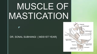
- 1. z MUSCLE OF MASTICATION DR. SONAL SUBHANGI { MDS1ST YEAR}
- 2. z CONTENT INTRODUCTION EMBRYOLOGY BLOOD SUPPLY AND LYMPHATICS NERVES MUSCLES CLINICAL SIGNIFICANCE PREVALENCE REFERENCES
- 3. z INTRODUCTION The primary muscles of mastication (chewing food) are the temporalis, medial pterygoid, lateral pterygoid, and masseter muscles. The four main muscles of mastication attach to the rami of the mandible and function to move the jaw (mandible). The cardinal mandibular movements of mastication are elevation, depression, protrusion, retraction, and side to side movement. To augment the process of eating, the muscles of mastication also move the mandible in a side to side motion to assist in the grinding of food. The muscles of mastication also function to approximate (bring together or close) the teeth. The superficial muscle of the neck, the platysma muscle, also assists with depression of the mandible against resistance.
- 4. z EMBRYOLOGY The muscles of mastication arise from the first pharyngeal arch. They are then differentiated into muscles starting the seventh week. The nerve supply to these muscles begins by the eighth week
- 5. z BLOOD SUPPLY AND LYMPHATICS The arterial supply to the muscles of mastication is via the maxillary artery, a branch of the external carotid artery.
- 6. z NERVES The four main muscles of mastication are all innervated by the anterior trunk of the mandibular nerve, which is the third division of the trigeminal nerve (CN V3). The mandibular nerve (CN V3) is the largest and inferior-most division of the trigeminal nerve (CN V). The trigeminal nerve (CN V) exits the skull via foramen ovale of the greater wing of the sphenoid bone. The mandibular nerve contains both sensory and motor fibers. The mandibular nerve is the only division of the trigeminal nerve that carries motor fibers. The mandibular nerve (CN V3) further subdivides as it innervates the four main muscles of mastication. The temporalis muscle receives innervation by deep temporal branches of the mandibular nerve. The medial pterygoid muscle receives innervation from the medial pterygoid nerve, a division of the mandibular nerve . The lateral pterygoid muscle gets its nerve supply from the lateral pterygoid nerves, divisions of the mandibular nerve. The masseter muscle receives nerve input from the masseteric nerve, a division of the mandibular nerve.
- 7. z MUSCLES The primary and accessory muscles of mastication work in a coordinated fashion to produce mandibular movement for chewing food. The accessory muscles of mastication are the buccinator, suprahyoid muscles (digastric muscle, mylohyoid muscle, and geniohyoid muscle), and infrahyoid muscles (the sternohyoid, sternothyroid, thyrohyoid and omohyoid muscle). The origin, insertion, and action of the main muscles of mastication, as well as a brief description of the accessory muscles of mastication, are as follows . Mainly 4 muscles: Masseter Temporalis Lateral pterygoid Medial pterygoid
- 8. z
- 9. z Temporalis Muscle The temporalis muscle is a fan-shaped muscle with anterior fibers that have a vertical orientation, mid fibers have an oblique orientation, and posterior fibers have a more of a horizontal orientation. ORIGIN INSERTION NERVE SUPPLY ACTIONS From whole length of temporal fossa. Fibres converge downwards. Anterior fibers – descend vertically . Intermediate – obliquely . Posterior fibers – horizontally to get inserted into the coronoid process & anterior margin of ramus of mandible . Some fibres also join masseter and pass on to mandible. By deep temporal branches of the anterior trunk of mandibular nerve. Elevates mandible . Posterior fibres retract the mandible after protraction . Helps in lateral sliding of mandible during grinding.
- 10. z Lateral Pterygoid The lateral pterygoid muscle is the primary muscle of the inferior temporal fossa. The lateral pterygoid has two parts: an upper head and a lower head . ORIGIN INSERTION NERVE SUPPLY ACTIONS upper head – arises from infra temporal surface and infra temporal crest of greater wings of sphenoid. Lower head – arises from lateral surface of lateral pterygoid plate of sphenoid . Depression in front of the neck of mandible. Articular disc of temporomandibular joint. By a branch of mandibular nerve. Protrusion of mandible along with medial ptergygoid.
- 11. zMedial Pterygoid The medial pterygoid muscle is a thick rectangular muscle with a superficial head and a deep head. ORIGIN INSERTION NERVE SUPPLY ACTION Superficial part – from the medial surface of lateral pterygoid plate . From the grooved surface of the pyramidal surface of palatine bone. Deep part – small slip originates from lateral surfaces of palatine bone and tuberosity of maxilla. into the lower and back part of the medial surfaces of angle and ramus of mandible as high as mandibular foramen above & nearly as forward as mylohyoid groove. By a branch of mandibular nerve. Assist in protrusion of mandible. Acting with lateral pterygoid alternatively , it produces a movement .
- 12. z Masseter The masseter muscle is a rectangularly shaped muscle with three layers (superficial, deep, and intermediate). ORIGIN INSERTION NERVE SUPPLY ACTION Superficial layer – its fibres originate from zygomatic process of maxilla in the form of a thick aponeurosis . Anterior 2/3rd of the lower border of zygomatic arch. Middle layer – deep surfaces of anterior 2/3rd of arch. Lower border of posterior 1/3rd of arch. Deep layer – from the deep surface of zygomatic arch. Superficial layer – downwards and backwards into the angle and lower half of the lateral surfaces of mandible. Middle layer- into the middle part of ramus. Deep layer – into the upper part of ramus and coronoid process. Masseteric nerve , branch of the anterior trunk of mandibular nerve. Elevation of mandible
- 13. z
- 14. z Accessory Muscles of Mastication The strap muscles are composed of the suprahyoid, and infrahyoid muscles are located on the side of the neck bilaterally. The strap muscles primarily function to raise and depress the hyoid bone and larynx. The strap muscles also assist with depression of the mandible when opening the mouth against an opposing force. The buccinator is a facial expression muscle that helps in mastication by keeping food pushed back within the oral cavity.
- 15. z CLINICAL SIGNIFICANCE Masticatory muscle disorders include myofascial pain and dysfunction, myositis, and neoplasms. Myofascial pain and dysfunction may result from several etiologies. The most common ones are nocturnal bruxism, habitual clenching of the mouth, and whiplash injuries during a trauma. Temporomandibular joint (TMJ) dysfunction can result from an imbalance of forces within the muscles of mastication. Grinding of teeth at night (bruxism) is a common cause of TMJ dysfunction secondary to a resultant imbalance in the muscle of mastication forces from excessive grinding of the teeth. Muscle spasm of the muscles of mastication (trismus) can be a symptom of tumor or infection.
- 16. z An infection like tetanus may present with "lockjaw" or trismus. Other infections or inflammation of the muscles may present as myositis or pain during the movement of the jaw. Tumors, although rare but may present in the masticator space, which is enveloped by the deep cervical facia. These tumors may have an extension from adjacent regions. The medial side of the fascia is attached to the skull base, and the lateral side extends to the temporalis muscle. Anteriorly it is attached to the body of the mandible at the level of the oblique line, and posteriorly it is attached to the ramus of the mandible.
- 17. z PREVALENCE The sample population included 199 participants (66% female and 34% male). The prevalence of TMD-related pain was 26.8% (n = 42); men and women did not differ statistically in their TMD-related pain. Alkhubaizi Q , Khalaf M E and Faridoun A . Prevalence of Temporomandibular Disorder-Related Pain among Adults Seeking Dental Care: A Cross-Sectional Study . Int J Dent . 2022 Sep ; 3186069.
- 18. z REFERENCES Standring S . Gray's Anatomy :The Anatomical Basis of Clinical Practice . 42th Edition. Selvakumari T L . Essentials of Anatomy for Dental Students . 1st Edition. Chaurasia B D & Garg K . B D Chaurasia's Human Anatomy . 9th Edition . Devi V S . Inderbir Singh's Textbook of Anatomy for Dental Students . 7TH Edition.
- 19. z