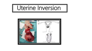
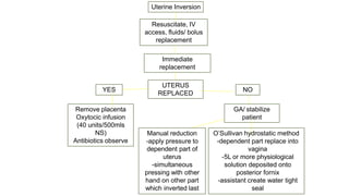
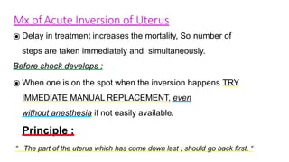
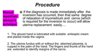
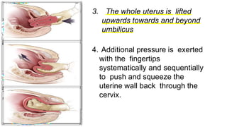
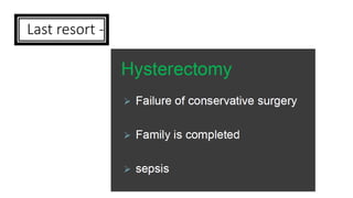
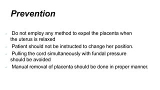
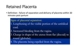
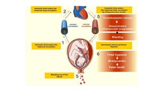
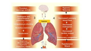
SlideShare a Scribd company logo
• Uterine Inversion
• Retained placenta
• Amniotic Fluid Embolism
Dr Sushma Sharma
Professor
Department of Obstetrics & Gynaecology
MIMER Medical College, Talegaon(D), Pune
Uterine Inversion
Dr
Introduction
• Uterine inversion occurs when the uterine fundus collapses into the
endometrial cavity, turning the uterus inside out.
• A rare complication of vaginal delivery /cesarean
• Life threatening obstetric emergency
• Needs prompt recognition and management
• Less than 5% maybe spontaneous
Classification
By the extent of inversion:
• First degree (Incomplete) – The fundus
is within the endometrial cavity
• Second Degree (Complete) – The
fundus protrudes through the os.
• Third Degree (Prolapsed) – The fundus
protrudes into or beyond the introitus
• Fourth Degree (Total) – Both the uterus
and vagina inverted
Classification-contd.
By time of occurrence:
• Acute – within 24 hours of delivery
• Subacute – more than 24 hours but less than four weeks post partum
• Chronic – more than one month post partum
Incidence -
Ranges from 1 in 3500 to 1 in 20,000
Pathogenesis -
Incompletely understood.
Maybe attributed to:
• Use of excessive cord traction
• Fundal pressure
• Crede’ manoeuvre
• Fundal implantation of placenta
• Likely that other factors play a role – eg connective tissue disorders like
Marfan’s syndrome
Risk factors –
• Short cord
• Prolonged labour
• Precipitate labour
• Macrosomia
• Use of uterine relaxants
• Uterine anomalies
• Retained placenta
• Placenta accreta spectrum
Clinical Features
Depends on extent and time of occurrence. May include:
• Mild to severe vaginal bleeding
• Mild to severe lower abdominal pain
• Mass protruding from cervix/ vagina
• Urinary retention
Commonest presentation of complete inversion is Shock- out of
proportion to blood loss- attributed to Neurogenic shock.
Clinical Features
On examination:
Features of shock:
• P/A- Atonic uterus and fundal notch
or dimple in the area of normally
globular fundus
• Local examination smooth round
mass protruding from cervix or
vagina
Diagnosis
Bases upon clinical findings:
• Imaging is rarely necessary
maybe used for confirmation in
subacute/ chronic variety
• Ultrasound, MRI maybe used
Differential Diagnosis
• Fibroid Polyp
• Cervical Polyp
• Uterine Prolapse
Management
Goals:
• Replace the fundus to its anatomical position.
• Manage PPH and shock
• Prevent recurrent inversion
Management
Uterine Inversion
Remove placenta
Oxytocic infusion
(40 units/500mls
NS)
Antibiotics observe
O’Sullivan hydrostatic method
-dependent part replace into
vagina
-5L or more physiological
solution deposited onto
posterior fornix
-assistant create water tight
seal
Manual reduction
-apply pressure to
dependent part of
uterus
-simultaneous
pressing with other
hand on other part
which inverted last
GA/ stabilize
patient
UTERUS
REPLACED
Immediate
replacement
Resuscitate, IV
access, fluids/ bolus
replacement
NO
YES
⦿Teamwork = resuscitation + uterine repositioning
simultaneously
⦿postpartum hemorrhage drill.
⦿The quickest way to treat neurogenic shock - to replace the
uterus.
⦿ Delay in treatment increases the mortality, So number of
steps are taken immediately and simultaneously.
Before shock develops :
⦿ When one is on the spot when the inversion happens TRY
IMMEDIATE MANUAL REPLACEMENT, even
without anesthesia if not easily available.
Principle :
“ The part of the uterus which has come down last , should go back first. “
Mx of Acute Inversion of Uterus
Manual
reposition-
Johnson’s
technique
Procedure
⦿ If the diagnosis is made immediately after the
inversion has occurred, then that same degree
of relaxation of myometrium and cervix (which
is required for the inversion to occur) will allow
uterine replacement easily…
1. The gloved hand is lubricated with suitable antiseptic cream
and placed inside the vagina.
2. The uterine fundus with or without the attached placenta, is
cupped in the palm of the hand. The fingers and thumb of the hand
are extended to identify margins of the cervix.
3. The whole uterus is lifted
upwards towards and beyond
umbilicus
4. Additional pressure is exerted
with the fingertips
systematically and sequentially
to push and squeeze the
uterine wall back through the
cervix.
5. Sustained pressure for 3-5 mins to achieve complete replacement
6. Apply counter support by the other hand placed on the abdomen
7. Once the fundus has been replaced keep the hand in the uterus
while rapid infusion of oxytocin is given to contract the uterus.
Initially, bimanual compression aids in control of further
hemorrhage until uterine tone is recovered.
8. When the uterus is felt contracting, the hand is slowly
withdrawn.
If placenta is attached, it is to be removed only after the uterus
becomes contracted.
If the placenta is partially attached , it should be peeled out
before replacement of uterus.
⦿ Tube passed into the posterior fornix
⦿ Assistant close vulva around
operator’s wrist
⦿ Warm saline run in until pressure
gradually restores position of uterus
O’Sullivan’s hydrostatic method
Surgical techniques
• When conservative management fails. Usually because the
constriction ring gets too tight
Surgery
Abdominal Vaginal
Huntington ; Haultain Kustner ; Spinelli
Modifications
of the classical
abdominal techniques
– use of ventouse cup
Last resort -
Prevention
 Do not employ any method to expel the placenta when
the uterus is relaxed
 Patient should not be instructed to change her position.
 Pulling the cord simultaneously with fundal pressure
should be avoided
 Manual removal of placenta should be done in proper manner.
Retained Placenta
• Definition : failure of separation and delivery of placenta within 30
minutes post partum
Aetiology
• Abnormality of lower segment / internal os behavior
• ? Premature injudicious oxytocics usage
• Adherent placenta – commonest , most dreaded. Also called morbidly
adherent placenta (MAP )
Adherent placenta occurs
when there is a defect in the decidua basalis ,
Resulting
in an abnormal invasion of the placenta
directly into the substance of the uterus.
Types
1 ) Simple Adherent Placenta.
2 ) Morbidly Adherent Placenta :
i ) Placenta Accreta
ii ) Placenta Increta
iii) Placenta Percreta
INCIDENCE
 It varies widely all over the world.
Increased dramatically over the last 3 decades
( Because of Increase in LSCS rate … ).
 A.C.O.G.  1 Per 2500 deliveries. Accreta : 75
-78 %
Increta : 15 – 18 %
Percreta : 5 -7 %
Associated Conditions :
 Placenta Previa
 Previous Surgeries such as …
- Cesarean Section
- Myomectomy
- Synecolysis
- D & C
- M.R.P.
- Cornual Resection
 Uterine Malformation
 Septic Endometritis
Risk Factors :
 High Parity
 Advanced Maternal Age
 Down Syndrome
 High level of Maternal Serum AFP.
 High level of Maternal free Beta HCG.
ETIOLOGY :
 Defective decidual formation :
- Partial / total absence of decidua basalis
- Imperfect development of fibrinoid layer (Nitabuch layer)
- Placental villi are attached to the myometrium
Significance :
 Increased Maternal Morbidity ( 2 – 7 % )
 Increased Maternal Mortality ( 7 – 10 % )
from,
- Severe Hemorrhage
- Infection
- Inversion of Uterus
DIAGNOSIS
Earliest diagnosis of Adherent Placenta is must to
avoid any catastrophic emergency in future.
Antenatal diagnosis is the single most important factor
in improving the outcome in MAP.
METHODS…
 Clinical suspicion
 Ultrasound
 Color Doppler
 MRI
 Biochemical Marker
 Histopathology
USG
First-line investigation for suspected
placental invasion of the myometrium.
• Both transabdominal and transvaginal
ultrasonography
USG CRITERIA
 1st Trimester :
G. Sac located in the lower uterine segment
(rather than the fundus), next to or lower than
the Prev. CS scar.
 2nd & 3rd Trimester :
 Presence of irregular lacunae within the placenta
 Loss of retro placental clear space
 Loss or disruption of the white line – Bladder line
Moth – eaten OR
Swiss Cheese
Appearance
• Obliteration of clear space between
placenta and uterine wall
Reliability :
• Sensitivity - 93%
• Specificity - 79%
The use of power Doppler, color Doppler, or three- dimensional
imaging does not significantly improve the diagnostic
sensitivity compared with that achieved by grayscale
Ultrasonography alone.
[ Chou MM, Ho ES, Lee YH. Prenatal diagnosis of placenta previa accreta by transabdominal color
Doppler ultrasound. Ultrasound Obstet Gynecol 2000;15:28–35. ]
3 D USG
Diagnostic Criteria :
Irregular intraplacental vascularization with
tortuous confluent vessels crossing placental
width.
Hypervascularity of uterine serosa– bladder
wall interface.
Colour Doppler
 Diffuse or focal
intraparenchymal lacunar
flow.
 Vascular lakes with
turbulent flow.
 Hypervascularity of serosa-
bladder interface.
 Prominent subplacental
venous complex.
M.R.I.
 No more sensitive than USG , But used as an adjunct
to USG , when there is strong clinical suspicion of
accreta.
 MRI achieves better images than Ultrasonography in
- Posteriorly sited MAP and
- With prior myomectomy,
( Because the ultrasound beam is impeded by the fetal
head in the former and by the scar tissue in the latter )
M.R.I. Criteria
Uterine bulging into the
bladder
Heterogeneous signal
intensity within the placenta
Presence of intra placental
bands on the T2W imaging
Abnormal placental vascularity
Focal interruption of the
myometrium
Laboratory Findings :
•
• Several series and case reports have reported an association
between placenta accreta and otherwise unexplained elevations in
second trimester MSAFP concentration (>2 or 2.5 multiples of the
median [MOM]).
Although an elevated MSAFP level supports an ultrasound-
based diagnosis of placenta accreta, it is an inconsistent finding and
is not useful by itself for diagnosis of accreta.
Histology
 Post Partum specimen shows :
Placental villi anchored directly on, or invading into
or through, the myometrium, without an intervening
decidual plate.
Treatment :
A multidisciplinary team approach is relevant
in managing these patients in order to reduce
morbidity and mortality associated with MAP.
Particular consideration should be given to
anticipation and management of massive
hemorrhage,
including
- availability of packed cells,
- platelets,
- fresh frozen plasma,
- cryoprecipitate, and
- activated factor VII.
Interventional Radiology and cell saver
technology are useful.
At present , placenta accrete can be managed in three ways:
( 1 ) Carry out a hysterectomy;
( 2 ) Leave the placenta in situ ; and
( 3 ) Resect the invaded tissues with the entire placenta
restoring uterine anatomy.
 Each one has weaknesses and strengths,
dependent on the condition itself and the specific
preferences taken by the surgeon and the team.
Women who have had a previous CS who also
have either placenta previa or an anterior placenta
underlying the old CS scar at 32 weeks of gestation
are at increased risk of placenta accreta and should
be managed as if they have placenta accreta, with
appropriate preparations for surgery made.
(RCOG 2011)
Elective delivery by caesarean section at 34–35
weeks of gestation for suspected placenta accreta
(ACOG 2012).
Conservative
/ moderate blood Loss / fertility to be
 In case of
( focal defect
preserved )
 Localized Resection with uterine repair
 Over sewing of the ut. Defect
 Blunt dissection followed by curetting the uterine cavity
• Uterus fails to contract (Multipara) :
Hysterectomy
Non Surgical
Leave the Placenta in situ to resorb with methotrexate
therapy
Ligation of the Ut. And Int. iliac artery
Fluoroscopic bilateral UAE
Argon beam coagulation for haemostasis
Insertion of occluding Balloons in the Int. iliac art. (Bilat)
Surgical
 Cesarean Hysterectomy.
Hysterectomy and partial / total resection of
bladder
Subtotal Hysterectomy with removal of large part
of placenta and Prophylactic occlusive Balloon
catheter in int. iliac art.
An Elective controlled condition is preferred rather
than an emergency condition without adequate
preparations.
A midline incision will facilitate better exposure, especially
if placenta Percreta is suspected.
Leaving the placenta undisturbed until completion of
the hysterectomy would prevent unnecessary
hemorrhage.
In cases where MAP is associated with placenta previa,
total hysterectomy is preferred to a subtotal hysterectomy.
Uterine Incision:
It is best to avoid cutting through a MAP because of the
possibility of massive haemorrhage.
Various modifications of the uterine
incision to avoid the placenta have been
reported…
- Classical incision,
- High transverse incision,
- Fundal incision,
- Fundal transverse incision
Remember

 The presence of pericervical or lower-segment varicose veins
proper of placenta praevia can be confused with the
neovascularization of placenta accreta.
Surgical exploration will make a differential diagnosis, thus
avoiding unnecessary hysterectomies.
Excision of placental site
 It is possible to "excise the placental site".
This is done by inverting the uterus in order to provide
good access to the placental site.
If the area of placental attachment is focal and the
majority of the placenta has been removed, then a
"wedge resection" of the area can be performed.
Balloon Catheterization
Pre-operative placement of arterial catheters in internal iliac
artery
After delivery balloons are inflated to achieve temporary
homeostasis
Selective arterial embolization (SAE) if necessary. . .
Bil. Int. iliac artery ligation is performed prior to peripartum
hysterectomy where Interventional Radiology is not
available.
Placement of occlusion balloon catheters
into both internal iliac arteries.
Methotrexate
 A folate antagonist, acts primarily against
rapidly dividing cells and therefore is effective
against proliferating trophoblasts.
First described by Arulkumaran et al in
1986. They reported administration 50 mg of
methotrexate as an intravenous infusion on
alternate days and the placental mass was
expelled on 11th postnatal day.
However, more recently, others have
argued that, after delivery of the fetus, the
placeta is no longer dividing and therefore,
methotrexate is of no value.
Methotrexate has been used in varying
doses and routes, however, there are no
randomized trials and no standard protocol
regarding its dosage.
The outcome when the placenta is left in
place after methotrexate administration varies
widely; it ranges from expulsion at 7 days to
progressive resorption in roughly 6 months.
Mtx – 50 mg IM + Folic Acid 6mg IM on
alternate day till β HCG comes to zero.
Other Modalities


Tamponade of the placental implantation site
with inflated Intra Uterine balloon catheter bags.
Lower Segment Compression Sutures
Pelvic pressure sponge packing.
Follow up…
1.- Ultrasound for Vascularity
2.- hCG titers weekly till it becomes Zero.
3.- Monitor for infection
4.- Bleeding
5.- Coagulation profile
Antibiotics till needed
99099 44160. 41
Resources Patient, clinical and
anatomic features
Decision Definitive treatment
Limited
experience
or expertise, poor
resources or no
facilities for safe
patient transfer
lower segment invasion
vaginal bleeding with high
suspicion of accreta
Possibility of percreta
Extraplacental
hysterotomy,
Placental left in
situ
Followed by
uterine closure
Delayed hysterectomy
or conservative procedure
according clinical
and surgical status
Qualified and
experienced
team, adequate
hospital
resources
No desire for future
pregnancy
Tissue destruction> 50% of
uterine circumference
Intractable haemorrhage
DIC
Resective surgery
Subtotal hysterectomy
for upper segment lesions
Total hysterectomy
for lower segment
and cervical involvement
Qualified and
experienced
team,
adequate
hospital
resou
22
r-c
De
ecs
-14
Desire for future
pregnancy
Destruction < 50% of
uterineaxial circumference
Minor coagulation
disorders
Dr Shashwat Jani.
Conservative
surgery
1Placenta in situ with or wi
MXT
2 One step surgery
OR
3- Two step surgery
Bladder Involvement
 Involve UROLOGIST.
Preoperative Ureteric
stenting aids in identifying
the ureters, which will
help reduce ureteric
injuries.
Care must be taken during
surgery not to attempt to
dissect the bladder off the
lower uterine segment
which results in torrential
bleeding.
Anterior bladder wall
incision is particularly
helpful in defining
dissection planes and the
location of the ureters.
Reality :
Even today, the ground reality is that a
majority of morbidly adherent placenta are
diagnosed during the third stage of labour
or during caesarean section and which
results in adverse consequences including
exanguinating haemorrhage.
To Conclude…
Caesarean hysterectomy was the
cornerstone in the management in the past.
Antenatal diagnosis permits effective and
safe conservative approaches today.
The use of methotrexate, monitoring with
serum hCG and follow up with USG is backed
only by conflicting evidence.
Amniotic Fluid Embolism
• Protects the unborn fetus from its surrounding environment.
• Isolated from the maternal intravascular compartment.
• When this isolation is disrupted in one way or another, a
systemic maternal reaction can ensue, affecting the heart,
lungs, and brain.
• This reaction can be lethal to both mother and fetus.
Amniotic Fluid
• The syndrome is know as amniotic fluid embolism (AFE)
and is one of the most serious complications of pregnancy
and delivery.
• Given the unpredictable and unexpected nature of AFE, every
obstetrician should be prepared to include this condition in
differential diagnosis to optimize the chances for survival of
both mother and infant.
Background
• The presence of fetal cellular debris in the maternal circulation,
associated with maternal complications, was reported for the first
time in the 1920s by Ricardo Meyer from Brazil.
• In an effort to better understand this condition, several animal
models have been developed.
• Studies to human beings cannot be easily made, since not all the
findings in animals are consistent with the clinical manifestations.
• Recently, the term Anaphylactoid syndrome of pregnancy has
been proposed instead of AFE.
Incidence
• The true incidence of AFE may not be known.
• The disparity between reported figures is large, with rates ranging
from 1 in 8,000 to 1 in 83,000 deliveries.
• A recent study from California reports an incidence of 1 per
20,646 deliveries.
Mortality
• AFE constitutes the leading cause of mortality during
labor and the first few postpartum hours.
• 5% and 18% of all maternal deaths are due to AFE, a rate
of about 7.8 to 12 deaths per million births.
• Maternal death usually occurs because of
*sudden cardiac arrest,
*hemorrhage due to coagulopathy,
*acute respiratory distress syndrome (ARDS)
*multiple organ failure.
• It’s rare enough that despite systematic research in an
autopsy series, no evidence of AFE was noted among 14
maternal deaths from 26,000 deliveries.
• For women diagnosed as having AFE, mortality rates ranging
from 26% to as high as 86% have been reported.
• Intensive care management of affected patients can
cause improvements.
Pathophysiology
• For years, it was thought that microscopic fetal debris
particles during an AFE episode caused acute occlusion
of a large part of the mother's pulmonary
microvasculature.
• This was assumed because of respiratory distress and
cyanosis were the manifestations.
The first hemodynamic assessments of patients with AFE
revealed
1. Elevated pulmonary capillary wedge pressure (PCWP),
2. Decreased left ventricular stroke work index (LVSWI),
3. Mild to moderate increases in mean pulmonary arterial
pressures (MPAP), and
4. Variable increases in central venous pressure (CVP).
• These findings are consistent with left-sided heart failure.
• The initial reaction of the pulmonary vasculature to
amniotic fluid exposure may be -transient vasospasm,
-pulmonary hypertension, and
-profound hypoxia.
• To date, however, no direct evidence of these reactions
has been documented, probably due to the delay between
the onset of symptoms.
• Clinically, the reaction to AFE comprises 3 distinct phases.
1. Respiratory, including respiratory distress and cyanosis;
2. Hemodynamic, with pulmonary edema and shock; and
3. Neurologic, including seizures, confusion or coma.
• These presentations can occur separately or in combination,
and in different degrees.
Cause
• AFE is very rare and complex.
• The disorder occurs during the last stages of labor when amniotic fluid
enters the circulatory system of the mother.
• When the fetal cells and amniotic fluid enters the
bloodstream, the immune system responds by releasing products that cause
an inflammatory reaction this causes severe changes in the mechanisms
that affect blood clotting.
• Intravascular coagulation (DIC) occurs and results in serious bleeding.
• The condition can also develop after elective
abortion, amniocentesis, cesarean delivery or
trauma.
• The use of drugs to induce labor, such as misoprostol,
nearly doubles the risk of AFE.
• A maternal age of 35 years or older is associated with AFE.
Diagnosis
• Until now, the diagnosis of AFE was made by a pathologist, based on a
finding in :- maternal tissue of epithelial squamous cells,
- fat derived from vernix caseosa,
- mucin derived from infant's intestinal mucus,
- bile derived from meconium.
• Increased awareness of the syndrome's existence has resulted in earlier
diagnoses, aggressive intervention, and better chance of survival.
• Diagnosis is also based upon the signs and symptoms observed
during the birth or procedures.
Symptoms & Signs
• Suspect AFE when confronted with any pregnant patient who has
sudden onset of - respiratory distress,
- cardiac collapse,
- seizures,
- unexplained fetal distress, and
- abnormal bleeding
• AFE is suspected when a woman giving birth experiences very
sudden insufficient oxygen to body tissues, low blood pressure,
and profuse bleeding due to defects in blood coagulation.
• They also can be entirely absent.
Clinical Conditions Associated With AFE
• The most frequently cited are
- onset of labor,
- fetal death, trauma, and
- uterine over distention by multiple gestation,
- polyhydramnios or fetal macrosomia.
• Cited risk factors are not consistent.
Management
• AFE is a rapidly lethal condition.
• To limit its associated mortality and morbidity,
supportive care must be initiated with the same
promptness and aggressiveness as the presentation of
AFE itself.
Medications
• Pharmacologic treatment
• The goals of therapy are:
(1) maintenance of systolic blood pressure above 90 mm Hg
(2) maintenance of arterial PO2 over 60 mm Hg
(3) correction of coagulation abnormalities.
• Use inotropics (ß adrenergics) and pressors (ephedrine,
dopamine, dobutamine, norepinephrine infusions).
Prognosis
• The neonatal survival rate after a delivery affected by
AFE was 79% according to the Registry, and only half
of the surviving infants were neurologically normal.
The infant's prognosis was adversely affected by
maternal cardiac arrest.
• National AFE Registry indicate a maternal mortality
rate of 61%.
Summary
• AFE is a sudden and unexpected complication of
pregnancy.
• It has a complex pathogenesis and serious implications
for both mother and infant.
• Continues to be associated with high rates of
morbidity and mortality.
• Obstetricians should be alert to the symptoms of AFE
and strive for prompt and aggressive treatment.
Inversion, retained placenta , afe

More Related Content

What's hot

ectopic pregnancy
ectopic pregnancyectopic pregnancy
ectopic pregnancy
Vishnu Narayanan
 
Normal and abnormal labor part 1
Normal and abnormal labor part 1Normal and abnormal labor part 1
Normal and abnormal labor part 1
Faculty of Medicine,Zagazig University,EGYPT
 
Forceps delivery and vacuum extraction
Forceps delivery and vacuum extractionForceps delivery and vacuum extraction
Forceps delivery and vacuum extraction
Dr ABU SURAIH SAKHRI
 
Induction of Labour: SOGC, 2013 WHO, 2011 NICE, 2008
Induction of Labour: SOGC, 2013 WHO, 2011 NICE, 2008Induction of Labour: SOGC, 2013 WHO, 2011 NICE, 2008
Induction of Labour: SOGC, 2013 WHO, 2011 NICE, 2008
Aboubakr Elnashar
 
MECONIUM STAINED AMNIOTIC FLUID
MECONIUM STAINED AMNIOTIC FLUIDMECONIUM STAINED AMNIOTIC FLUID
MECONIUM STAINED AMNIOTIC FLUID
Niranjan Chavan
 
BREECH DELIVERY By Odokoyero Abdalah Fadhil and Nanfuka Mariam
BREECH DELIVERY By Odokoyero Abdalah Fadhil and Nanfuka MariamBREECH DELIVERY By Odokoyero Abdalah Fadhil and Nanfuka Mariam
BREECH DELIVERY By Odokoyero Abdalah Fadhil and Nanfuka Mariam
Odokonyerofadhil
 
Aph Antepartum hemorrhage
Aph Antepartum hemorrhageAph Antepartum hemorrhage
Aph Antepartum hemorrhage
HuzaifaMD
 
Management of hyperemesis gravidarum rcog 2016
Management of hyperemesis gravidarum  rcog 2016Management of hyperemesis gravidarum  rcog 2016
Management of hyperemesis gravidarum rcog 2016
Dr Meenakshi Sharma
 
Malpresentation & Malpositioning
Malpresentation & MalpositioningMalpresentation & Malpositioning
Malpresentation & Malpositioning
Ahmed Ali
 
Obstructed labor management
Obstructed labor managementObstructed labor management
Obstructed labor management
Beka Aberra
 
Vacuum extraction (ventouse)
Vacuum extraction (ventouse)Vacuum extraction (ventouse)
Vacuum extraction (ventouse)raj kumar
 
High risk pregnancy delfin 202
High risk pregnancy delfin 202High risk pregnancy delfin 202
High risk pregnancy delfin 202
shenell delfin
 
Inversion of uterus by Sunil Kumar Daha
Inversion of uterus by Sunil Kumar DahaInversion of uterus by Sunil Kumar Daha
Inversion of uterus by Sunil Kumar Daha
sunil kumar daha
 
Mellss obg y3 breech
Mellss obg y3 breechMellss obg y3 breech
Mellss obg y3 breech
nur amalina aminuddin baki
 
Ectopic pregnancy
Ectopic pregnancyEctopic pregnancy
Ectopic pregnancy
OM VERMA
 
MANAGEMENT OF ACUTE UTERINE INVERSION BY DR SHASHWAT JANI
MANAGEMENT OF ACUTE UTERINE INVERSION BY DR SHASHWAT JANIMANAGEMENT OF ACUTE UTERINE INVERSION BY DR SHASHWAT JANI
MANAGEMENT OF ACUTE UTERINE INVERSION BY DR SHASHWAT JANI
DR SHASHWAT JANI
 
Transverse lie
Transverse lie Transverse lie
Transverse lie
Chandrima Karki
 
Operative vaginal delivery - forceps , vacuum delivery.
Operative vaginal delivery  - forceps , vacuum delivery.Operative vaginal delivery  - forceps , vacuum delivery.
Operative vaginal delivery - forceps , vacuum delivery.
dr. gokul reshmi mariappan
 
Instrumental vaginaldelivery...
Instrumental  vaginaldelivery...Instrumental  vaginaldelivery...
Instrumental vaginaldelivery...
imanswati
 

What's hot (20)

ectopic pregnancy
ectopic pregnancyectopic pregnancy
ectopic pregnancy
 
Normal and abnormal labor part 1
Normal and abnormal labor part 1Normal and abnormal labor part 1
Normal and abnormal labor part 1
 
Forceps delivery and vacuum extraction
Forceps delivery and vacuum extractionForceps delivery and vacuum extraction
Forceps delivery and vacuum extraction
 
Induction of Labour: SOGC, 2013 WHO, 2011 NICE, 2008
Induction of Labour: SOGC, 2013 WHO, 2011 NICE, 2008Induction of Labour: SOGC, 2013 WHO, 2011 NICE, 2008
Induction of Labour: SOGC, 2013 WHO, 2011 NICE, 2008
 
MECONIUM STAINED AMNIOTIC FLUID
MECONIUM STAINED AMNIOTIC FLUIDMECONIUM STAINED AMNIOTIC FLUID
MECONIUM STAINED AMNIOTIC FLUID
 
BREECH DELIVERY By Odokoyero Abdalah Fadhil and Nanfuka Mariam
BREECH DELIVERY By Odokoyero Abdalah Fadhil and Nanfuka MariamBREECH DELIVERY By Odokoyero Abdalah Fadhil and Nanfuka Mariam
BREECH DELIVERY By Odokoyero Abdalah Fadhil and Nanfuka Mariam
 
Amniotic fluid
Amniotic fluidAmniotic fluid
Amniotic fluid
 
Aph Antepartum hemorrhage
Aph Antepartum hemorrhageAph Antepartum hemorrhage
Aph Antepartum hemorrhage
 
Management of hyperemesis gravidarum rcog 2016
Management of hyperemesis gravidarum  rcog 2016Management of hyperemesis gravidarum  rcog 2016
Management of hyperemesis gravidarum rcog 2016
 
Malpresentation & Malpositioning
Malpresentation & MalpositioningMalpresentation & Malpositioning
Malpresentation & Malpositioning
 
Obstructed labor management
Obstructed labor managementObstructed labor management
Obstructed labor management
 
Vacuum extraction (ventouse)
Vacuum extraction (ventouse)Vacuum extraction (ventouse)
Vacuum extraction (ventouse)
 
High risk pregnancy delfin 202
High risk pregnancy delfin 202High risk pregnancy delfin 202
High risk pregnancy delfin 202
 
Inversion of uterus by Sunil Kumar Daha
Inversion of uterus by Sunil Kumar DahaInversion of uterus by Sunil Kumar Daha
Inversion of uterus by Sunil Kumar Daha
 
Mellss obg y3 breech
Mellss obg y3 breechMellss obg y3 breech
Mellss obg y3 breech
 
Ectopic pregnancy
Ectopic pregnancyEctopic pregnancy
Ectopic pregnancy
 
MANAGEMENT OF ACUTE UTERINE INVERSION BY DR SHASHWAT JANI
MANAGEMENT OF ACUTE UTERINE INVERSION BY DR SHASHWAT JANIMANAGEMENT OF ACUTE UTERINE INVERSION BY DR SHASHWAT JANI
MANAGEMENT OF ACUTE UTERINE INVERSION BY DR SHASHWAT JANI
 
Transverse lie
Transverse lie Transverse lie
Transverse lie
 
Operative vaginal delivery - forceps , vacuum delivery.
Operative vaginal delivery  - forceps , vacuum delivery.Operative vaginal delivery  - forceps , vacuum delivery.
Operative vaginal delivery - forceps , vacuum delivery.
 
Instrumental vaginaldelivery...
Instrumental  vaginaldelivery...Instrumental  vaginaldelivery...
Instrumental vaginaldelivery...
 

Similar to Inversion, retained placenta , afe

A brief introduction to c section and how its done.
A brief introduction to c section and how its done.A brief introduction to c section and how its done.
A brief introduction to c section and how its done.
JudeMusoke1
 
A brief introduction to c section and how its done.
A brief introduction to c section and how its done.A brief introduction to c section and how its done.
A brief introduction to c section and how its done.
JudeMusoke1
 
PLACENTA PREVIA. a disorder of Pregnancy
PLACENTA PREVIA. a disorder of PregnancyPLACENTA PREVIA. a disorder of Pregnancy
PLACENTA PREVIA. a disorder of Pregnancy
virengeeta
 
operative obstetrics emergency.pptx
operative obstetrics emergency.pptxoperative obstetrics emergency.pptx
operative obstetrics emergency.pptx
MesfinShifara
 
Ectopic Pregnancy-1.pptx
Ectopic Pregnancy-1.pptxEctopic Pregnancy-1.pptx
Ectopic Pregnancy-1.pptx
ImranKhan127540
 
abnormallabour-130512231046-phpapp02 (2).pptx
abnormallabour-130512231046-phpapp02 (2).pptxabnormallabour-130512231046-phpapp02 (2).pptx
abnormallabour-130512231046-phpapp02 (2).pptx
Mohammad Naik
 
CS by Berihu.Gebre yohannes.ppt presentations
CS by Berihu.Gebre yohannes.ppt presentationsCS by Berihu.Gebre yohannes.ppt presentations
CS by Berihu.Gebre yohannes.ppt presentations
RedaeMaldey
 
Breech presentation
Breech presentationBreech presentation
Breech presentation
Ali S. Mayali
 
Ectopic Pregnancy
Ectopic PregnancyEctopic Pregnancy
Ectopic Pregnancy
DJ CrissCross
 
Complications of 3 rd stage of the Labour
Complications of 3 rd stage of the LabourComplications of 3 rd stage of the Labour
Complications of 3 rd stage of the Labour
SREEVIDYA UMMADISETTI
 
Late pregnancy bleeding
Late pregnancy bleedingLate pregnancy bleeding
Late pregnancy bleeding
Eneutron
 
BREECH PRESENTATION, TYPES, DELIVERY.pptx
BREECH PRESENTATION, TYPES, DELIVERY.pptxBREECH PRESENTATION, TYPES, DELIVERY.pptx
BREECH PRESENTATION, TYPES, DELIVERY.pptx
PhilemonChizororo
 
LOWER SEGMENT CAESAREAN SECTION.pptx
LOWER SEGMENT CAESAREAN SECTION.pptxLOWER SEGMENT CAESAREAN SECTION.pptx
LOWER SEGMENT CAESAREAN SECTION.pptx
Subi Babu
 
RH 2 LECTURE 1.pptx
RH 2 LECTURE 1.pptxRH 2 LECTURE 1.pptx
RH 2 LECTURE 1.pptx
Lilian523287
 
Ectopic pregnancy
Ectopic pregnancy Ectopic pregnancy
Ectopic pregnancy
Dr. Darayus P. Gazder
 
ectopicpregnancydpg-170824070854.pdf
ectopicpregnancydpg-170824070854.pdfectopicpregnancydpg-170824070854.pdf
ectopicpregnancydpg-170824070854.pdf
AugustusCaesar7
 
Abruptio Placenta (Original)
Abruptio Placenta (Original)Abruptio Placenta (Original)
Abruptio Placenta (Original)boblhen
 
Obg seminar
Obg seminarObg seminar

Similar to Inversion, retained placenta , afe (20)

A brief introduction to c section and how its done.
A brief introduction to c section and how its done.A brief introduction to c section and how its done.
A brief introduction to c section and how its done.
 
A brief introduction to c section and how its done.
A brief introduction to c section and how its done.A brief introduction to c section and how its done.
A brief introduction to c section and how its done.
 
Cesarean section
Cesarean sectionCesarean section
Cesarean section
 
PLACENTA PREVIA. a disorder of Pregnancy
PLACENTA PREVIA. a disorder of PregnancyPLACENTA PREVIA. a disorder of Pregnancy
PLACENTA PREVIA. a disorder of Pregnancy
 
operative obstetrics emergency.pptx
operative obstetrics emergency.pptxoperative obstetrics emergency.pptx
operative obstetrics emergency.pptx
 
Ectopic Pregnancy-1.pptx
Ectopic Pregnancy-1.pptxEctopic Pregnancy-1.pptx
Ectopic Pregnancy-1.pptx
 
abnormallabour-130512231046-phpapp02 (2).pptx
abnormallabour-130512231046-phpapp02 (2).pptxabnormallabour-130512231046-phpapp02 (2).pptx
abnormallabour-130512231046-phpapp02 (2).pptx
 
CS by Berihu.Gebre yohannes.ppt presentations
CS by Berihu.Gebre yohannes.ppt presentationsCS by Berihu.Gebre yohannes.ppt presentations
CS by Berihu.Gebre yohannes.ppt presentations
 
Abnormal+labour
Abnormal+labourAbnormal+labour
Abnormal+labour
 
Breech presentation
Breech presentationBreech presentation
Breech presentation
 
Ectopic Pregnancy
Ectopic PregnancyEctopic Pregnancy
Ectopic Pregnancy
 
Complications of 3 rd stage of the Labour
Complications of 3 rd stage of the LabourComplications of 3 rd stage of the Labour
Complications of 3 rd stage of the Labour
 
Late pregnancy bleeding
Late pregnancy bleedingLate pregnancy bleeding
Late pregnancy bleeding
 
BREECH PRESENTATION, TYPES, DELIVERY.pptx
BREECH PRESENTATION, TYPES, DELIVERY.pptxBREECH PRESENTATION, TYPES, DELIVERY.pptx
BREECH PRESENTATION, TYPES, DELIVERY.pptx
 
LOWER SEGMENT CAESAREAN SECTION.pptx
LOWER SEGMENT CAESAREAN SECTION.pptxLOWER SEGMENT CAESAREAN SECTION.pptx
LOWER SEGMENT CAESAREAN SECTION.pptx
 
RH 2 LECTURE 1.pptx
RH 2 LECTURE 1.pptxRH 2 LECTURE 1.pptx
RH 2 LECTURE 1.pptx
 
Ectopic pregnancy
Ectopic pregnancy Ectopic pregnancy
Ectopic pregnancy
 
ectopicpregnancydpg-170824070854.pdf
ectopicpregnancydpg-170824070854.pdfectopicpregnancydpg-170824070854.pdf
ectopicpregnancydpg-170824070854.pdf
 
Abruptio Placenta (Original)
Abruptio Placenta (Original)Abruptio Placenta (Original)
Abruptio Placenta (Original)
 
Obg seminar
Obg seminarObg seminar
Obg seminar
 

More from Sushma Sharma

Iud sushma
Iud sushmaIud sushma
Iud sushma
Sushma Sharma
 
Stress and health
Stress and health  Stress and health
Stress and health
Sushma Sharma
 
The power to destress lies within your own
The power to destress lies within your ownThe power to destress lies within your own
The power to destress lies within your own
Sushma Sharma
 
Myths and safe abortion practices (1)
Myths and safe abortion practices (1)Myths and safe abortion practices (1)
Myths and safe abortion practices (1)
Sushma Sharma
 
Presentation how to write a research protocol
Presentation how to write a research protocolPresentation how to write a research protocol
Presentation how to write a research protocol
Sushma Sharma
 
Bleeding in early pregnancy
Bleeding in early pregnancyBleeding in early pregnancy
Bleeding in early pregnancySushma Sharma
 

More from Sushma Sharma (7)

Iud sushma
Iud sushmaIud sushma
Iud sushma
 
Stress and health
Stress and health  Stress and health
Stress and health
 
The power to destress lies within your own
The power to destress lies within your ownThe power to destress lies within your own
The power to destress lies within your own
 
Myths and safe abortion practices (1)
Myths and safe abortion practices (1)Myths and safe abortion practices (1)
Myths and safe abortion practices (1)
 
Presentation how to write a research protocol
Presentation how to write a research protocolPresentation how to write a research protocol
Presentation how to write a research protocol
 
Gestational dm
Gestational dmGestational dm
Gestational dm
 
Bleeding in early pregnancy
Bleeding in early pregnancyBleeding in early pregnancy
Bleeding in early pregnancy
 

Recently uploaded

BENIGN PROSTATIC HYPERPLASIA.BPH. BPHpdf
BENIGN PROSTATIC HYPERPLASIA.BPH. BPHpdfBENIGN PROSTATIC HYPERPLASIA.BPH. BPHpdf
BENIGN PROSTATIC HYPERPLASIA.BPH. BPHpdf
DR SETH JOTHAM
 
Triangles of Neck and Clinical Correlation by Dr. RIG.pptx
Triangles of Neck and Clinical Correlation by Dr. RIG.pptxTriangles of Neck and Clinical Correlation by Dr. RIG.pptx
Triangles of Neck and Clinical Correlation by Dr. RIG.pptx
Dr. Rabia Inam Gandapore
 
The hemodynamic and autonomic determinants of elevated blood pressure in obes...
The hemodynamic and autonomic determinants of elevated blood pressure in obes...The hemodynamic and autonomic determinants of elevated blood pressure in obes...
The hemodynamic and autonomic determinants of elevated blood pressure in obes...
Catherine Liao
 
HOT NEW PRODUCT! BIG SALES FAST SHIPPING NOW FROM CHINA!! EU KU DB BK substit...
HOT NEW PRODUCT! BIG SALES FAST SHIPPING NOW FROM CHINA!! EU KU DB BK substit...HOT NEW PRODUCT! BIG SALES FAST SHIPPING NOW FROM CHINA!! EU KU DB BK substit...
HOT NEW PRODUCT! BIG SALES FAST SHIPPING NOW FROM CHINA!! EU KU DB BK substit...
GL Anaacs
 
ARTIFICIAL INTELLIGENCE IN HEALTHCARE.pdf
ARTIFICIAL INTELLIGENCE IN  HEALTHCARE.pdfARTIFICIAL INTELLIGENCE IN  HEALTHCARE.pdf
ARTIFICIAL INTELLIGENCE IN HEALTHCARE.pdf
Anujkumaranit
 
How to Give Better Lectures: Some Tips for Doctors
How to Give Better Lectures: Some Tips for DoctorsHow to Give Better Lectures: Some Tips for Doctors
How to Give Better Lectures: Some Tips for Doctors
LanceCatedral
 
Alcohol_Dr. Jeenal Mistry MD Pharmacology.pdf
Alcohol_Dr. Jeenal Mistry MD Pharmacology.pdfAlcohol_Dr. Jeenal Mistry MD Pharmacology.pdf
Alcohol_Dr. Jeenal Mistry MD Pharmacology.pdf
Dr Jeenal Mistry
 
24 Upakrama.pptx class ppt useful in all
24 Upakrama.pptx class ppt useful in all24 Upakrama.pptx class ppt useful in all
24 Upakrama.pptx class ppt useful in all
DrSathishMS1
 
Non-respiratory Functions of the Lungs.pdf
Non-respiratory Functions of the Lungs.pdfNon-respiratory Functions of the Lungs.pdf
Non-respiratory Functions of the Lungs.pdf
MedicoseAcademics
 
Physiology of Special Chemical Sensation of Taste
Physiology of Special Chemical Sensation of TastePhysiology of Special Chemical Sensation of Taste
Physiology of Special Chemical Sensation of Taste
MedicoseAcademics
 
Ophthalmology Clinical Tests for OSCE exam
Ophthalmology Clinical Tests for OSCE examOphthalmology Clinical Tests for OSCE exam
Ophthalmology Clinical Tests for OSCE exam
KafrELShiekh University
 
The Normal Electrocardiogram - Part I of II
The Normal Electrocardiogram - Part I of IIThe Normal Electrocardiogram - Part I of II
The Normal Electrocardiogram - Part I of II
MedicoseAcademics
 
Cervical & Brachial Plexus By Dr. RIG.pptx
Cervical & Brachial Plexus By Dr. RIG.pptxCervical & Brachial Plexus By Dr. RIG.pptx
Cervical & Brachial Plexus By Dr. RIG.pptx
Dr. Rabia Inam Gandapore
 
Couples presenting to the infertility clinic- Do they really have infertility...
Couples presenting to the infertility clinic- Do they really have infertility...Couples presenting to the infertility clinic- Do they really have infertility...
Couples presenting to the infertility clinic- Do they really have infertility...
Sujoy Dasgupta
 
Factory Supply Best Quality Pmk Oil CAS 28578–16–7 PMK Powder in Stock
Factory Supply Best Quality Pmk Oil CAS 28578–16–7 PMK Powder in StockFactory Supply Best Quality Pmk Oil CAS 28578–16–7 PMK Powder in Stock
Factory Supply Best Quality Pmk Oil CAS 28578–16–7 PMK Powder in Stock
rebeccabio
 
Maxilla, Mandible & Hyoid Bone & Clinical Correlations by Dr. RIG.pptx
Maxilla, Mandible & Hyoid Bone & Clinical Correlations by Dr. RIG.pptxMaxilla, Mandible & Hyoid Bone & Clinical Correlations by Dr. RIG.pptx
Maxilla, Mandible & Hyoid Bone & Clinical Correlations by Dr. RIG.pptx
Dr. Rabia Inam Gandapore
 
Charaka Samhita Sutra Sthana 9 Chapter khuddakachatuspadadhyaya
Charaka Samhita Sutra Sthana 9 Chapter khuddakachatuspadadhyayaCharaka Samhita Sutra Sthana 9 Chapter khuddakachatuspadadhyaya
Charaka Samhita Sutra Sthana 9 Chapter khuddakachatuspadadhyaya
Dr KHALID B.M
 
ANATOMY AND PHYSIOLOGY OF URINARY SYSTEM.pptx
ANATOMY AND PHYSIOLOGY OF URINARY SYSTEM.pptxANATOMY AND PHYSIOLOGY OF URINARY SYSTEM.pptx
ANATOMY AND PHYSIOLOGY OF URINARY SYSTEM.pptx
Swetaba Besh
 
BRACHYTHERAPY OVERVIEW AND APPLICATORS
BRACHYTHERAPY OVERVIEW  AND  APPLICATORSBRACHYTHERAPY OVERVIEW  AND  APPLICATORS
BRACHYTHERAPY OVERVIEW AND APPLICATORS
Krishan Murari
 
Lung Cancer: Artificial Intelligence, Synergetics, Complex System Analysis, S...
Lung Cancer: Artificial Intelligence, Synergetics, Complex System Analysis, S...Lung Cancer: Artificial Intelligence, Synergetics, Complex System Analysis, S...
Lung Cancer: Artificial Intelligence, Synergetics, Complex System Analysis, S...
Oleg Kshivets
 

Recently uploaded (20)

BENIGN PROSTATIC HYPERPLASIA.BPH. BPHpdf
BENIGN PROSTATIC HYPERPLASIA.BPH. BPHpdfBENIGN PROSTATIC HYPERPLASIA.BPH. BPHpdf
BENIGN PROSTATIC HYPERPLASIA.BPH. BPHpdf
 
Triangles of Neck and Clinical Correlation by Dr. RIG.pptx
Triangles of Neck and Clinical Correlation by Dr. RIG.pptxTriangles of Neck and Clinical Correlation by Dr. RIG.pptx
Triangles of Neck and Clinical Correlation by Dr. RIG.pptx
 
The hemodynamic and autonomic determinants of elevated blood pressure in obes...
The hemodynamic and autonomic determinants of elevated blood pressure in obes...The hemodynamic and autonomic determinants of elevated blood pressure in obes...
The hemodynamic and autonomic determinants of elevated blood pressure in obes...
 
HOT NEW PRODUCT! BIG SALES FAST SHIPPING NOW FROM CHINA!! EU KU DB BK substit...
HOT NEW PRODUCT! BIG SALES FAST SHIPPING NOW FROM CHINA!! EU KU DB BK substit...HOT NEW PRODUCT! BIG SALES FAST SHIPPING NOW FROM CHINA!! EU KU DB BK substit...
HOT NEW PRODUCT! BIG SALES FAST SHIPPING NOW FROM CHINA!! EU KU DB BK substit...
 
ARTIFICIAL INTELLIGENCE IN HEALTHCARE.pdf
ARTIFICIAL INTELLIGENCE IN  HEALTHCARE.pdfARTIFICIAL INTELLIGENCE IN  HEALTHCARE.pdf
ARTIFICIAL INTELLIGENCE IN HEALTHCARE.pdf
 
How to Give Better Lectures: Some Tips for Doctors
How to Give Better Lectures: Some Tips for DoctorsHow to Give Better Lectures: Some Tips for Doctors
How to Give Better Lectures: Some Tips for Doctors
 
Alcohol_Dr. Jeenal Mistry MD Pharmacology.pdf
Alcohol_Dr. Jeenal Mistry MD Pharmacology.pdfAlcohol_Dr. Jeenal Mistry MD Pharmacology.pdf
Alcohol_Dr. Jeenal Mistry MD Pharmacology.pdf
 
24 Upakrama.pptx class ppt useful in all
24 Upakrama.pptx class ppt useful in all24 Upakrama.pptx class ppt useful in all
24 Upakrama.pptx class ppt useful in all
 
Non-respiratory Functions of the Lungs.pdf
Non-respiratory Functions of the Lungs.pdfNon-respiratory Functions of the Lungs.pdf
Non-respiratory Functions of the Lungs.pdf
 
Physiology of Special Chemical Sensation of Taste
Physiology of Special Chemical Sensation of TastePhysiology of Special Chemical Sensation of Taste
Physiology of Special Chemical Sensation of Taste
 
Ophthalmology Clinical Tests for OSCE exam
Ophthalmology Clinical Tests for OSCE examOphthalmology Clinical Tests for OSCE exam
Ophthalmology Clinical Tests for OSCE exam
 
The Normal Electrocardiogram - Part I of II
The Normal Electrocardiogram - Part I of IIThe Normal Electrocardiogram - Part I of II
The Normal Electrocardiogram - Part I of II
 
Cervical & Brachial Plexus By Dr. RIG.pptx
Cervical & Brachial Plexus By Dr. RIG.pptxCervical & Brachial Plexus By Dr. RIG.pptx
Cervical & Brachial Plexus By Dr. RIG.pptx
 
Couples presenting to the infertility clinic- Do they really have infertility...
Couples presenting to the infertility clinic- Do they really have infertility...Couples presenting to the infertility clinic- Do they really have infertility...
Couples presenting to the infertility clinic- Do they really have infertility...
 
Factory Supply Best Quality Pmk Oil CAS 28578–16–7 PMK Powder in Stock
Factory Supply Best Quality Pmk Oil CAS 28578–16–7 PMK Powder in StockFactory Supply Best Quality Pmk Oil CAS 28578–16–7 PMK Powder in Stock
Factory Supply Best Quality Pmk Oil CAS 28578–16–7 PMK Powder in Stock
 
Maxilla, Mandible & Hyoid Bone & Clinical Correlations by Dr. RIG.pptx
Maxilla, Mandible & Hyoid Bone & Clinical Correlations by Dr. RIG.pptxMaxilla, Mandible & Hyoid Bone & Clinical Correlations by Dr. RIG.pptx
Maxilla, Mandible & Hyoid Bone & Clinical Correlations by Dr. RIG.pptx
 
Charaka Samhita Sutra Sthana 9 Chapter khuddakachatuspadadhyaya
Charaka Samhita Sutra Sthana 9 Chapter khuddakachatuspadadhyayaCharaka Samhita Sutra Sthana 9 Chapter khuddakachatuspadadhyaya
Charaka Samhita Sutra Sthana 9 Chapter khuddakachatuspadadhyaya
 
ANATOMY AND PHYSIOLOGY OF URINARY SYSTEM.pptx
ANATOMY AND PHYSIOLOGY OF URINARY SYSTEM.pptxANATOMY AND PHYSIOLOGY OF URINARY SYSTEM.pptx
ANATOMY AND PHYSIOLOGY OF URINARY SYSTEM.pptx
 
BRACHYTHERAPY OVERVIEW AND APPLICATORS
BRACHYTHERAPY OVERVIEW  AND  APPLICATORSBRACHYTHERAPY OVERVIEW  AND  APPLICATORS
BRACHYTHERAPY OVERVIEW AND APPLICATORS
 
Lung Cancer: Artificial Intelligence, Synergetics, Complex System Analysis, S...
Lung Cancer: Artificial Intelligence, Synergetics, Complex System Analysis, S...Lung Cancer: Artificial Intelligence, Synergetics, Complex System Analysis, S...
Lung Cancer: Artificial Intelligence, Synergetics, Complex System Analysis, S...
 

Inversion, retained placenta , afe

  • 1. • Uterine Inversion • Retained placenta • Amniotic Fluid Embolism Dr Sushma Sharma Professor Department of Obstetrics & Gynaecology MIMER Medical College, Talegaon(D), Pune
  • 3. Introduction • Uterine inversion occurs when the uterine fundus collapses into the endometrial cavity, turning the uterus inside out. • A rare complication of vaginal delivery /cesarean • Life threatening obstetric emergency • Needs prompt recognition and management • Less than 5% maybe spontaneous
  • 4. Classification By the extent of inversion: • First degree (Incomplete) – The fundus is within the endometrial cavity • Second Degree (Complete) – The fundus protrudes through the os. • Third Degree (Prolapsed) – The fundus protrudes into or beyond the introitus • Fourth Degree (Total) – Both the uterus and vagina inverted
  • 5. Classification-contd. By time of occurrence: • Acute – within 24 hours of delivery • Subacute – more than 24 hours but less than four weeks post partum • Chronic – more than one month post partum
  • 6. Incidence - Ranges from 1 in 3500 to 1 in 20,000 Pathogenesis - Incompletely understood. Maybe attributed to: • Use of excessive cord traction • Fundal pressure • Crede’ manoeuvre • Fundal implantation of placenta • Likely that other factors play a role – eg connective tissue disorders like Marfan’s syndrome
  • 7.
  • 8. Risk factors – • Short cord • Prolonged labour • Precipitate labour • Macrosomia • Use of uterine relaxants • Uterine anomalies • Retained placenta • Placenta accreta spectrum
  • 9. Clinical Features Depends on extent and time of occurrence. May include: • Mild to severe vaginal bleeding • Mild to severe lower abdominal pain • Mass protruding from cervix/ vagina • Urinary retention Commonest presentation of complete inversion is Shock- out of proportion to blood loss- attributed to Neurogenic shock.
  • 10. Clinical Features On examination: Features of shock: • P/A- Atonic uterus and fundal notch or dimple in the area of normally globular fundus • Local examination smooth round mass protruding from cervix or vagina
  • 11. Diagnosis Bases upon clinical findings: • Imaging is rarely necessary maybe used for confirmation in subacute/ chronic variety • Ultrasound, MRI maybe used
  • 12. Differential Diagnosis • Fibroid Polyp • Cervical Polyp • Uterine Prolapse
  • 13. Management Goals: • Replace the fundus to its anatomical position. • Manage PPH and shock • Prevent recurrent inversion
  • 14. Management Uterine Inversion Remove placenta Oxytocic infusion (40 units/500mls NS) Antibiotics observe O’Sullivan hydrostatic method -dependent part replace into vagina -5L or more physiological solution deposited onto posterior fornix -assistant create water tight seal Manual reduction -apply pressure to dependent part of uterus -simultaneous pressing with other hand on other part which inverted last GA/ stabilize patient UTERUS REPLACED Immediate replacement Resuscitate, IV access, fluids/ bolus replacement NO YES
  • 15. ⦿Teamwork = resuscitation + uterine repositioning simultaneously ⦿postpartum hemorrhage drill. ⦿The quickest way to treat neurogenic shock - to replace the uterus.
  • 16. ⦿ Delay in treatment increases the mortality, So number of steps are taken immediately and simultaneously. Before shock develops : ⦿ When one is on the spot when the inversion happens TRY IMMEDIATE MANUAL REPLACEMENT, even without anesthesia if not easily available. Principle : “ The part of the uterus which has come down last , should go back first. “ Mx of Acute Inversion of Uterus
  • 17. Manual reposition- Johnson’s technique Procedure ⦿ If the diagnosis is made immediately after the inversion has occurred, then that same degree of relaxation of myometrium and cervix (which is required for the inversion to occur) will allow uterine replacement easily… 1. The gloved hand is lubricated with suitable antiseptic cream and placed inside the vagina. 2. The uterine fundus with or without the attached placenta, is cupped in the palm of the hand. The fingers and thumb of the hand are extended to identify margins of the cervix.
  • 18. 3. The whole uterus is lifted upwards towards and beyond umbilicus 4. Additional pressure is exerted with the fingertips systematically and sequentially to push and squeeze the uterine wall back through the cervix.
  • 19. 5. Sustained pressure for 3-5 mins to achieve complete replacement 6. Apply counter support by the other hand placed on the abdomen 7. Once the fundus has been replaced keep the hand in the uterus while rapid infusion of oxytocin is given to contract the uterus. Initially, bimanual compression aids in control of further hemorrhage until uterine tone is recovered.
  • 20. 8. When the uterus is felt contracting, the hand is slowly withdrawn. If placenta is attached, it is to be removed only after the uterus becomes contracted. If the placenta is partially attached , it should be peeled out before replacement of uterus.
  • 21. ⦿ Tube passed into the posterior fornix ⦿ Assistant close vulva around operator’s wrist ⦿ Warm saline run in until pressure gradually restores position of uterus O’Sullivan’s hydrostatic method
  • 22.
  • 23. Surgical techniques • When conservative management fails. Usually because the constriction ring gets too tight Surgery Abdominal Vaginal Huntington ; Haultain Kustner ; Spinelli
  • 24. Modifications of the classical abdominal techniques – use of ventouse cup
  • 26. Prevention  Do not employ any method to expel the placenta when the uterus is relaxed  Patient should not be instructed to change her position.  Pulling the cord simultaneously with fundal pressure should be avoided  Manual removal of placenta should be done in proper manner.
  • 27. Retained Placenta • Definition : failure of separation and delivery of placenta within 30 minutes post partum
  • 28. Aetiology • Abnormality of lower segment / internal os behavior • ? Premature injudicious oxytocics usage • Adherent placenta – commonest , most dreaded. Also called morbidly adherent placenta (MAP )
  • 29. Adherent placenta occurs when there is a defect in the decidua basalis , Resulting in an abnormal invasion of the placenta directly into the substance of the uterus.
  • 30. Types 1 ) Simple Adherent Placenta. 2 ) Morbidly Adherent Placenta : i ) Placenta Accreta ii ) Placenta Increta iii) Placenta Percreta
  • 31. INCIDENCE  It varies widely all over the world. Increased dramatically over the last 3 decades ( Because of Increase in LSCS rate … ).  A.C.O.G.  1 Per 2500 deliveries. Accreta : 75 -78 % Increta : 15 – 18 % Percreta : 5 -7 %
  • 32. Associated Conditions :  Placenta Previa  Previous Surgeries such as … - Cesarean Section - Myomectomy - Synecolysis - D & C - M.R.P. - Cornual Resection  Uterine Malformation  Septic Endometritis
  • 33. Risk Factors :  High Parity  Advanced Maternal Age  Down Syndrome  High level of Maternal Serum AFP.  High level of Maternal free Beta HCG.
  • 34. ETIOLOGY :  Defective decidual formation : - Partial / total absence of decidua basalis - Imperfect development of fibrinoid layer (Nitabuch layer) - Placental villi are attached to the myometrium
  • 35. Significance :  Increased Maternal Morbidity ( 2 – 7 % )  Increased Maternal Mortality ( 7 – 10 % ) from, - Severe Hemorrhage - Infection - Inversion of Uterus
  • 36. DIAGNOSIS Earliest diagnosis of Adherent Placenta is must to avoid any catastrophic emergency in future. Antenatal diagnosis is the single most important factor in improving the outcome in MAP.
  • 37. METHODS…  Clinical suspicion  Ultrasound  Color Doppler  MRI  Biochemical Marker  Histopathology
  • 38. USG First-line investigation for suspected placental invasion of the myometrium. • Both transabdominal and transvaginal ultrasonography
  • 39. USG CRITERIA  1st Trimester : G. Sac located in the lower uterine segment (rather than the fundus), next to or lower than the Prev. CS scar.  2nd & 3rd Trimester :  Presence of irregular lacunae within the placenta  Loss of retro placental clear space  Loss or disruption of the white line – Bladder line
  • 40. Moth – eaten OR Swiss Cheese Appearance • Obliteration of clear space between placenta and uterine wall
  • 41. Reliability : • Sensitivity - 93% • Specificity - 79% The use of power Doppler, color Doppler, or three- dimensional imaging does not significantly improve the diagnostic sensitivity compared with that achieved by grayscale Ultrasonography alone. [ Chou MM, Ho ES, Lee YH. Prenatal diagnosis of placenta previa accreta by transabdominal color Doppler ultrasound. Ultrasound Obstet Gynecol 2000;15:28–35. ]
  • 42. 3 D USG Diagnostic Criteria : Irregular intraplacental vascularization with tortuous confluent vessels crossing placental width. Hypervascularity of uterine serosa– bladder wall interface.
  • 43. Colour Doppler  Diffuse or focal intraparenchymal lacunar flow.  Vascular lakes with turbulent flow.  Hypervascularity of serosa- bladder interface.  Prominent subplacental venous complex.
  • 44. M.R.I.  No more sensitive than USG , But used as an adjunct to USG , when there is strong clinical suspicion of accreta.  MRI achieves better images than Ultrasonography in - Posteriorly sited MAP and - With prior myomectomy, ( Because the ultrasound beam is impeded by the fetal head in the former and by the scar tissue in the latter )
  • 45. M.R.I. Criteria Uterine bulging into the bladder Heterogeneous signal intensity within the placenta Presence of intra placental bands on the T2W imaging Abnormal placental vascularity Focal interruption of the myometrium
  • 46. Laboratory Findings : • • Several series and case reports have reported an association between placenta accreta and otherwise unexplained elevations in second trimester MSAFP concentration (>2 or 2.5 multiples of the median [MOM]). Although an elevated MSAFP level supports an ultrasound- based diagnosis of placenta accreta, it is an inconsistent finding and is not useful by itself for diagnosis of accreta.
  • 47. Histology  Post Partum specimen shows : Placental villi anchored directly on, or invading into or through, the myometrium, without an intervening decidual plate.
  • 48. Treatment : A multidisciplinary team approach is relevant in managing these patients in order to reduce morbidity and mortality associated with MAP.
  • 49. Particular consideration should be given to anticipation and management of massive hemorrhage, including - availability of packed cells, - platelets, - fresh frozen plasma, - cryoprecipitate, and - activated factor VII. Interventional Radiology and cell saver technology are useful.
  • 50. At present , placenta accrete can be managed in three ways: ( 1 ) Carry out a hysterectomy; ( 2 ) Leave the placenta in situ ; and ( 3 ) Resect the invaded tissues with the entire placenta restoring uterine anatomy.  Each one has weaknesses and strengths, dependent on the condition itself and the specific preferences taken by the surgeon and the team.
  • 51. Women who have had a previous CS who also have either placenta previa or an anterior placenta underlying the old CS scar at 32 weeks of gestation are at increased risk of placenta accreta and should be managed as if they have placenta accreta, with appropriate preparations for surgery made. (RCOG 2011) Elective delivery by caesarean section at 34–35 weeks of gestation for suspected placenta accreta (ACOG 2012).
  • 52. Conservative / moderate blood Loss / fertility to be  In case of ( focal defect preserved )  Localized Resection with uterine repair  Over sewing of the ut. Defect  Blunt dissection followed by curetting the uterine cavity • Uterus fails to contract (Multipara) : Hysterectomy
  • 53. Non Surgical Leave the Placenta in situ to resorb with methotrexate therapy Ligation of the Ut. And Int. iliac artery Fluoroscopic bilateral UAE Argon beam coagulation for haemostasis Insertion of occluding Balloons in the Int. iliac art. (Bilat)
  • 54. Surgical  Cesarean Hysterectomy. Hysterectomy and partial / total resection of bladder Subtotal Hysterectomy with removal of large part of placenta and Prophylactic occlusive Balloon catheter in int. iliac art.
  • 55. An Elective controlled condition is preferred rather than an emergency condition without adequate preparations. A midline incision will facilitate better exposure, especially if placenta Percreta is suspected. Leaving the placenta undisturbed until completion of the hysterectomy would prevent unnecessary hemorrhage. In cases where MAP is associated with placenta previa, total hysterectomy is preferred to a subtotal hysterectomy.
  • 56. Uterine Incision: It is best to avoid cutting through a MAP because of the possibility of massive haemorrhage.
  • 57. Various modifications of the uterine incision to avoid the placenta have been reported… - Classical incision, - High transverse incision, - Fundal incision, - Fundal transverse incision
  • 58. Remember   The presence of pericervical or lower-segment varicose veins proper of placenta praevia can be confused with the neovascularization of placenta accreta. Surgical exploration will make a differential diagnosis, thus avoiding unnecessary hysterectomies.
  • 59. Excision of placental site  It is possible to "excise the placental site". This is done by inverting the uterus in order to provide good access to the placental site. If the area of placental attachment is focal and the majority of the placenta has been removed, then a "wedge resection" of the area can be performed.
  • 60. Balloon Catheterization Pre-operative placement of arterial catheters in internal iliac artery After delivery balloons are inflated to achieve temporary homeostasis Selective arterial embolization (SAE) if necessary. . . Bil. Int. iliac artery ligation is performed prior to peripartum hysterectomy where Interventional Radiology is not available.
  • 61. Placement of occlusion balloon catheters into both internal iliac arteries.
  • 62. Methotrexate  A folate antagonist, acts primarily against rapidly dividing cells and therefore is effective against proliferating trophoblasts. First described by Arulkumaran et al in 1986. They reported administration 50 mg of methotrexate as an intravenous infusion on alternate days and the placental mass was expelled on 11th postnatal day. However, more recently, others have argued that, after delivery of the fetus, the placeta is no longer dividing and therefore, methotrexate is of no value.
  • 63. Methotrexate has been used in varying doses and routes, however, there are no randomized trials and no standard protocol regarding its dosage. The outcome when the placenta is left in place after methotrexate administration varies widely; it ranges from expulsion at 7 days to progressive resorption in roughly 6 months. Mtx – 50 mg IM + Folic Acid 6mg IM on alternate day till β HCG comes to zero.
  • 64. Other Modalities   Tamponade of the placental implantation site with inflated Intra Uterine balloon catheter bags. Lower Segment Compression Sutures Pelvic pressure sponge packing.
  • 65. Follow up… 1.- Ultrasound for Vascularity 2.- hCG titers weekly till it becomes Zero. 3.- Monitor for infection 4.- Bleeding 5.- Coagulation profile Antibiotics till needed
  • 66. 99099 44160. 41 Resources Patient, clinical and anatomic features Decision Definitive treatment Limited experience or expertise, poor resources or no facilities for safe patient transfer lower segment invasion vaginal bleeding with high suspicion of accreta Possibility of percreta Extraplacental hysterotomy, Placental left in situ Followed by uterine closure Delayed hysterectomy or conservative procedure according clinical and surgical status Qualified and experienced team, adequate hospital resources No desire for future pregnancy Tissue destruction> 50% of uterine circumference Intractable haemorrhage DIC Resective surgery Subtotal hysterectomy for upper segment lesions Total hysterectomy for lower segment and cervical involvement Qualified and experienced team, adequate hospital resou 22 r-c De ecs -14 Desire for future pregnancy Destruction < 50% of uterineaxial circumference Minor coagulation disorders Dr Shashwat Jani. Conservative surgery 1Placenta in situ with or wi MXT 2 One step surgery OR 3- Two step surgery
  • 67. Bladder Involvement  Involve UROLOGIST. Preoperative Ureteric stenting aids in identifying the ureters, which will help reduce ureteric injuries.
  • 68. Care must be taken during surgery not to attempt to dissect the bladder off the lower uterine segment which results in torrential bleeding. Anterior bladder wall incision is particularly helpful in defining dissection planes and the location of the ureters.
  • 69. Reality : Even today, the ground reality is that a majority of morbidly adherent placenta are diagnosed during the third stage of labour or during caesarean section and which results in adverse consequences including exanguinating haemorrhage.
  • 70. To Conclude… Caesarean hysterectomy was the cornerstone in the management in the past. Antenatal diagnosis permits effective and safe conservative approaches today. The use of methotrexate, monitoring with serum hCG and follow up with USG is backed only by conflicting evidence.
  • 71. Amniotic Fluid Embolism • Protects the unborn fetus from its surrounding environment. • Isolated from the maternal intravascular compartment. • When this isolation is disrupted in one way or another, a systemic maternal reaction can ensue, affecting the heart, lungs, and brain. • This reaction can be lethal to both mother and fetus. Amniotic Fluid
  • 72. • The syndrome is know as amniotic fluid embolism (AFE) and is one of the most serious complications of pregnancy and delivery. • Given the unpredictable and unexpected nature of AFE, every obstetrician should be prepared to include this condition in differential diagnosis to optimize the chances for survival of both mother and infant.
  • 73. Background • The presence of fetal cellular debris in the maternal circulation, associated with maternal complications, was reported for the first time in the 1920s by Ricardo Meyer from Brazil. • In an effort to better understand this condition, several animal models have been developed. • Studies to human beings cannot be easily made, since not all the findings in animals are consistent with the clinical manifestations. • Recently, the term Anaphylactoid syndrome of pregnancy has been proposed instead of AFE.
  • 74. Incidence • The true incidence of AFE may not be known. • The disparity between reported figures is large, with rates ranging from 1 in 8,000 to 1 in 83,000 deliveries. • A recent study from California reports an incidence of 1 per 20,646 deliveries.
  • 75. Mortality • AFE constitutes the leading cause of mortality during labor and the first few postpartum hours. • 5% and 18% of all maternal deaths are due to AFE, a rate of about 7.8 to 12 deaths per million births. • Maternal death usually occurs because of *sudden cardiac arrest, *hemorrhage due to coagulopathy, *acute respiratory distress syndrome (ARDS) *multiple organ failure.
  • 76. • It’s rare enough that despite systematic research in an autopsy series, no evidence of AFE was noted among 14 maternal deaths from 26,000 deliveries. • For women diagnosed as having AFE, mortality rates ranging from 26% to as high as 86% have been reported. • Intensive care management of affected patients can cause improvements.
  • 77. Pathophysiology • For years, it was thought that microscopic fetal debris particles during an AFE episode caused acute occlusion of a large part of the mother's pulmonary microvasculature. • This was assumed because of respiratory distress and cyanosis were the manifestations.
  • 78. The first hemodynamic assessments of patients with AFE revealed 1. Elevated pulmonary capillary wedge pressure (PCWP), 2. Decreased left ventricular stroke work index (LVSWI), 3. Mild to moderate increases in mean pulmonary arterial pressures (MPAP), and 4. Variable increases in central venous pressure (CVP). • These findings are consistent with left-sided heart failure.
  • 79. • The initial reaction of the pulmonary vasculature to amniotic fluid exposure may be -transient vasospasm, -pulmonary hypertension, and -profound hypoxia. • To date, however, no direct evidence of these reactions has been documented, probably due to the delay between the onset of symptoms.
  • 80. • Clinically, the reaction to AFE comprises 3 distinct phases. 1. Respiratory, including respiratory distress and cyanosis; 2. Hemodynamic, with pulmonary edema and shock; and 3. Neurologic, including seizures, confusion or coma. • These presentations can occur separately or in combination, and in different degrees.
  • 81.
  • 82.
  • 83. Cause • AFE is very rare and complex. • The disorder occurs during the last stages of labor when amniotic fluid enters the circulatory system of the mother. • When the fetal cells and amniotic fluid enters the bloodstream, the immune system responds by releasing products that cause an inflammatory reaction this causes severe changes in the mechanisms that affect blood clotting. • Intravascular coagulation (DIC) occurs and results in serious bleeding.
  • 84. • The condition can also develop after elective abortion, amniocentesis, cesarean delivery or trauma. • The use of drugs to induce labor, such as misoprostol, nearly doubles the risk of AFE. • A maternal age of 35 years or older is associated with AFE.
  • 85. Diagnosis • Until now, the diagnosis of AFE was made by a pathologist, based on a finding in :- maternal tissue of epithelial squamous cells, - fat derived from vernix caseosa, - mucin derived from infant's intestinal mucus, - bile derived from meconium. • Increased awareness of the syndrome's existence has resulted in earlier diagnoses, aggressive intervention, and better chance of survival. • Diagnosis is also based upon the signs and symptoms observed during the birth or procedures.
  • 86. Symptoms & Signs • Suspect AFE when confronted with any pregnant patient who has sudden onset of - respiratory distress, - cardiac collapse, - seizures, - unexplained fetal distress, and - abnormal bleeding • AFE is suspected when a woman giving birth experiences very sudden insufficient oxygen to body tissues, low blood pressure, and profuse bleeding due to defects in blood coagulation. • They also can be entirely absent.
  • 87.
  • 88.
  • 89. Clinical Conditions Associated With AFE • The most frequently cited are - onset of labor, - fetal death, trauma, and - uterine over distention by multiple gestation, - polyhydramnios or fetal macrosomia. • Cited risk factors are not consistent.
  • 90. Management • AFE is a rapidly lethal condition. • To limit its associated mortality and morbidity, supportive care must be initiated with the same promptness and aggressiveness as the presentation of AFE itself.
  • 91.
  • 92. Medications • Pharmacologic treatment • The goals of therapy are: (1) maintenance of systolic blood pressure above 90 mm Hg (2) maintenance of arterial PO2 over 60 mm Hg (3) correction of coagulation abnormalities. • Use inotropics (ß adrenergics) and pressors (ephedrine, dopamine, dobutamine, norepinephrine infusions).
  • 93. Prognosis • The neonatal survival rate after a delivery affected by AFE was 79% according to the Registry, and only half of the surviving infants were neurologically normal. The infant's prognosis was adversely affected by maternal cardiac arrest. • National AFE Registry indicate a maternal mortality rate of 61%.
  • 94. Summary • AFE is a sudden and unexpected complication of pregnancy. • It has a complex pathogenesis and serious implications for both mother and infant. • Continues to be associated with high rates of morbidity and mortality. • Obstetricians should be alert to the symptoms of AFE and strive for prompt and aggressive treatment.