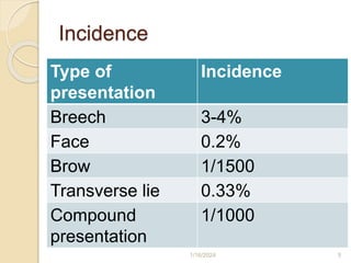
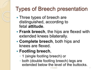
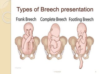
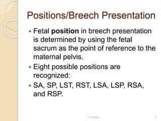
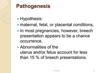
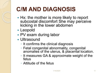
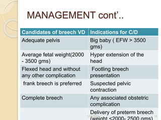
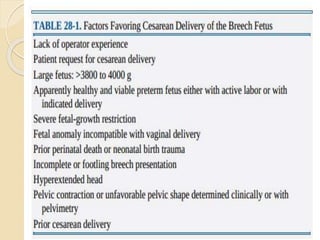
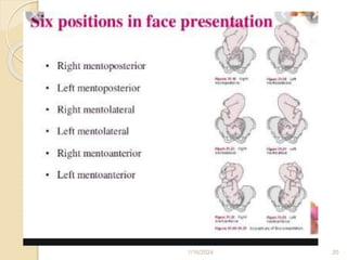
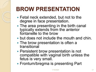
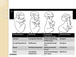
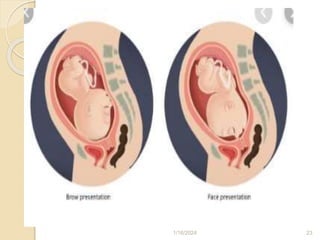
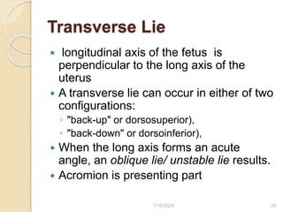
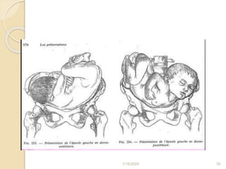
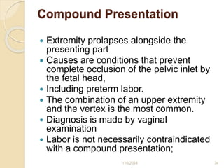
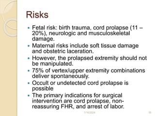
2. Definition
Any presentation other than the vertex
.
Incidence: up to 5% of term
pregnancies
Incidence is high with preterms
Pathophysiology
1/16/2024 2
6. Breech Presentation
The commonest malpresentation
Complicates 3–4% of all pregnancies,
Occurs when the fetal pelvis or lower
extremities engage the maternal pelvic
inlet.
Has higher perinatal mortality and
morbidty
Causes of breech presentation-
◦ prematurity, multiple pregnancy, fetal
malformation, uterine malformation,
hydramnious…
1/16/2024 6
7. Types of Breech presentation
Three types of breech are
distinguished, according to
fetal attitude.
Frank breech, the hips are flexed with
extended knees bilaterally.
Complete breech, both hips and
knees are flexed.
Footling breech,
◦ 1 (single footling breech) or
◦ both (double footling breech) legs are
extended below the level of the buttocks.
1/16/2024 7
9. Positions/Breech Presentation
Fetal position in breech presentation
is determined by using the fetal
sacrum as the point of reference to the
maternal pelvis.
Eight possible positions are
recognized:
SA, SP, LST, RST, LSA, LSP, RSA,
and RSP.
1/16/2024 9
10. Pathogenesis
Hypothesis:
maternal, fetal, or placental conditions,
In most pregnancies, however, breech
presentation appears to be a chance
occurrence.
Abnormalities of the
uterus and/or fetus account for less
than 15 % of breech presentations.
1/16/2024 10
11. C/M AND DIAGNOSIS
Hx: the mother is more likely to report
subcostal discomfort She may perceive
kicking in the lower abdomen
Leopold
PV exam during labor
Ultrasound
◦ It confirms the clinical diagnosis
◦ Fetal congenital abnormality, congenital
anomalies of the uterus, & placental location.
◦ It measures GA & approximate weight of the
fetus
◦ Attitude of the fetus
1/16/2024 11
12. MANAGEMENT OF BREECH
PRESENTATION AT TERM
Management options
(1) external cephalic version
(2) elective caesarean section
(3) trial of vaginal delivery
1/16/2024 12
13. MANAGEMENT cont’..
Candidates of breech VD Indications for C/D
Adequate pelvis Big baby ( EFW > 3500
gms)
Average fetal weight(2000
- 3500 gms)
Hyper extension of the
head
Flexed head and without
any other complication
Footling breech
presentation
frank breech is preferred Suspected pelvic
contraction
Complete breech Any associated obstetric
complication
Delivery of preterm breech
1/16/2024 13
15. Types OF Breech Vaginal
Delivery
Spontaneous Breech Delivery
Partial Breech Extraction
Total Breech Extraction
1/16/2024 15
16. Planned C/D vs planned VD
Term breech trail
WHO study
PREMODA
1/16/2024 16
17. Delivery Complications
Cord prolapse
Birth trauma
Trauma is more common with vaginal
births, but fetal trauma is also seen
with C/D.
Entrapment of after coming head
Genital tract laceration
1/16/2024 17
19. Face Presentation
The neck is hyperextended so that the
occiput is in contact with the fetal back.
The chin (mentum) is presenting part.
Most MP convert spontaneously to MA
even in late labor, some persist.
If not, the fetal brow (bregma) is pressed
against the maternal symphysis pubis.
This position precludes flexion of the fetal
head necessary to negotiate the birth
canal.
1/16/2024 19
21. BROW PRESENTATION
Fetal neck extended, but not to the
degree in face presentation.
The area presenting in the birth canal
typically extends from the anterior
fontanelle to the brow
but does not include the mouth and chin.
The brow presentation is often a
transitional
Persistent brow presentation is not
compatible with vaginal birth unless the
fetus is very small.
Frontum/bregma is presenting Part
1/16/2024 21
26. Diagnosis
Face and brow presentations are diagnosed
by vaginal examination.
A breech may be mistaken for a face
presentation.
Namely, the anus vs mouth, and the ischial
tuberosities vs malar prominences.
Landmarks indicating brow presentation are
the ability to palpate the forehead, saddle of
the nose, and orbits.
Face presentation is excluded because the
mouth and chin are not palpable.
incidence of 1 in 600, or 0.17 percent. Face
1/16/2024 26
27. Etiology
include conditions that favor extension or
prevent head flexion.
In exceptional instances, marked
enlargement of the neck or coils of cord
around the neck may cause extension.
Othes …..
◦ Fetal malformations, hydramnios, contracted
pelvis, High parity,
1/16/2024 27
29. Transverse Lie
longitudinal axis of the fetus is
perpendicular to the long axis of the
uterus
A transverse lie can occur in either of two
configurations:
◦ "back-up" or dorsosuperior),
◦ "back-down" or dorsoinferior),
When the long axis forms an acute
angle, an oblique lie/ unstable lie results.
Acromion is presenting part
1/16/2024 29
31. ETIOLOGY
1. Abdominal wall relaxation from high
parity,
2. Preterm fetus,
3. Placenta previa,
4. Abnormal uterine anatomy,
5. Hydramnios, and
6. Contracted pelvis.
1/16/2024 31
32. Complications
Increased operative delivery
Fetal injury
◦ Fracture of long bones
Cord prolapse(20%)
Obstructed labor and uterine rupture if
not managed properly
High perinatal mortality and morbidity
1/16/2024 32
34. Compound Presentation
Extremity prolapses alongside the
presenting part
Causes are conditions that prevent
complete occlusion of the pelvic inlet by
the fetal head,
Including preterm labor.
The combination of an upper extremity
and the vertex is the most common.
Diagnosis is made by vaginal
examination
Labor is not necessarily contraindicated
with a compound presentation;
1/16/2024 34
35. Risks
Fetal risk: birth trauma, cord prolapse (11 –
20%), neurologic and musculoskeletal
damage.
Maternal risks include soft tissue damage
and obstetric laceration.
However, the prolapsed extremity should not
be manipulated.
75% of vertex/upper extremity combinations
deliver spontaneously.
Occult or undetected cord prolapse is
possible
The primary indications for surgical
intervention are cord prolapse, non-
reassuring FHR, and arrest of labor.
1/16/2024 35
It is hypothesized that a normally proportioned active fetus in a normal volume of amniotic fluid adopts the cephalic presentation near term because this position is the best fit in the intrauterine space. If any of these variables are disrupted by underlying maternal, fetal, or placental conditions, then breech presentation becomes more likely. In most pregnancies, however, breech presentation appears to be a chance occurrence. Abnormalities of the uterus and/or fetus account for less than 15 percent of breech presentations.
Further neck extension leads to a face presentation or neck flexion results in vertex presentation in 50% of cases.
The area presenting in the birth canal typically extends from the anterior fontanelle to the brow (orbital ridge),
The accompanying extremity may retract as the major presenting part descends.
Occult or undetected cord prolapse is possible, and therefore, continuous electronic (one to-one Pinnard stethoscope) fetal heart rate monitoring is recommended.